View Featured Offers >>
12581
Glucose-6-Phosphate Dehydrogenase (G6PD) Activity Assay Kit
Cellular Assay Kits
Assay Kit

Glucose-6-Phosphate Dehydrogenase (G6PD) Activity Assay Kit #12581

Citations (0)
Figure 1. Schematic diagram of glucose-6-phosphate dehydrogenase (G6PD) assay. Glucose-6-phosphate (G6P) is oxidized by G6PD in the presence of NADP, which generates 6-phosphogluconolactone and NADPH. The generated NADPH is then amplified by the diaphorase-cycling system to produce highly fluorescent resorufin molecules.
Figure 2. Each assay component is individually omitted from the assay system and the resultant RFU is compared to that of a control test that contains all of the assay components.
Figure 3. The relationship between the protein concentration of lysates from untreated and G6PD inhibitor DHEA (0.5 mM) treated Jurkat cells and relative fluorescence (RFU) is shown. The G6PD inhibitor DHEA can effectively inhibit this chain reaction as shown in this figure.
To Purchase # 12581
Cat. # Size Qty. Price
12581S
1 Kit  (100 assays (96 well format))

Product Includes Quantity Solution Color Cap Color
Tris Assay Buffer 25 ml
G6PDH Substrate (40X) 250 µl Blue
G6PDH Cofactor (100X) 100 µl Yellow Yellow
NADP+ (100X) 100 µl White
G6PDH Developer (100X) 100 µl Blue Brown
G6PDH Positive Control (100X) 50 µl Brown
PathScan® Sandwich ELISA Lysis Buffer (1X) 7018 30 ml

Protocol

PRINT

View >Collapse >

#12581 Glucose-6-Phosphate Dehydrogenase (G6PD) Activity Assay Kit Protocol

A. Solutions and Reagents

NOTE: Prepare solutions with deionized/purified water or equivalent.

  1. Tris Assay Buffer: Bring to room temperature before use.
  2. G6PDH Substrate (40X): Reconstitute lyophilized component with 250µl dH2O.
  3. G6PDH Developer (100X): Reconstitute lyophilized component with 100µl dH2O.
  4. G6PDH Positive Control (100X): Reconstitute lyophilized component with 50µl dH2O.
  5. NADP+ (100X): Reconstitute lyophilized component with 100µl dH2O.
  6. G6PDH Cofactor (100X): Reconstitute lyophilized component with 100µl dH2O.
  7. PathScan® Sandwich ELISA Lysis Buffer (1X) (#7018): Thaw and keep on ice.

Working Solution Preparation

  1. Calculate the number of tests: number of samples + positive controls.
  2. Dilute Total Detection Solution for the calculated number of tests.
    1. 70 µl/well is recommended for a 96-well plate.
    2. 20 µl/well is recommended for a 384-well plate.

    Example: Number of tests = 10 samples (n=3) + 3 positive controls

    1. 96-Well Plate: 70 µl/well x 33 samples = 2310 µl + 10% = ~2500 µl
    2. 384-Well Plate: 20 µl/well x 33 samples = 660 µl + 10% = ~750 µl
  3. Make Negative Control Solution (See Table).

    Note: DO NOT add G6PD substrate to the Negative Control Solution.

  4. Make Positive Control Solution (See Table).

    Table 1: Example of calculation for 10 samples in a 96-well plate (All triplicates)

    Total Detection Solution (µl) Negative Control Solution (µl) Positive Control Solution (µl)
    G6PD Substrate (40X) 62.5 0 0
    G6PD Developer (100X) 25 3.3 0
    G6PDH Cofactor (100X) 25 3.3 0
    G6PD Positive Control (100X) 0 0 1
    NADP+ (100X) 25 3.3 0
    Tris Assay Buffer 2362.5 320 99
    Total (µl) (with ~10% extra volume) 2500 (30 samples + 3 positive controls) 330 100

    Table 2: Example calculation for 10 samples in a 384-plate (All triplicates)

    Total Detection Solution (µl) Negative Control Solution (µl) Positive Control Solution (µl)
    G6PD Substrate (40X) 19 0 0
    G6PD Developer (100X) 7.5 1 0
    G6PDH Cofactor (100X) 7.5 1 0
    G6PD Positive Control (100X) 0 0 1
    NADP+ (100X) 7.5 1 0
    Tris Assay Buffer 708.5 97 99
    Total (µl) (with ~10% extra volume) 750 (30 samples + 3 positive controls) 100 100

B. Preparing Cell Lysates

For adherent cells

  1. Grow target cells to 80–90% confluence and aspirate media.
  2. Add fresh media containing regulator for desired time.
  3. Aspirate media and rinse cells once with ice-cold 1X PBS (#9872).
  4. Aspirate PBS and add 0.5 ml ice-cold 1X Cell Lysis Buffer plus 1 mM PMSF to each plate (10 cm diameter).
  5. Incubate the plate on ice for 5 min.
  6. Scrape cells off the plate and transfer to an appropriate tube. Keep on ice.
  7. Sonicate lysates on ice.
  8. Microcentrifuge for 10 min (x14,000 rpm) at 4°C and transfer the supernatant to a new tube. The supernatant is the cell lysate.
  9. Store at −80°C in single-use aliquots.

For suspension cells

  1. Remove media by low speed centrifugation (~1200 rpm) when the culture reaches 0.5–1.0 x 10⁶ viable cells/ml. Treat cells by adding fresh media containing regulator for desired time.
  2. Collect cells by low speed centrifugation (~1200 rpm) and wash once with 5–10 ml ice-cold 1X PBS.
  3. Cells harvested from 50 ml of growth media can be lysed in 2.0 ml of 1X Cell Lysis Buffer plus 1 mM PMSF.
  4. Sonicate lysates on ice.
  5. Microcentrifuge for 10 min (x14,000 rpm) at 4°C and transfer the supernatant to a new tube. The supernatant is the cell lysate. Store at −80°C in single-use aliquots.

C. Test Procedure

  1. Dilute sample in G6PD Assay Buffer (1X) to desired concentration.
  2. Add 70 µl of Total Detection Solution and 30 µl of sample in a black 96-well plate. Mix well. (Alternatively, add 20 µl of Total Detection Solution and 10 µl of sample for a 384-well plate.)
  3. Negative control: Add 100 µl of Negative Control Solution to three wells. (Add 30 µl of Negative Control Solution for a 384-well plate.)
  4. Positive control: Add 70 µl of Total Detection Solution and 30 µl Positive Control to three wells. Mix well. (Add 20 µl of Total Detection Solution and 10 µl of Positive Control for a 384-well plate.)
  5. Incubate at 37°C for 15-30 min.
  6. Read RFU on a plate reader with excitation around 540 nm and emission around 590 nm.

posted June 2020

Protocol Id: 2051

Product Description

The Glucose-6-Phosphate Dehydrogenase (G6PD) Activity Assay Kit contains the necessary reagents for rapid, sensitive, and simple detection of G6PD activity in various samples. In the assay, glucose-6-phosphate (G6P), in the presence of NADP, is oxidized by G6PD to generate 6-phosphogluconolactone and NADPH. The generated NADPH is then amplified by the diaphorase-cycling system to produce highly fluorescent resorufin molecules (see Figure 1). The relative fluorescent units (RFU) can then be determined using a plate reader with excitation about 540 nm and emission about 590 nm. The magnitude of RFU is proportional to G6PD activity in the sample.

Specificity / Sensitivity

The Glucose-6-Phosphate Dehydrogenase (G6PD) Activity Assay Kit detects sample G6PD activity. The presence of NADH and NADPH may interfere with the assay.

Species Reactivity:

All Species Expected

Background

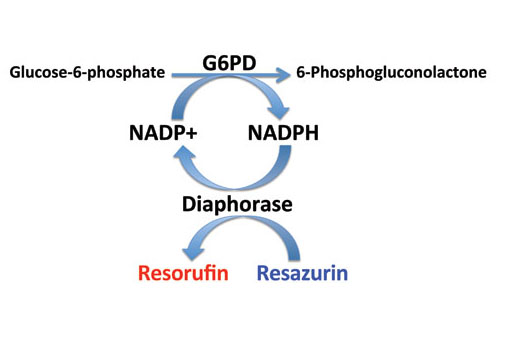
Glucose-6-phosphate dehydrogenase (G6PD) catalyses the first, and rate-limiting, step of the pentose phosphate pathway (1). The NADPH generated from this reaction is essential to protect cells from oxidative stress (1). Research studies have shown that p53 interacts with G6PD and inhibits its activity, therefore suppressing glucose consumption through the pentose phosphate pathway (2). In cancer cells with p53 mutations, the increased glucose consumption is directed towards increased biosynthesis, which is critical for cancer cell proliferation (2).

Limited Uses

Except as otherwise expressly agreed in a writing signed by a legally authorized representative of CST, the following terms apply to Products provided by CST, its affiliates or its distributors. Any Customer's terms and conditions that are in addition to, or different from, those contained herein, unless separately accepted in writing by a legally authorized representative of CST, are rejected and are of no force or effect.

Products are labeled with For Research Use Only or a similar labeling statement and have not been approved, cleared, or licensed by the FDA or other regulatory foreign or domestic entity, for any purpose. Customer shall not use any Product for any diagnostic or therapeutic purpose, or otherwise in any manner that conflicts with its labeling statement. Products sold or licensed by CST are provided for Customer as the end-user and solely for research and development uses. Any use of Product for diagnostic, prophylactic or therapeutic purposes, or any purchase of Product for resale (alone or as a component) or other commercial purpose, requires a separate license from CST. Customer shall (a) not sell, license, loan, donate or otherwise transfer or make available any Product to any third party, whether alone or in combination with other materials, or use the Products to manufacture any commercial products, (b) not copy, modify, reverse engineer, decompile, disassemble or otherwise attempt to discover the underlying structure or technology of the Products, or use the Products for the purpose of developing any products or services that would compete with CST products or services, (c) not alter or remove from the Products any trademarks, trade names, logos, patent or copyright notices or markings, (d) use the Products solely in accordance with CST Product Terms of Sale and any applicable documentation, and (e) comply with any license, terms of service or similar agreement with respect to any third party products or services used by Customer in connection with the Products.

For Research Use Only. Not for Use in Diagnostic Procedures.
Cell Signaling Technology is a trademark of Cell Signaling Technology, Inc.
PathScan is a registered trademark of Cell Signaling Technology, Inc.
All other trademarks are the property of their respective owners. Visit our Trademark Information page.