Product Information
| Molecular Formula | Peptide substrate, Biotin-FGFR-3 (Tyr724): 1,789 Daltons. GST-EphB4 Kinase: 77 kDa. |
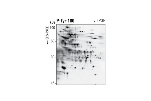
The Eph receptors are the largest known family of receptor tyrosine kinases (RTKs). They can be divided into two groups based on sequence similarity and on their preference for a subset of ligands: EphA receptors bind to a glycosylphosphatidylinositol-anchored ephrin A ligand; EphB receptors bind to ephrin B proteins that have a transmembrane and cytoplasmic domain (1,2). Research studies have shown that Eph receptors and ligands may be involved in many diseases including cancer (3). Both ephrin A and B ligands have dual functions. As RTK ligands, ephrins stimulate the kinase activity of Eph receptors and activate signaling pathways in receptor-expressing cells. The ephrin extracellular domain is sufficient for this function as long as it is clustered (4). The second function of ephrins has been described as "reverse signaling", whereby the cytoplasmic domain becomes tyrosine phosphorylated, allowing interactions with other proteins that may activate signaling pathways in the ligand-expressing cells (5). Various stimuli can induce tyrosine phosphorylation of ephrin B, including binding to EphB receptors, activation of Src kinase, and stimulation by PDGF and FGF (6). Tyr324 and Tyr327 have been identified as major phosphorylation sites of ephrin B1 in vivo (7).
Except as otherwise expressly agreed in a writing signed by a legally authorized representative of CST, the following terms apply to Products provided by CST, its affiliates or its distributors. Any Customer's terms and conditions that are in addition to, or different from, those contained herein, unless separately accepted in writing by a legally authorized representative of CST, are rejected and are of no force or effect.
Products are labeled with For Research Use Only or a similar labeling statement and have not been approved, cleared, or licensed by the FDA or other regulatory foreign or domestic entity, for any purpose. Customer shall not use any Product for any diagnostic or therapeutic purpose, or otherwise in any manner that conflicts with its labeling statement. Products sold or licensed by CST are provided for Customer as the end-user and solely for research and development uses. Any use of Product for diagnostic, prophylactic or therapeutic purposes, or any purchase of Product for resale (alone or as a component) or other commercial purpose, requires a separate license from CST. Customer shall (a) not sell, license, loan, donate or otherwise transfer or make available any Product to any third party, whether alone or in combination with other materials, or use the Products to manufacture any commercial products, (b) not copy, modify, reverse engineer, decompile, disassemble or otherwise attempt to discover the underlying structure or technology of the Products, or use the Products for the purpose of developing any products or services that would compete with CST products or services, (c) not alter or remove from the Products any trademarks, trade names, logos, patent or copyright notices or markings, (d) use the Products solely in accordance with CST Product Terms of Sale and any applicable documentation, and (e) comply with any license, terms of service or similar agreement with respect to any third party products or services used by Customer in connection with the Products.