View Featured Offers >>
50929
PathScan® Phospho-Histone H2A.X (Ser139) Sandwich ELISA Kit
ELISA Kits
ELISA Kit

PathScan® Phospho-Histone H2A.X (Ser139) Sandwich ELISA Kit #50929

Citations (0)
Figure 1: Phospho-Histone H2A.X (Ser139) protein from untreated (-) and UV-treated (+) 293 cells is detected by PathScan® Phospho-Histone H2A.X (Ser139) Sandwich ELISA kit. A lower signal of phospho-Histone H2A.X protein is seen in the untreated lysate. However, similar levels of total Histone H2A.X protein from both untreated and treated lysates are shown by western analysis. The absorbance readings at 450 nm are shown in the top figure, while the corresponding western blot using Phospho-Histone H2A.X (Ser139) (20E3) Rabbit mAb #9718 is shown in the bottom figure.
Figure 2: The relationship between protein concentration of lysates from untreated and UV-treated 293 cells and the absorbance at 450 nm as detected by the PathScan® Phospho-Histone H2A.X Ser139 Sandwich ELISA Kit is shown.

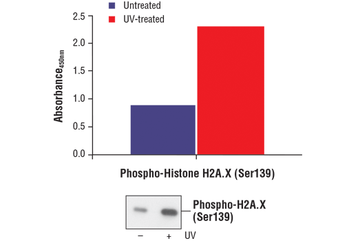
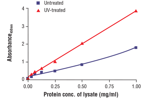
Unstarved 293 cells (80-90% confluence) were treated with 100 mJ/cm2 UV with 1 hr recovery at 37ºC, and then lysed with Cell Lysis Buffer (#9803).

To Purchase # 50929
Cat. # Size Qty. Price
50929C
1 Kit  (96 assays)

Important Ordering Details

Custom Ordering Details:

When ordering five or more kits, please contact us for processing time and pricing.

Looking for this ELISA kit in a 384-well format? Inquire for availability, processing time, and pricing.

Supporting Data

REACTIVITY H

Application Key:

  • WB-Western Blot
  • IP-Immunoprecipitation
  • IHC-Immunohistochemistry
  • ChIP-Chromatin Immunoprecipitation
  • C&R-CUT&RUN
  • C&T-CUT&Tag
  • DB-Dot Blot
  • eCLIP-eCLIP
  • IF-Immunofluorescence
  • F-Flow Cytometry

Species Cross-Reactivity Key:

  • H-Human
  • M-Mouse
  • R-Rat
  • Hm-Hamster
  • Mk-Monkey
  • Vir-Virus
  • Mi-Mink
  • C-Chicken
  • Dm-D. melanogaster
  • X-Xenopus
  • Z-Zebrafish
  • B-Bovine
  • Dg-Dog
  • Pg-Pig
  • Sc-S. cerevisiae
  • Ce-C. elegans
  • Hr-Horse
  • GP-Guinea Pig
  • Rab-Rabbit
  • All-All Species Expected
Product Includes Volume Solution Color
Histone H2A.X Rabbit mAb Coated Microwells 96 tests
P-Histone H2A.X (Ser139) Mouse Detection mAb 1 ea Green (Lyophilized)
Anti-mouse IgG, HRP-linked Antibody (ELISA Formulated) 1 ea Red (Lyophilized)
Detection Antibody Diluent 11 ml Green
HRP Diluent 11 ml Red
TMB Substrate 7004 11 ml
STOP Solution 7002 11 ml
Sealing Tape 2 ea
ELISA Wash Buffer (20X) 9801 25 ml
ELISA Sample Diluent 25 ml Blue
Cell Lysis Buffer (10X) 9803 15 ml

Product Description

The PathScan® Phospho-Histone H2A.X (Ser139) Sandwich ELISA Kit is a solid phase sandwich enzyme-linked immunosorbent assay (ELISA) that detects endogenous levels of histone H2A.X protein phosphorylated at Ser139. A histone H2A.X rabbit antibody has been coated onto the microwells. After incubation with cell lysates, both phospho- and non-phospho-histone H2A.X proteins are captured by the coated antibody. Following extensive washing, a phospho-histone H2A.X (Ser139) mouse antibody is added to detect the captured phospho-histone H2A.X protein. Anti-mouse IgG, HRP-linked Antibody is then used to recognize the bound detection antibody. HRP substrate, TMB, is added to develop color. The magnitude of the absorbance for this developed color is proportional to the quantity of histone H2A.X phosphorylated at Ser139.

*Antibodies in this kit are custom formulations specific to kit.

Protocol

PRINT

View >Collapse >

ELISA Colorimetric (Lyophilized)

A. Solutions and Reagents

NOTE: Prepare solutions with purified water.

  1. Microwell strips: Bring all to room temperature before use.
  2. Detection Antibody: Supplied lyophilized as a green colored cake or powder. Add 1.0 ml of Detection Antibody Diluent (green solution) to yield a concentrated stock solution. Incubate at room temperature for 5 min with occasional gentle mixing to fully reconstitute. To make the final working solution, add the full 1.0 ml volume of reconstituted Detection Antibody to 10.0 ml of Detection Antibody Diluent in a clean tube and gently mix. Unused working solution may be stored for 4 weeks at 4°C.
  3. HRP-Linked Antibody*: Supplied lyophilized as a red colored cake or powder. Add 1.0 ml of HRP Diluent (red solution) to yield a concentrated stock solution. Incubate at room temperature for 5 min with occasional gentle mixing to fully reconstitute. To make the final working solution, add the full 1.0 ml volume of reconstituted HRP-Linked Antibody to 10.0 ml of HRP Diluent in a clean tube and gently mix. Unused working solution may be stored for 4 weeks at 4°C.
  4. Detection Antibody Diluent: Green colored diluent for reconstitution and dilution of the detection antibody (11 ml provided).
  5. HRP Diluent: Red colored diluent for reconstitution and dilution of the HRP‑Linked Antibody (11 ml provided).
  6. Sample Diluent: Blue colored diluent provided for dilution of cell lysates.
  7. 1X Wash Buffer: Prepare by diluting 20X Wash Buffer (included in each PathScan® Sandwich ELISA Kit) in purified water.
  8. Cell Lysis Buffer: 10X Cell Lysis Buffer #9803: This buffer can be stored at 4°C for short-term use (1–2 weeks). Recommended: Add 1 mM phenylmethylsulfonyl fluoride (PMSF) immediately before use.
  9. TMB Substrate (#7004).
  10. STOP Solution (#7002).

*NOTE: Some PathScan® ELISA Kits may include HRP-Linked Streptavidin in place of HRP-Linked Antibody.

B. Preparing Cell Lysates

For adherent cells.

  1. Aspirate media when the culture reaches 80–90% confluence. Treat cells by adding fresh media containing regulator for desired time.
  2. Remove media and rinse cells once with ice-cold 1X PBS.
  3. Remove PBS and add 0.5 ml ice-cold 1X Cell Lysis Buffer plus 1 mM PMSF to each plate (10 cm diameter) and incubate the plate on ice for 5 min.
  4. Scrape cells off the plate and transfer to an appropriate tube. Keep on ice.
  5. Sonicate lysates on ice.
  6. Microcentrifuge for 10 min (x14,000 rpm) at 4°C and transfer the supernatant to a new tube. The supernatant is the cell lysate. Store at −80°C in single-use aliquots.

For suspension cells

  1. Remove media by low speed centrifugation (~1200 rpm) when the culture reaches 0.5–1.0 x 10⁶ viable cells/ml. Treat cells by adding fresh media containing regulator for desired time.
  2. Collect cells by low speed centrifugation (~1200 rpm) and wash once with 5–10 ml ice-cold 1X PBS.
  3. Cells harvested from 50 ml of growth media can be lysed in 2.0 ml of 1X Cell Lysis Buffer plus 1 mM PMSF.
  4. Sonicate lysates on ice.
  5. Microcentrifuge for 10 min (x14,000 rpm) at 4°C and transfer the supernatant to a new tube. The supernatant is the cell lysate. Store at −80°C in single-use aliquots.

C. Test Procedure

  1. After the microwell strips have reached room temperature, break off the required number of microwells. Place the microwells in the strip holder. Unused microwells must be resealed and stored at 4°C immediately.
  2. Cell lysates can be undiluted or diluted with Sample Diluent (supplied in each PathScan® Sandwich ELISA Kit, blue color). Individual datasheets for each kit provide a sensitivity curve that serves as a reference for selection of an appropriate starting lysate concentration. The sensitivity curve shows typical kit assay results across a range of lysate concentration points.
  3. Add 100 µl of each undiluted or diluted cell lysate to the appropriate well. Seal with tape and press firmly onto top of microwells. Incubate the plate for 2 hr at 37°C. Alternatively, the plate can be incubated overnight at 4°C.
  4. Gently remove the tape and wash wells:
    1. Discard plate contents into a receptacle.
    2. Wash 4 times with 1X Wash Buffer, 200 µl each time for each well.
    3. For each wash, strike plates on fresh towels hard enough to remove the residual solution in each well, but do not allow wells to completely dry at any time.
    4. Clean the underside of all wells with a lint-free tissue.
  5. Add 100 µl of reconstituted Detection Antibody (green color) to each well (refer to Section A, Step 2). Seal with tape and incubate the plate at 37°C for 1 hr.
  6. Repeat wash procedure (Section C, Step 4).
  7. Add 100 µl of reconstituted HRP-Linked secondary antibody (red color) to each well (refer to Section A, Step 3). Seal with tape and incubate the plate for 30 min at 37°C.
  8. Repeat wash procedure (Section C, Step 4).
  9. Add 100 µl of TMB Substrate to each well. Seal with tape and incubate the plate for 10 min at 37°C or 30 min at 25°C.
  10. Add 100 µl of STOP Solution to each well. Shake gently for a few seconds.

NOTE: Initial color of positive reaction is blue, which changes to yellow upon addition of STOP Solution.

  1. Read results.
    1. Visual Determination: Read within 30 min after adding STOP Solution.
    2. Spectrophotometric Determination: Wipe underside of wells with a lint-free tissue. Read absorbance at 450 nm within 30 min after adding STOP Solution.

posted November 2013

Protocol Id: 204

Specificity / Sensitivity

PathScan® Phospho-Histone H2A.X (Ser139) Sandwich ELISA Kit detects endogenous levels of histone H2A.X protein phosphorylated at Ser139 in human cells, as shown in Figure 1. The kit sensitivity is shown in Figure 2. This kit detects proteins from the indicated species, as determined through in-house testing, but may also detect homologous proteins from other species.

Background

Histone H2A.X is a variant histone that represents approximately 10% of the total H2A histone proteins in normal human fibroblasts (1). H2A.X is required for checkpoint-mediated cell cycle arrest and DNA repair following double-stranded DNA breaks (1). DNA damage, caused by ionizing radiation, UV-light, or radiomimetic agents, results in rapid phosphorylation of H2A.X at Ser139 by PI3K-like kinases, including ATM, ATR, and DNA-PK (2,3). Within minutes following DNA damage, H2A.X is phosphorylated at Ser139 at sites of DNA damage (4). This very early event in the DNA-damage response is required for recruitment of a multitude of DNA-damage response proteins, including MDC1, NBS1, RAD50, MRE11, 53BP1, and BRCA1 (1). In addition to its role in DNA-damage repair, H2A.X is required for DNA fragmentation during apoptosis and is phosphorylated by various kinases in response to apoptotic signals. H2A.X is phosphorylated at Ser139 by DNA-PK in response to cell death receptor activation, c-Jun N-terminal Kinase (JNK1) in response to UV-A irradiation, and p38 MAPK in response to serum starvation (5-8). H2A.X is constitutively phosphorylated on Tyr142 in undamaged cells by WSTF (Williams-Beuren syndrome transcription factor) (9,10). Upon DNA damage, and concurrent with phosphorylation of Ser139, Tyr142 is dephosphorylated at sites of DNA damage by recruited EYA1 and EYA3 phosphatases (9). While phosphorylation at Ser139 facilitates the recruitment of DNA repair proteins and apoptotic proteins to sites of DNA damage, phosphorylation at Tyr142 appears to determine which set of proteins are recruited. Phosphorylation of H2A.X at Tyr142 inhibits the recruitment of DNA repair proteins and promotes binding of pro-apoptotic factors such as JNK1 (9). Mouse embryonic fibroblasts expressing only mutant H2A.X Y142F, which favors recruitment of DNA repair proteins over apoptotic proteins, show a reduced apoptotic response to ionizing radiation (9). Thus, it appears that the balance of H2A.X Tyr142 phosphorylation and dephosphorylation provides a switch mechanism to determine cell fate after DNA damage.

Limited Uses

Except as otherwise expressly agreed in a writing signed by a legally authorized representative of CST, the following terms apply to Products provided by CST, its affiliates or its distributors. Any Customer's terms and conditions that are in addition to, or different from, those contained herein, unless separately accepted in writing by a legally authorized representative of CST, are rejected and are of no force or effect.

Products are labeled with For Research Use Only or a similar labeling statement and have not been approved, cleared, or licensed by the FDA or other regulatory foreign or domestic entity, for any purpose. Customer shall not use any Product for any diagnostic or therapeutic purpose, or otherwise in any manner that conflicts with its labeling statement. Products sold or licensed by CST are provided for Customer as the end-user and solely for research and development uses. Any use of Product for diagnostic, prophylactic or therapeutic purposes, or any purchase of Product for resale (alone or as a component) or other commercial purpose, requires a separate license from CST. Customer shall (a) not sell, license, loan, donate or otherwise transfer or make available any Product to any third party, whether alone or in combination with other materials, or use the Products to manufacture any commercial products, (b) not copy, modify, reverse engineer, decompile, disassemble or otherwise attempt to discover the underlying structure or technology of the Products, or use the Products for the purpose of developing any products or services that would compete with CST products or services, (c) not alter or remove from the Products any trademarks, trade names, logos, patent or copyright notices or markings, (d) use the Products solely in accordance with CST Product Terms of Sale and any applicable documentation, and (e) comply with any license, terms of service or similar agreement with respect to any third party products or services used by Customer in connection with the Products.

For Research Use Only. Not for Use in Diagnostic Procedures.
Cell Signaling Technology is a trademark of Cell Signaling Technology, Inc.
PathScan is a registered trademark of Cell Signaling Technology, Inc.
All other trademarks are the property of their respective owners. Visit our Trademark Information page.