| Cat. # | Size | Qty. | Price |
|---|---|---|---|
| 7936C | 1 Kit (96 assays) |
|
When ordering five or more kits, please contact us for processing time and pricing.
Looking for this ELISA kit in a 384-well format? Inquire for availability, processing time, and pricing.
| REACTIVITY | H |
| Product Includes | Volume | Solution Color | |||
|---|---|---|---|---|---|
| LAT Rabbit mAb Coated Microwells | 96 tests | ||||
| Phospho-LAT (Tyr220) Rabbit Detection mAb | 1 ea | Red (Lyophilized) | |||
| HRP Diluent | 5.5 ml | Red | |||
| TMB Substrate 7004 | 11 ml | ||||
| STOP Solution 7002 | 11 ml | ||||
| Sealing Tape | 2 ea | ||||
| ELISA Wash Buffer (20X) 9801 | 25 ml | ||||
| Cell Lysis Buffer (10X) 9803 | 15 ml |
Product Information
The rapid protocol (RP) PathScan® RP Phospho-LAT (Tyr220) Sandwich ELISA Kit is a solid phase sandwich enzyme-linked immunosorbent assay (ELISA) that detects endogenous levels of phospho-LAT (Tyr220) in a reduced assay time of 1.5 hours. Incubation of cell lysate and detection antibody on the capture antibody coated microwell plate forms a sandwich with LAT protein phosphorylated at Tyr220 in a single step. The plate is then extensively washed and TMB reagent is added for signal development. The magnitude of absorbance for the developed color is proportional to the quantity of LAT protein phosphorylated at Tyr220. Learn more about all of your ELISA kit options here.
*Antibodies in this kit are custom formulations specific to kit.
NOTE: This protocol is for PathScan® kits that use an HRP directly conjugated to the detection antibody (Rapid Protocol), rather than a 2-step method where the detection antibody and a secondary-HRP are added sequentially.
NOTE: Prepare solutions with deionized/purified water or equivalent.
For adherent cells
For suspension cells
NOTE: Equilibrate all materials and prepared reagents to room temperature prior to running the assay.
NOTE: Initial color of positive reaction is blue, which changes to yellow upon addition of STOP Solution.
created July 2020
Protocol Id: 2144
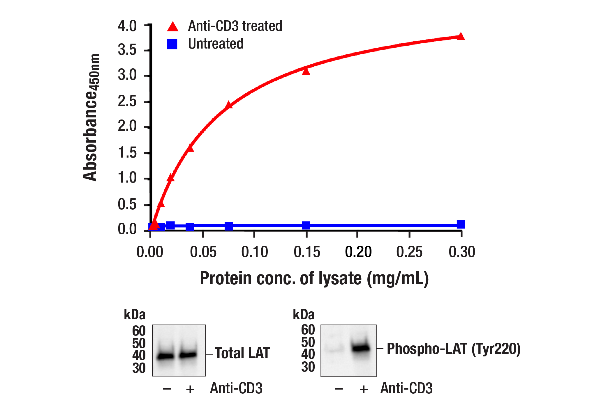
The PathScan® RP Phospho-LAT (Tyr220) Sandwich ELISA Kit #7936 detects endogenous levels of phospho-LAT when phosphorylated at Tyr191. The kit sensitivity is shown in Figure 1. This kit detects proteins from the indicated species, as determined through in-house testing, but may also detect homologous proteins from other species.
LAT, a transmembrane adaptor protein expressed in T, NK, and mast cells, is an important mediator for T cell receptor (TCR) signaling (1). Upon TCR engagement, activated Zap-70 phosphorylates LAT at multiple conserved tyrosine residues within SH2 binding motifs, exposing these motifs as the docking sites for downstream signaling targets (2,3). The phosphorylation of LAT at Tyr171 and Tyr220 enables the binding of Grb2, Gads/SLP-76, PLCγ1, and PI3 kinase through their SH2 domain and translocates them to the membrane. This process eventually leads to activation of the corresponding signaling pathways (1-4).
Explore pathways related to this product.
STRING - Known and Predicted Protein-Protein Interactions.
Except as otherwise expressly agreed in a writing signed by a legally authorized representative of CST, the following terms apply to Products provided by CST, its affiliates or its distributors. Any Customer's terms and conditions that are in addition to, or different from, those contained herein, unless separately accepted in writing by a legally authorized representative of CST, are rejected and are of no force or effect.
Products are labeled with For Research Use Only or a similar labeling statement and have not been approved, cleared, or licensed by the FDA or other regulatory foreign or domestic entity, for any purpose. Customer shall not use any Product for any diagnostic or therapeutic purpose, or otherwise in any manner that conflicts with its labeling statement. Products sold or licensed by CST are provided for Customer as the end-user and solely for research and development uses. Any use of Product for diagnostic, prophylactic or therapeutic purposes, or any purchase of Product for resale (alone or as a component) or other commercial purpose, requires a separate license from CST. Customer shall (a) not sell, license, loan, donate or otherwise transfer or make available any Product to any third party, whether alone or in combination with other materials, or use the Products to manufacture any commercial products, (b) not copy, modify, reverse engineer, decompile, disassemble or otherwise attempt to discover the underlying structure or technology of the Products, or use the Products for the purpose of developing any products or services that would compete with CST products or services, (c) not alter or remove from the Products any trademarks, trade names, logos, patent or copyright notices or markings, (d) use the Products solely in accordance with CST Product Terms of Sale and any applicable documentation, and (e) comply with any license, terms of service or similar agreement with respect to any third party products or services used by Customer in connection with the Products.