Revision 13
#88918
Store at +4C
PathScan® RP Phospho-RIP (Ser166) Chemiluminescent Sandwich ELISA Kit
1 Kit
(96 assays)
Species Cross Reactivity:
H M
UniProt ID:
#Q13546
Entrez-Gene Id:
#8737
877-616-CELL (2355)
877-678-TECH (8324)
3 Trask Lane | Danvers | Massachusetts | 01923 | USA
For Research Use Only. Not for Use in Diagnostic Procedures.
| Product Includes | Product # | Quantity | Color | Storage Temp |
|---|---|---|---|---|
| RIP Rabbit Monoclonal Antibody Coated Microwells | 94588 | 96 tests | +4C | |
| Phospho-RIP (Ser166) Rabbit Detection Monoclonal Antibody | 24352 | 1 ea | Red (Lyophilized) | +4C |
| HRP Diluent | 13515 | 5.5 ml | Red | +4C |
| Luminol/Enhancer Solution | 84850 | 3 ml | RT | |
| Stable Peroxide Buffer | 42552 | 3 ml | RT | |
| Sealing Tape | 54503 | 2 ea | +4C | |
| ELISA Wash Buffer (20X) | 9801 | 25 ml | +4C | |
| Cell Lysis Buffer (10X) | 9803 | 15 ml | -20C |
Kit contents scale proportionally with size, except sealing tape.
Example: The V1 kit contains 5X the listed quantities above, but will exclude the sealing tape.
For the “C” and “V” kits, the supplied 96-well strip plate consists of twelve 8-well strips in a support frame. This enables custom plate configurations.
Description
The rapid protocol (RP) PathScan® RP Phospho-RIP (Ser166) Chemiluminescent Sandwich ELISA Kit is a solid phase sandwich enzyme-linked immunosorbent assay (ELISA) that detects endogenous levels of RIP protein phosphorylated at Ser166 in a reduced assay time of 1.5 hours. This chemiluminescent ELISA, which is offered in low volume microplates, shows increased signal and sensitivity while using a smaller sample size. Incubation of cell lysates and detection antibody on the coated microwell plate forms a sandwich with phospho-RIP (Ser166) protein in a single step. The plate is then extensively washed and chemiluminescent reagent is added for signal development. The magnitude of light emission, measured in relative light units (RLU), is proportional to the quantity of RIP protein phosphorylated at Ser166. Learn more about all of your ELISA kit options here.
*Antibodies in this kit are custom formulations specific to kit.
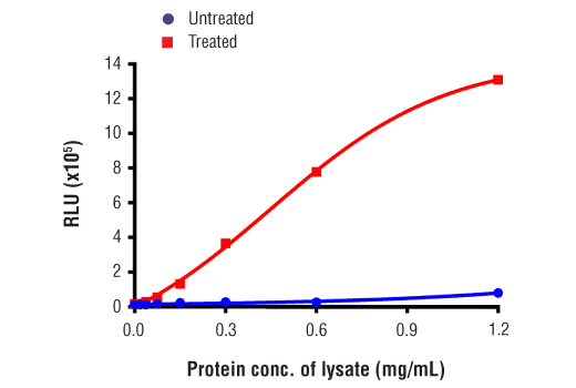
Specificity/Sensitivity
Background
Necroptosis, a regulated pathway for necrotic cell death, is triggered by a number of inflammatory signals including cytokines in the tumor necrosis factor (TNF) family, pathogen sensors such as toll-like receptors (TLRs), and ischemic injury (9,10). The process is negatively regulated by caspases and is initiated through a complex containing the RIP and RIP3 kinases, typically referred to as the necrosome. Necroptosis is inhibited by a small molecule inhibitor of RIP, necrostatin-1 (Nec-1) (11). Research studies show that necroptosis contributes to a number of pathological conditions, and Nec-1 has been shown to provide neuroprotection in models such as ischemic brain injury (12). RIP is phosphorylated at several sites within the kinase domain that are sensitive to Nec-1, including Ser14, Ser15, Ser161, and Ser166 (13).
Background References
- Meylan, E. and Tschopp, J. (2005) Trends Biochem Sci 30, 151-9.
- Hsu, H. et al. (1996) Immunity 4, 387-96.
- Stanger, B.Z. et al. (1995) Cell 81, 513-23.
- Ting, A.T. et al. (1996) EMBO J 15, 6189-96.
- Kelliher, M.A. et al. (1998) Immunity 8, 297-303.
- Devin, A. et al. (2000) Immunity 12, 419-29.
- Zhang, S.Q. et al. (2000) Immunity 12, 301-11.
- Lin, Y. et al. (1999) Genes Dev 13, 2514-26.
- Christofferson, D.E. and Yuan, J. (2010) Curr Opin Cell Biol 22, 263-8.
- Kaczmarek, A. et al. (2013) Immunity 38, 209-23.
- Degterev, A. et al. (2008) Nat Chem Biol 4, 313-21.
- Degterev, A. et al. (2005) Nat Chem Biol 1, 112-9.
- Ofengeim, D. and Yuan, J. (2013) Nat Rev Mol Cell Biol 14, 727-36.
Trademarks and Patents
Cell Signaling Technology is a trademark of Cell Signaling Technology, Inc.
PathScan is a registered trademark of Cell Signaling Technology, Inc.
All other trademarks are the property of their respective owners. Visit cellsignal.com/trademarks for more information.
Limited Uses
Except as otherwise expressly agreed in a writing signed by a legally authorized representative of CST, the following terms apply to Products provided by CST, its affiliates or its distributors. Any Customer's terms and conditions that are in addition to, or different from, those contained herein, unless separately accepted in writing by a legally authorized representative of CST, are rejected and are of no force or effect.
Products are labeled with For Research Use Only or a similar labeling statement and have not been approved, cleared, or licensed by the FDA or other regulatory foreign or domestic entity, for any purpose. Customer shall not use any Product for any diagnostic or therapeutic purpose, or otherwise in any manner that conflicts with its labeling statement. Products sold or licensed by CST are provided for Customer as the end-user and solely for research and development uses. Any use of Product for diagnostic, prophylactic or therapeutic purposes, or any purchase of Product for resale (alone or as a component) or other commercial purpose, requires a separate license from CST. Customer shall (a) not sell, license, loan, donate or otherwise transfer or make available any Product to any third party, whether alone or in combination with other materials, or use the Products to manufacture any commercial products, (b) not copy, modify, reverse engineer, decompile, disassemble or otherwise attempt to discover the underlying structure or technology of the Products, or use the Products for the purpose of developing any products or services that would compete with CST products or services, (c) not alter or remove from the Products any trademarks, trade names, logos, patent or copyright notices or markings, (d) use the Products solely in accordance with CST Product Terms of Sale and any applicable documentation, and (e) comply with any license, terms of service or similar agreement with respect to any third party products or services used by Customer in connection with the Products.
Revision 13