| Cat. # | Size | Qty. | Price |
|---|---|---|---|
| 7038C | 1 Kit (96 assays) |
|
When ordering five or more kits, please contact us for processing time and pricing.
Looking for this ELISA kit in a 384-well format? Inquire for availability, processing time, and pricing.
| REACTIVITY | H M |
| Product Includes | Volume | Solution Color | |||
|---|---|---|---|---|---|
| p70 S6 Kinase Rabbit Antibody Coated Microwells | 96 tests | ||||
| p70 S6 Kinase Rabbit Detection mAb (Biotinylated) | 1 ea | Green (Lyophilized) | |||
| HRP-Linked Streptavidin (ELISA Formulated) | 1 ea | Red (Lyophilized) | |||
| Detection Antibody Diluent | 11 ml | Green | |||
| HRP Diluent | 11 ml | Red | |||
| TMB Substrate 7004 | 11 ml | ||||
| STOP Solution 7002 | 11 ml | ||||
| Sealing Tape | 2 ea | ||||
| ELISA Wash Buffer (20X) 9801 | 25 ml | ||||
| ELISA Sample Diluent | 25 ml | Blue | |||
| Cell Lysis Buffer (10X) 9803 | 15 ml |
Product Information
NOTE: Prepare solutions with purified water.
*NOTE: Some PathScan® ELISA Kits may include HRP-Linked Streptavidin in place of HRP-Linked Antibody.
NOTE: Initial color of positive reaction is blue, which changes to yellow upon addition of STOP Solution.
posted November 2013
Protocol Id: 204
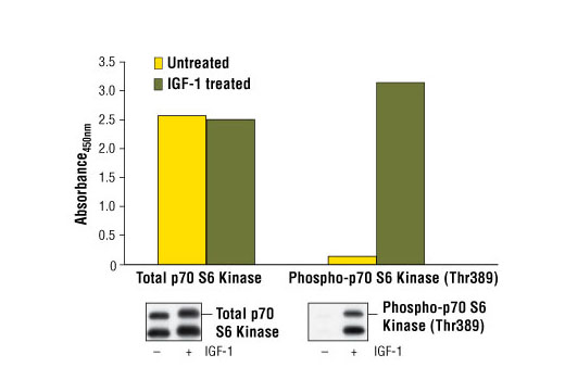
p70 S6 kinase is a mitogen activated Ser/Thr protein kinase that is required for cell growth and G1 cell cycle progression (1,2). p70 S6 kinase phosphorylates the S6 protein of the 40S ribosomal subunit and is involved in translational control of 5' oligopyrimidine tract mRNAs (1). A second isoform, p85 S6 kinase, is derived from the same gene and is identical to p70 S6 kinase except for 23 extra residues at the amino terminus, which encode a nuclear localizing signal (1). Both isoforms lie on a mitogen activated signaling pathway downstream of phosphoinositide-3 kinase (PI-3K) and the target of rapamycin, FRAP/mTOR, a pathway distinct from the Ras/MAP kinase cascade (1). The activity of p70 S6 kinase is controlled by multiple phosphorylation events located within the catalytic, linker and pseudosubstrate domains (1). Phosphorylation of Thr229 in the catalytic domain and Thr389 in the linker domain are most critical for kinase function (1). Phosphorylation of Thr389, however, most closely correlates with p70 kinase activity in vivo (3). Prior phosphorylation of Thr389 is required for the action of phosphoinositide 3-dependent protein kinase 1 (PDK1) on Thr229 (4,5). Phosphorylation of this site is stimulated by growth factors such as insulin, EGF and FGF, as well as by serum and some G-protein-coupled receptor ligands, and is blocked by wortmannin, LY294002 (PI-3K inhibitor) and rapamycin (FRAP/mTOR inhibitor) (1,6,7). Ser411, Thr421 and Ser424 lie within a Ser-Pro-rich region located in the pseudosubstrate region (1). Phosphorylation at these sites is thought to activate p70 S6 kinase via relief of pseudosubstrate suppression (1,2). Another LY294002 and rapamycin sensitive phosphorylation site, Ser371, is an in vitro substrate for mTOR and correlates well with the activity of a partially rapamycin resistant mutant p70 S6 kinase (8).
Explore pathways related to this product.
STRING - Known and Predicted Protein-Protein Interactions.
Except as otherwise expressly agreed in a writing signed by a legally authorized representative of CST, the following terms apply to Products provided by CST, its affiliates or its distributors. Any Customer's terms and conditions that are in addition to, or different from, those contained herein, unless separately accepted in writing by a legally authorized representative of CST, are rejected and are of no force or effect.
Products are labeled with For Research Use Only or a similar labeling statement and have not been approved, cleared, or licensed by the FDA or other regulatory foreign or domestic entity, for any purpose. Customer shall not use any Product for any diagnostic or therapeutic purpose, or otherwise in any manner that conflicts with its labeling statement. Products sold or licensed by CST are provided for Customer as the end-user and solely for research and development uses. Any use of Product for diagnostic, prophylactic or therapeutic purposes, or any purchase of Product for resale (alone or as a component) or other commercial purpose, requires a separate license from CST. Customer shall (a) not sell, license, loan, donate or otherwise transfer or make available any Product to any third party, whether alone or in combination with other materials, or use the Products to manufacture any commercial products, (b) not copy, modify, reverse engineer, decompile, disassemble or otherwise attempt to discover the underlying structure or technology of the Products, or use the Products for the purpose of developing any products or services that would compete with CST products or services, (c) not alter or remove from the Products any trademarks, trade names, logos, patent or copyright notices or markings, (d) use the Products solely in accordance with CST Product Terms of Sale and any applicable documentation, and (e) comply with any license, terms of service or similar agreement with respect to any third party products or services used by Customer in connection with the Products.