| Cat. # | Size | Qty. | Price |
|---|---|---|---|
| 12724T | 1 Kit (8 x 20 microliters) |
|
| Product Includes | Quantity | Applications | Reactivity | MW(kDa) | Isotype |
|---|---|---|---|---|---|
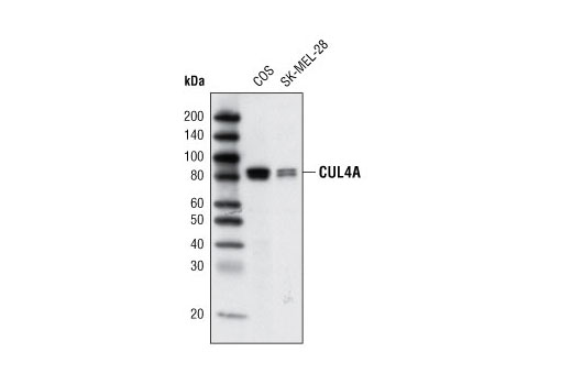
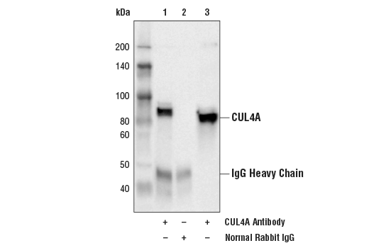
| CUL4A Antibody 2699 | 20 µl |
|
H Mk | 80, 82 | Rabbit |
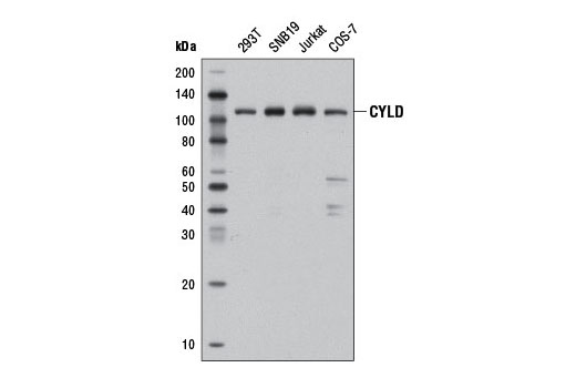
| CYLD (D6O5O) Rabbit mAb 12797 | 20 µl |
|
H M R Hm Mk | 109 | Rabbit IgG |
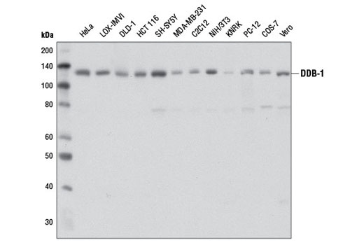
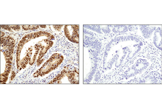
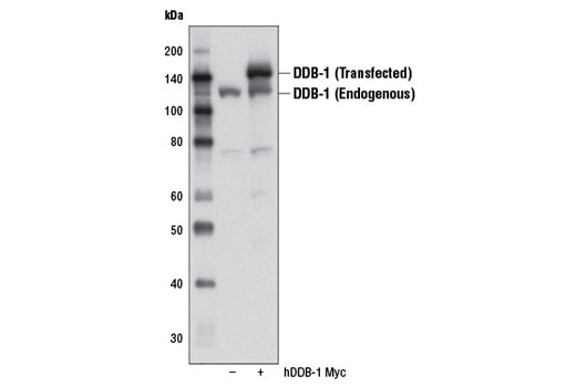
| DDB-1 (D4C8) Rabbit mAb 6998 | 20 µl |
|
H M R Mk | 127 | Rabbit IgG |
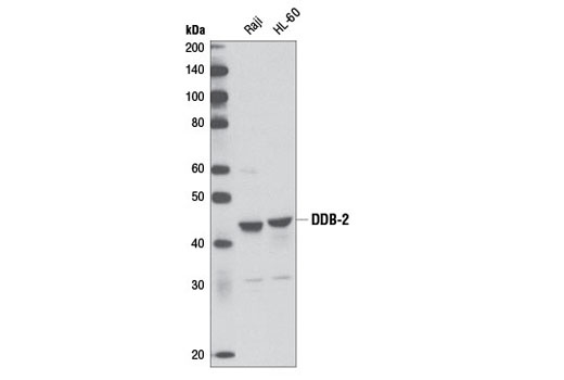
| DDB-2 (D4C4) Rabbit mAb 5416 | 20 µl |
|
H M | 43 | Rabbit IgG |
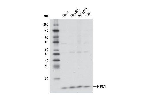
| RBX1 (D3J5I) Rabbit mAb 11922 | 20 µl |
|
H M R Mk | 13 | Rabbit IgG |
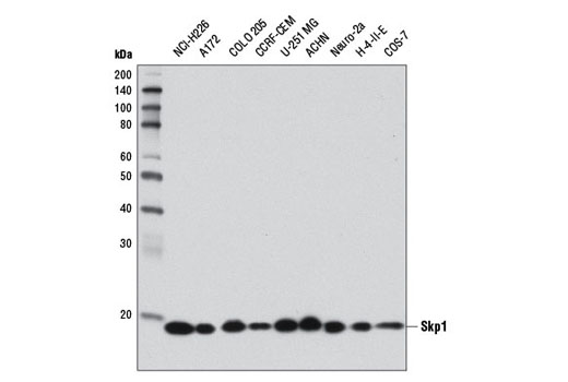
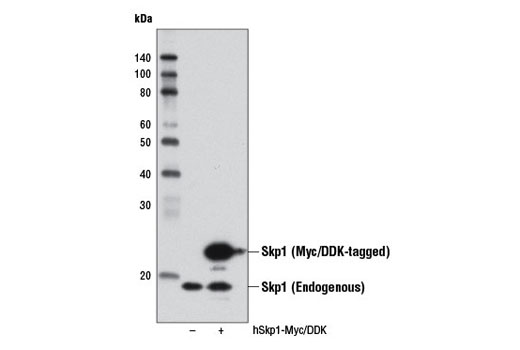
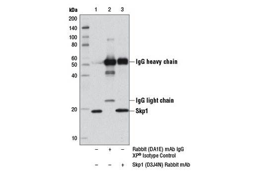
| Skp1 (D3J4N) Rabbit mAb 12248 | 20 µl |
|
H M R Mk | 19 | Rabbit IgG |
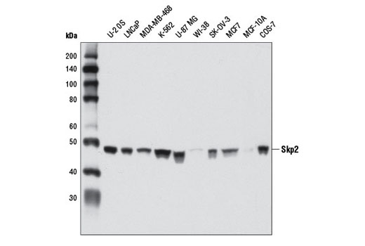
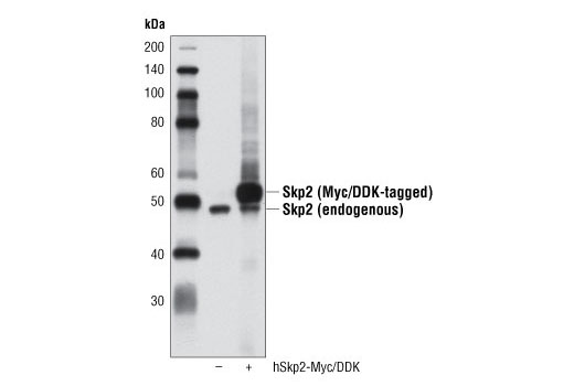
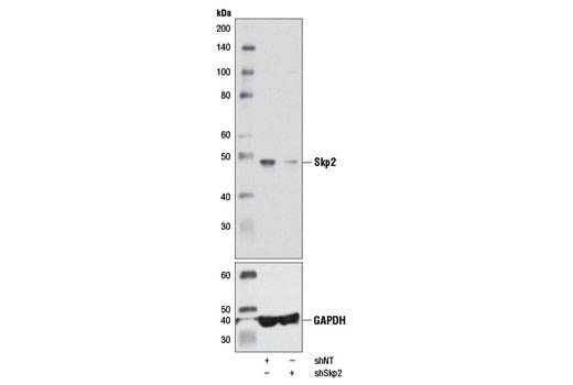
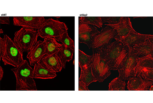
| Skp2 (D3G5) XP® Rabbit mAb 2652 | 20 µl |
|
H Mk | 48 | Rabbit IgG |
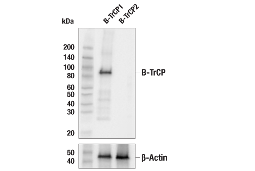
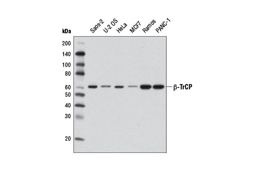
| β-TrCP (D12C8) Rabbit mAb 11984 | 20 µl |
|
H M R Mk | 62 | Rabbit IgG |
| Anti-rabbit IgG, HRP-linked Antibody 7074 | 100 µl |
|
Goat |
Product Information
Monoclonal antibodies are produced by immunizing animals with a recombinant protein specific to human CYLD protein, a synthetic peptide corresponding to residues surrounding Gly832 of human DDB-1 protein, to residues surrounding Ala174 of human DDB-2 protein, to the carboxy terminus of human RBX1 protein, to the carboxy terminus of human Skp1 protein, to the amino terminus of human Skp2 protein, and with a recombinant protein specific to human β-TrCP protein. Polyclonal antibodies are produced by immunizing animals with a synthetic peptide corresponding to residues surrounding Ser12 of human CUL4A protein. Polyclonal antibodies are purified by protein A and peptide affinity chromatography.
Ubiquitin can be covalently linked to many cellular proteins by the ubiquitination process, which targets proteins for degradation by the 26S proteasome. Ubiquitin is first activated by forming a thiolester complex with the activation component E1. The activated ubiquitin is subsequently transferred to the ubiquitin-carrier protein E2, and then from E2 to ubiquitin ligase E3 for final delivery to the ε-NH2 of the target protein lysine residue (1-3). Research studies suggest that activated E2 associates transiently with E3, and the dissociation is a critical step for ubiquitination (4).
S phase kinase-associated protein 1 (Skp1) is a critical scaffold protein of the Skp1/CUL1/F-box (SCF) E3 ubiquitin ligase protein complex. Various F-box proteins (e.g. β-TrCP, Skp2) mediate an interaction with Skp1 via their defining and conserved domain of 40 amino acids and with substrates to be ubiquitinated (5). RING-box protein 1 (RBX1 or ROC1) is another essential component of the SCF complex (6). RBX1 mediates the neddylation of CUL1, which activates SCF E3 ligase by facilitating the ubiquitin transfer from E2 to substrates (7-9). The RING finger domain of RBX1 is required for ubiquitin ligation (10).
Cullin-4 (CUL4) is a member of the cullin family of related ubiquitin ligases (11). The carboxy-terminal domain of CUL4 interacts with Rbx1 and E2 enzyme while the amino-terminal CUL4 domain interacts with BPB domain of UV-damaged DNA binding protein DDB-1 to form a CUL4-DDB1 ubiquitin ligase complex (12). Damaged DNA-Binding Protein (DDB) consists of a 127 kDa subunit (DDB-1) and a 48 kDa subunit (DDB-2) that contribute to the formation of the UV-damaged DNA-binding protein complex (UV-DDB) (13-15). In conjunction with CUL4A and RBX1, the UV-DDB complex forms an E3 ubiquitin ligase that recognizes a broad spectrum of DNA lesions. The complex polyubiquitinates components of the nucleotide excision repair pathway (16-18).
Explore pathways related to this product.
STRING - Known and Predicted Protein-Protein Interactions.
Except as otherwise expressly agreed in a writing signed by a legally authorized representative of CST, the following terms apply to Products provided by CST, its affiliates or its distributors. Any Customer's terms and conditions that are in addition to, or different from, those contained herein, unless separately accepted in writing by a legally authorized representative of CST, are rejected and are of no force or effect.
Products are labeled with For Research Use Only or a similar labeling statement and have not been approved, cleared, or licensed by the FDA or other regulatory foreign or domestic entity, for any purpose. Customer shall not use any Product for any diagnostic or therapeutic purpose, or otherwise in any manner that conflicts with its labeling statement. Products sold or licensed by CST are provided for Customer as the end-user and solely for research and development uses. Any use of Product for diagnostic, prophylactic or therapeutic purposes, or any purchase of Product for resale (alone or as a component) or other commercial purpose, requires a separate license from CST. Customer shall (a) not sell, license, loan, donate or otherwise transfer or make available any Product to any third party, whether alone or in combination with other materials, or use the Products to manufacture any commercial products, (b) not copy, modify, reverse engineer, decompile, disassemble or otherwise attempt to discover the underlying structure or technology of the Products, or use the Products for the purpose of developing any products or services that would compete with CST products or services, (c) not alter or remove from the Products any trademarks, trade names, logos, patent or copyright notices or markings, (d) use the Products solely in accordance with CST Product Terms of Sale and any applicable documentation, and (e) comply with any license, terms of service or similar agreement with respect to any third party products or services used by Customer in connection with the Products.