| Cat. # | Size | Qty. | Price |
|---|---|---|---|
| 4607S | 100 µl |
|
| REACTIVITY | H |
| SENSITIVITY | Endogenous |
| MW (kDa) | 140 |
| Source/Isotype | Rabbit IgG |
Product Information
| Application | Dilution |
|---|---|
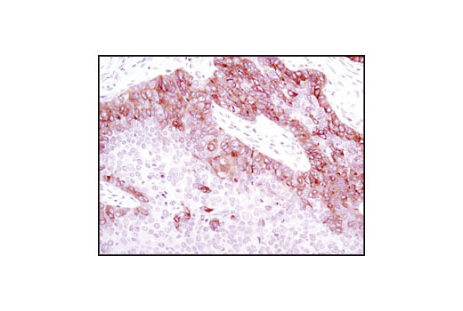
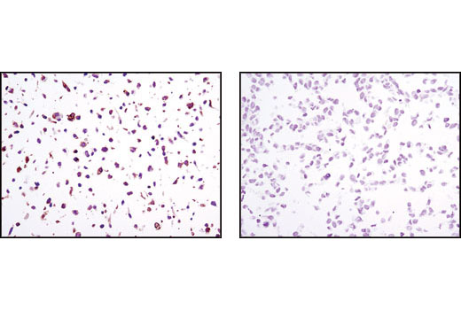
| Immunohistochemistry (Paraffin) | 1:2560 |
NOTE: Prepare solutions with reverse osmosis deionized (RODI) or equivalent grade water.
NOTE: Do not allow slides to dry at any time during this procedure.
For Citrate: Heat slides in a microwave submersed in 1X citrate unmasking solution until boiling is initiated; follow with 10 min at a sub-boiling temperature (95°-98°C). Cool slides on bench top for 30 min.
|
RECOMMENDED DETECTION REAGENTS |
SignalStain® Boost IHC Detection Reagent (HRP, Rabbit) #8114 | SignalStain® Boost IHC Detection Reagent (AP, Rabbit) #18653 |
|---|---|---|
|
COMPATIBLE CHROMOGEN |
SignalStain® DAB Substrate Kit #8059 | SignalStain® Vibrant Red Alkaline Phosphatase Substrate Kit #76713 |
| SignalStain® Vivid Purple Peroxidase Substrate Kit #96632 | SignalStain® Ultra Blue Alkaline Phosphatase Substrate Kit #12824 | |
| SignalStain® Deep Black Peroxidase Substrate Kit #72986 | ||
| SignalStain® Radiant Yellow Peroxidase Substrate Kit #69644 |
NOTE: Use of detection reagents other than those specified in this protocol may require further optimization of the primary antibody to account for the different sensitivities of the detection reagents.
posted February 2010
revised June 2020
Protocol Id: 283
Human
Mouse, Rat
Monoclonal antibody is produced by immunizing animals with a synthetic peptide surrounding Pro50 of human TrkB.
The family of Trk receptor tyrosine kinases consists of TrkA, TrkB, and TrkC. While the sequence of these family members is highly conserved, they are activated by different neurotrophins: TrkA by NGF, TrkB by BDNF or NT4, and TrkC by NT3 (1). Neurotrophin signaling through these receptors regulates a number of physiological processes, such as cell survival, proliferation, neural development, and axon and dendrite growth and patterning (1). In the adult nervous system, the Trk receptors regulate synaptic strength and plasticity. TrkA regulates proliferation and is important for development and maturation of the nervous system (2). Phosphorylation at Tyr490 is required for Shc association and activation of the Ras-MAP kinase cascade (3,4). Residues Tyr674/675 lie within the catalytic domain, and phosphorylation at these sites reflects TrkA kinase activity (3-6). Point mutations, deletions, and chromosomal rearrangements (chimeras) cause ligand-independent receptor dimerization and activation of TrkA (7-10). TrkA is activated in many malignancies including breast, ovarian, prostate, and thyroid carcinomas (8-13). Research studies suggest that expression of TrkA in neuroblastomas may be a good prognostic marker as TrkA signals growth arrest and differentiation of cells originating from the neural crest (10).
The phosphorylation sites are conserved between TrkA and TrkB: Tyr490 of TrkA corresponds to Tyr512 in TrkB, and Tyr674/675 of TrkA to Tyr706/707 in TrkB of the human sequence (14). TrkB is overexpressed in tumors, such as neuroblastoma, prostate adenocarcinoma, and pancreatic ductal adenocarcinoma (15). Research studies have shown that in neuroblastomas, overexpression of TrkB correlates with an unfavorable disease outcome when autocrine loops signaling tumor survival are potentiated by additional overexpression of brain-derived neurotrophic factor (BDNF) (16-18). An alternatively spliced truncated TrkB isoform lacking the kinase domain is overexpressed in Wilms’ tumors and this isoform may act as a dominant-negative regulator of TrkB signaling (17).
Explore pathways related to this product.
STRING - Known and Predicted Protein-Protein Interactions.
Except as otherwise expressly agreed in a writing signed by a legally authorized representative of CST, the following terms apply to Products provided by CST, its affiliates or its distributors. Any Customer's terms and conditions that are in addition to, or different from, those contained herein, unless separately accepted in writing by a legally authorized representative of CST, are rejected and are of no force or effect.
Products are labeled with For Research Use Only or a similar labeling statement and have not been approved, cleared, or licensed by the FDA or other regulatory foreign or domestic entity, for any purpose. Customer shall not use any Product for any diagnostic or therapeutic purpose, or otherwise in any manner that conflicts with its labeling statement. Products sold or licensed by CST are provided for Customer as the end-user and solely for research and development uses. Any use of Product for diagnostic, prophylactic or therapeutic purposes, or any purchase of Product for resale (alone or as a component) or other commercial purpose, requires a separate license from CST. Customer shall (a) not sell, license, loan, donate or otherwise transfer or make available any Product to any third party, whether alone or in combination with other materials, or use the Products to manufacture any commercial products, (b) not copy, modify, reverse engineer, decompile, disassemble or otherwise attempt to discover the underlying structure or technology of the Products, or use the Products for the purpose of developing any products or services that would compete with CST products or services, (c) not alter or remove from the Products any trademarks, trade names, logos, patent or copyright notices or markings, (d) use the Products solely in accordance with CST Product Terms of Sale and any applicable documentation, and (e) comply with any license, terms of service or similar agreement with respect to any third party products or services used by Customer in connection with the Products.