| Cat. # | Size | Qty. | Price |
|---|---|---|---|
| 9955T | 1 Kit (6 x 20 microliters) |
|
| Product Includes | Quantity | Applications | Reactivity | MW(kDa) | Isotype |
|---|---|---|---|---|---|
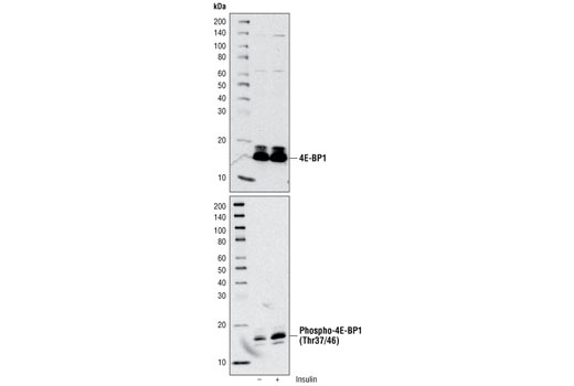
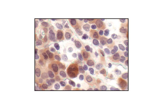
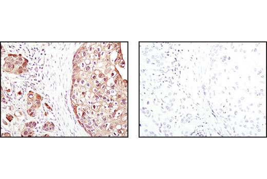
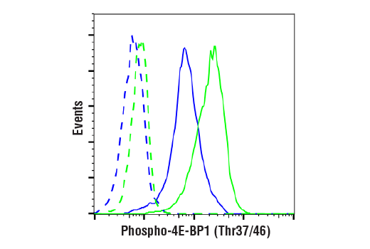
| Phospho-4E-BP1 (Thr37/46) (236B4) Rabbit mAb 2855 | 20 µl |
|
H M R Mk Dm | 15 to 20 | Rabbit IgG |
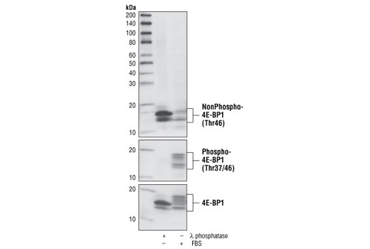
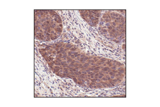
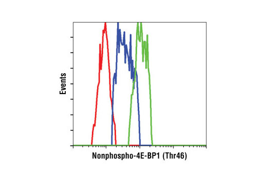
| Non-phospho-4E-BP1 (Thr46) (87D12) Rabbit mAb 4923 | 20 µl |
|
H M R Mk | 15-20 | Rabbit IgG |
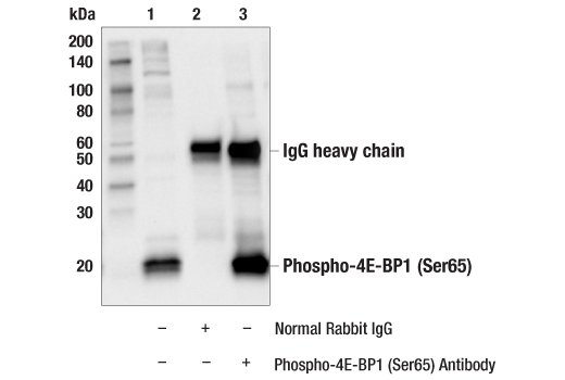
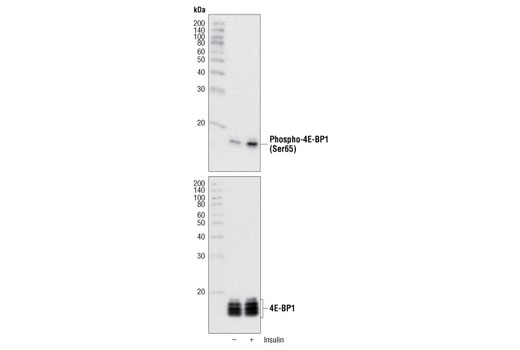
| Phospho-4E-BP1 (Ser65) Antibody 9451 | 20 µl |
|
H M R Mk | 15 to 20 | Rabbit |
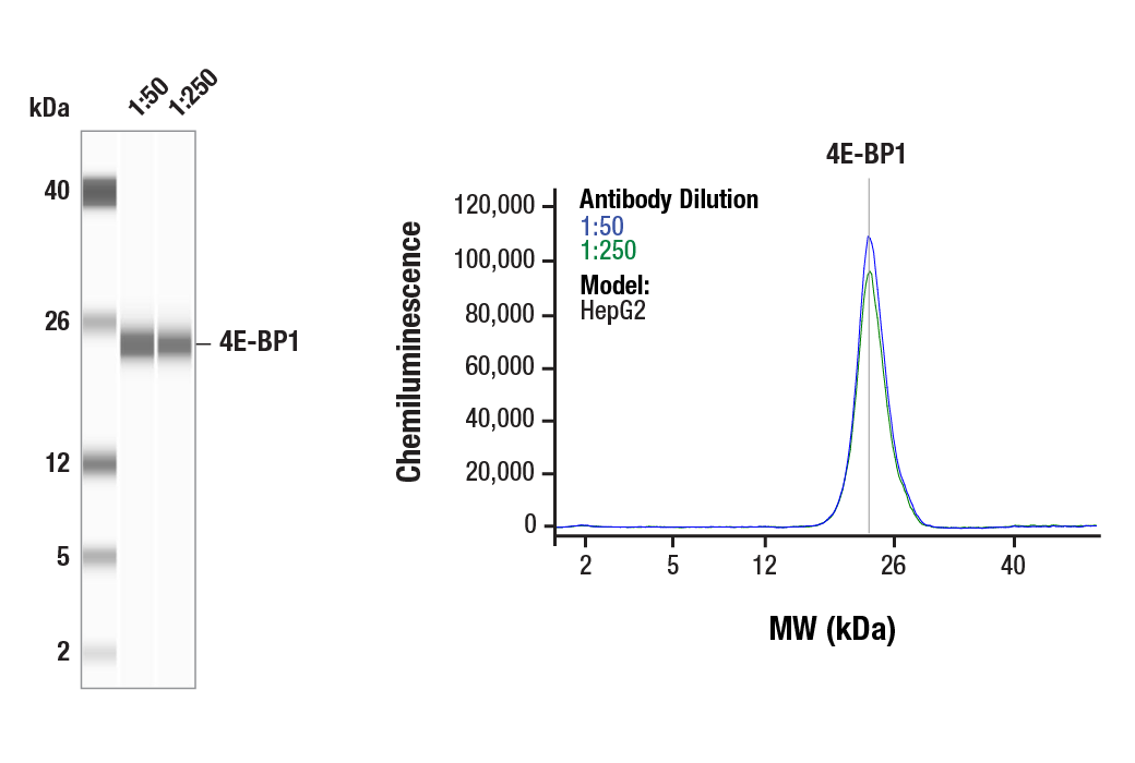
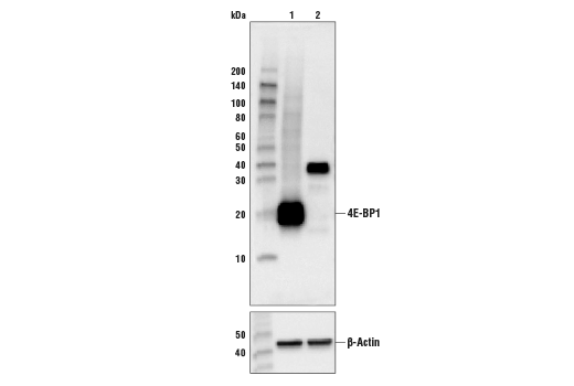
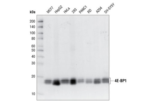
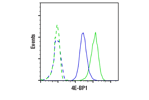
| 4E-BP1 (53H11) Rabbit mAb 9644 | 20 µl |
|
H M R Mk | 15-20 | Rabbit IgG |
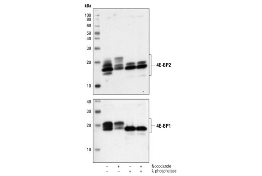
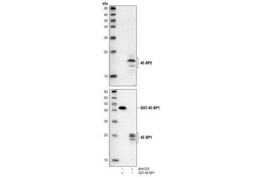
| 4E-BP2 Antibody 2845 | 20 µl |
|
H M R Mk B | 15 to 20 | Rabbit |
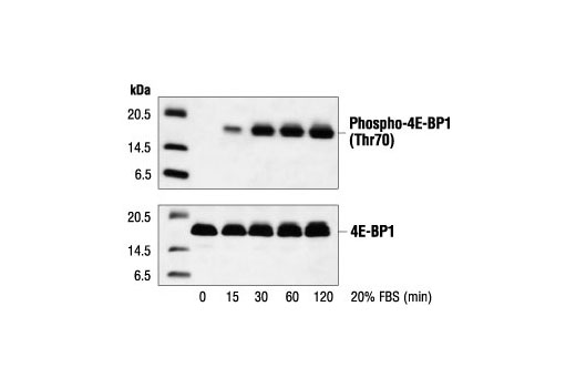
| Phospho-4E-BP1 (Thr70) Antibody 9455 | 20 µl |
|
H M R Mk | 15 to 20 | Rabbit |
| Anti-rabbit IgG, HRP-linked Antibody 7074 | 100 µl |
|
Goat |
Product Information
Monoclonal antibody is produced by immunizing animals with a synthetic phosphopeptide corresponding to residues surrounding Thr37 and Thr46 of mouse 4E-BP1, residues surrounding Thr46 of human 4E-BP1, or Ser112 of human 4E-BP1. Polyclonal antibodies are produced by immunizing animals with a synthetic peptide corresponding to the residues at the carboxy-terminus of human 4E-BP2 (#2845), or phosphopeptides surrounding mouse Ser65 (#9451) and human Thr70 (#5078) 4E-BP1. Polyclonal antibodies were purified by protein A and peptide affinity chromatography.
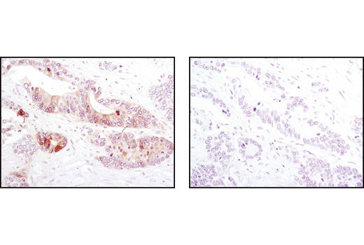
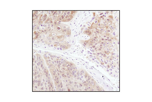
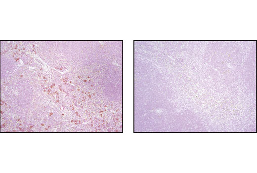
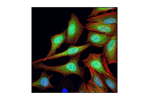
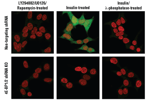
Translation repressor protein 4E-BP1 (also known as PHAS-1) inhibits cap-dependent translation by binding to the translation initiation factor eIF4E. Hyperphosphorylation of 4E-BP1 disrupts this interaction and results in activation of cap-dependent translation (1). Both the PI3 kinase/Akt pathway and FRAP/mTOR kinase regulate 4E-BP1 activity (2,3). Multiple 4E-BP1 residues are phosphorylated in vivo (4). While phosphorylation by FRAP/mTOR at Thr37 and Thr46 does not prevent the binding of 4E-BP1 to eIF4E, it is thought to prime 4E-BP1 for subsequent phosphorylation at Ser65 and Thr70 (5).
4E-BP2 and 4E-BP3 share high sequence homology with 4E-BP1, including conservation of the major FRAP/mTOR-dependent phosphorylation sites. Preliminary data suggests that phosphorylation of 4E-BP2 is regulated in a similar manner to that of 4E-BP1, although phosphorylation of this protein has not been as extensively studied (6).
Explore pathways related to this product.
STRING - Known and Predicted Protein-Protein Interactions.
Except as otherwise expressly agreed in a writing signed by a legally authorized representative of CST, the following terms apply to Products provided by CST, its affiliates or its distributors. Any Customer's terms and conditions that are in addition to, or different from, those contained herein, unless separately accepted in writing by a legally authorized representative of CST, are rejected and are of no force or effect.
Products are labeled with For Research Use Only or a similar labeling statement and have not been approved, cleared, or licensed by the FDA or other regulatory foreign or domestic entity, for any purpose. Customer shall not use any Product for any diagnostic or therapeutic purpose, or otherwise in any manner that conflicts with its labeling statement. Products sold or licensed by CST are provided for Customer as the end-user and solely for research and development uses. Any use of Product for diagnostic, prophylactic or therapeutic purposes, or any purchase of Product for resale (alone or as a component) or other commercial purpose, requires a separate license from CST. Customer shall (a) not sell, license, loan, donate or otherwise transfer or make available any Product to any third party, whether alone or in combination with other materials, or use the Products to manufacture any commercial products, (b) not copy, modify, reverse engineer, decompile, disassemble or otherwise attempt to discover the underlying structure or technology of the Products, or use the Products for the purpose of developing any products or services that would compete with CST products or services, (c) not alter or remove from the Products any trademarks, trade names, logos, patent or copyright notices or markings, (d) use the Products solely in accordance with CST Product Terms of Sale and any applicable documentation, and (e) comply with any license, terms of service or similar agreement with respect to any third party products or services used by Customer in connection with the Products.