Recombinant antibodies offer several key advantages compared to traditional antibodies. These include superior lot-to-lot consistency, continuous supply, and animal-free manufacturing. As such, recombinant antibodies are seeing increased use for scientific research, especially as a means of addressing the ongoing reproducibility crisis.

Traditional polyclonal and monoclonal antibodies are the product of normal B cell development and genetic recombination. They are generated by immunizing an animal with an antigen to elicit an immune response. While polyclonal antibodies are secreted by many different B cell clones and recognize multiple antigenic epitopes, monoclonals originate from a single B cell clone and are specific for just one epitope.
Recombinant antibodies are monoclonal, but their production involves in vitro genetic manipulation. After cloning the antibody genes into an expression vector, this is then transfected into an appropriate host cell line for antibody expression. Mammalian cell lines are most commonly used for recombinant antibody production, although cell lines of bacterial, yeast, or insect origin are also suitable.
Because recombinant antibody production involves sequencing the antibody light and heavy chains, it is a highly controlled and reliable process. In contrast, hybridoma-based systems for producing monoclonal antibodies are subject to genetic drift and instability, increasing the potential for lot-to-lot variability or loss of antibody expression. Recombinant antibodies are highly consistent from lot to lot, thereby ensuring reproducible experimental results.
In vitro methods for producing antibodies are amenable to large-scale production, meaning antibody availability is unlikely to become a limiting factor. Moreover, since the recombinant antibody sequence is known, continuity of supply is assured; in situations where an antibody will be used to support large, long-term studies, this can be an especially critical factor.
Unlike traditional methods for antibody production, recombinant approaches avoid the need to use animals. Where polyclonal antibodies are purified directly from the serum of the immunized host, and monoclonals are purified from either hybridoma-derived tissue culture supernatant or ascites, recombinant antibodies are instead purified from the tissue culture supernatants of transfected host cell lines. Regardless of whether an antibody is polyclonal, monoclonal or recombinant, it must always be properly validated in the intended application prior to experimental use. At CST, we adhere to the Hallmarks of Antibody Validation™, six complementary strategies for determining the specificity, sensitivity, and functionality of an antibody in any given assay. By carefully tailoring these strategies to each antibody product, we guarantee that CST antibodies will work as expected, to help you achieve results you can trust.

| Cat. # | Size | Qty. | Price |
|---|---|---|---|
| 28692S | 100 µl |
|
| REACTIVITY | All |
| SENSITIVITY | Endogenous |
| MW (kDa) | |
| Source/Isotype | Rabbit IgG |
Product Information
| Application | Dilution |
|---|---|
| Immunofluorescence (Immunocytochemistry) | 1:1600 |
| DNA Dot Blot | 1:1000 |
IMPORTANT: This protocol employs an atypical fixation and denaturation strategy with which only certain targets are compatible. Where appropriate, this protocol will be linked to its validated antibody under the Product Information banner on the product-specific webpage.
NOTE: Prepare solutions with reverse osmosis deionized (RODI) or equivalently purified water.
NOTE: All subsequent incubations should be carried out at room temperature (20-25°C) unless noted otherwise.
Rinse three times in 1X PBS for 5 min each.
NOTE: If using a fluorochrome-conjugated primary antibody, then skip to Section C, Step 8.
Counterstain as appropriate.
NOTE: When including fluorescent cellular dyes in your experiment (DNA dyes, etc.), please refer to the dye product page for its recommended protocol. View our listing of cellular dyes validated for use in immunofluorescence.
posted December 2015
revised December 2020
Protocol Id: 865
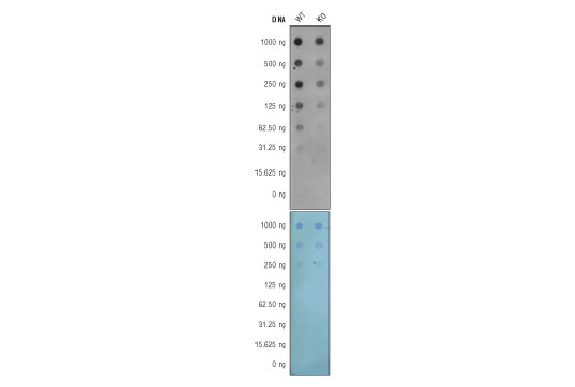
Note: This protocol is written for spotting fragmented, purified genomic DNA (titration of 1000 ng, 500 ng, 250 ng, 125 ng, 62.5 ng, 31.25 ng, and 15.625 ng) onto a positively charged nylon membrane using a 96-well dot blotting apparatus. Depending on the source and type of DNA, more or less DNA may be required for detection with the antibody.
Before Starting:
• Purify genomic DNA using a genomic DNA purification protocol or kit and sonicate
genomic DNA to generate fragments between 200 and 500 bp. DNA fragment size
can be analyzed by gel electrophoresis on a 1% agarose gel with a 100 bp DNA
marker.
• Cut a piece of nylon membrane to the size of the dot blot manifold.
• Wet nylon membrane with 10x SSC Buffer.
• Dry membrane by placing it in 96-well dot blot apparatus and applying vacuum.
NOTE: Due to the kinetics of the detection reaction, signal is most intense immediately following incubation and declines over the following 2 hr
posted November 2015
Protocol Id: 804
All Species Expected
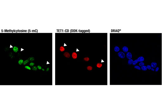
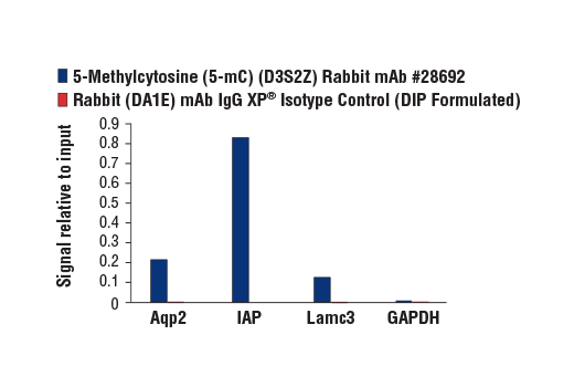
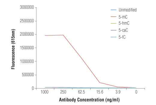
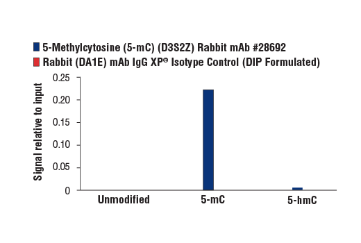
Monoclonal antibody is produced by immunizing animals with 5-methylcytidine.
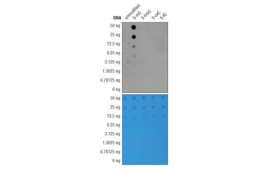
Methylation of DNA at cytosine residues is a heritable, epigenetic modification that is critical for proper regulation of gene expression, genomic imprinting, and mammalian development (1,2). 5-methylcytosine is a repressive epigenetic mark established de novo by two enzymes, DNMT3a and DNMT3b, and is maintained by DNMT1 (3, 4). 5-methylcytosine was originally thought to be passively depleted during DNA replication. However, subsequent studies have shown that Ten-Eleven Translocation (TET) proteins TET1, TET2, and TET3 can catalyze the oxidation of methylated cytosine to 5-hydroxymethylcytosine (5-hmC) (5). Additionally, TET proteins can further oxidize 5-hmC to form 5-formylcytosine (5-fC) and 5-carboxylcytosine (5-caC), both of which are excised by thymine-DNA glycosylase (TDG), effectively linking cytosine oxidation to the base excision repair pathway and supporting active cytosine demethylation (6,7).
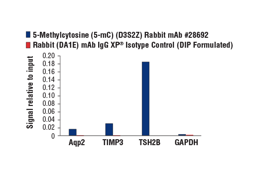
Normally DNA methylation occurs in a bimodal fashion, such that CpG dinucleotides are largely methylated across the genome, except in short stretches of CpG-rich sequences associated with gene promoters, known as CpG-islands, where methylation is virtually absent (8). Cancer cell genomes often undergo global hypomethylation, while CpG-islands become hypermethylated, causing their associated promoters to become repressed (9). There is evidence that a number of aberrantly hypermethylated CpG-islands found in carcinomas occur at tumor suppressor genes such as RB1, MLH1, and BRCA1 (10).
Except as otherwise expressly agreed in a writing signed by a legally authorized representative of CST, the following terms apply to Products provided by CST, its affiliates or its distributors. Any Customer's terms and conditions that are in addition to, or different from, those contained herein, unless separately accepted in writing by a legally authorized representative of CST, are rejected and are of no force or effect.
Products are labeled with For Research Use Only or a similar labeling statement and have not been approved, cleared, or licensed by the FDA or other regulatory foreign or domestic entity, for any purpose. Customer shall not use any Product for any diagnostic or therapeutic purpose, or otherwise in any manner that conflicts with its labeling statement. Products sold or licensed by CST are provided for Customer as the end-user and solely for research and development uses. Any use of Product for diagnostic, prophylactic or therapeutic purposes, or any purchase of Product for resale (alone or as a component) or other commercial purpose, requires a separate license from CST. Customer shall (a) not sell, license, loan, donate or otherwise transfer or make available any Product to any third party, whether alone or in combination with other materials, or use the Products to manufacture any commercial products, (b) not copy, modify, reverse engineer, decompile, disassemble or otherwise attempt to discover the underlying structure or technology of the Products, or use the Products for the purpose of developing any products or services that would compete with CST products or services, (c) not alter or remove from the Products any trademarks, trade names, logos, patent or copyright notices or markings, (d) use the Products solely in accordance with CST Product Terms of Sale and any applicable documentation, and (e) comply with any license, terms of service or similar agreement with respect to any third party products or services used by Customer in connection with the Products.