Recombinant antibodies offer several key advantages compared to traditional antibodies. These include superior lot-to-lot consistency, continuous supply, and animal-free manufacturing. As such, recombinant antibodies are seeing increased use for scientific research, especially as a means of addressing the ongoing reproducibility crisis.

Traditional polyclonal and monoclonal antibodies are the product of normal B cell development and genetic recombination. They are generated by immunizing an animal with an antigen to elicit an immune response. While polyclonal antibodies are secreted by many different B cell clones and recognize multiple antigenic epitopes, monoclonals originate from a single B cell clone and are specific for just one epitope.
Recombinant antibodies are monoclonal, but their production involves in vitro genetic manipulation. After cloning the antibody genes into an expression vector, this is then transfected into an appropriate host cell line for antibody expression. Mammalian cell lines are most commonly used for recombinant antibody production, although cell lines of bacterial, yeast, or insect origin are also suitable.
Because recombinant antibody production involves sequencing the antibody light and heavy chains, it is a highly controlled and reliable process. In contrast, hybridoma-based systems for producing monoclonal antibodies are subject to genetic drift and instability, increasing the potential for lot-to-lot variability or loss of antibody expression. Recombinant antibodies are highly consistent from lot to lot, thereby ensuring reproducible experimental results.
In vitro methods for producing antibodies are amenable to large-scale production, meaning antibody availability is unlikely to become a limiting factor. Moreover, since the recombinant antibody sequence is known, continuity of supply is assured; in situations where an antibody will be used to support large, long-term studies, this can be an especially critical factor.
Unlike traditional methods for antibody production, recombinant approaches avoid the need to use animals. Where polyclonal antibodies are purified directly from the serum of the immunized host, and monoclonals are purified from either hybridoma-derived tissue culture supernatant or ascites, recombinant antibodies are instead purified from the tissue culture supernatants of transfected host cell lines. Regardless of whether an antibody is polyclonal, monoclonal or recombinant, it must always be properly validated in the intended application prior to experimental use. At CST, we adhere to the Hallmarks of Antibody Validation™, six complementary strategies for determining the specificity, sensitivity, and functionality of an antibody in any given assay. By carefully tailoring these strategies to each antibody product, we guarantee that CST antibodies will work as expected, to help you achieve results you can trust.
| Cat. # | Size | Qty. | Price |
|---|---|---|---|
| 85403T | 20 µl |
|
|
| 85403S | 100 µl |
|
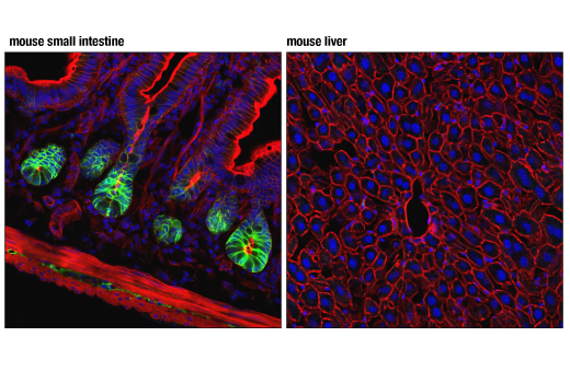
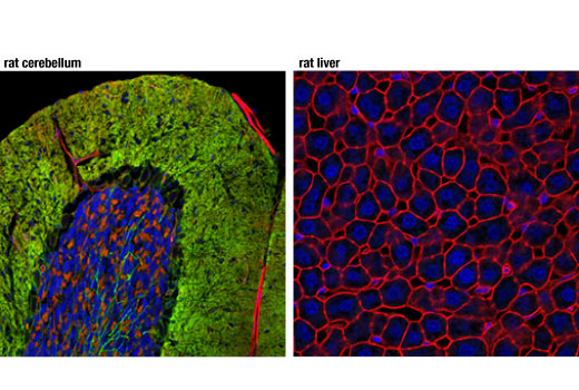
| REACTIVITY | M R |
| SENSITIVITY | Endogenous |
| MW (kDa) | |
| Source/Isotype | Rabbit IgG |
Product Information
| Application | Dilution |
|---|---|
| Immunofluorescence (Frozen) | 1:200 |
NOTE: Prepare solutions with reverse osmosis deionized (RODI) or equivalent grade water.
Recommended Fluorochrome-conjugated Anti-Rabbit secondary antibodies:
NOTE: When using any primary or fluorochrome-conjugated secondary antibody for the first time, titrate the antibody to determine which dilution allows for the strongest specific signal with the least background for your sample.
Cover sections with 4% formaldehyde dilute in 1X PBS.
NOTE: Formaldehyde is toxic, use only in fume hood.
NOTE: All subsequent incubations should be carried out at room temperature unless otherwise noted in a humid light-tight box or covered dish/plate to prevent drying and fluorochrome fading.
posted November 2006
revised July 2016
Protocol Id: 151
Mouse, Rat
Monoclonal antibody is produced by immunizing animals with a synthetic peptide corresponding to residues near the amino terminus of mouse NKCC1 protein.
The electroneutral cation-chloride-coupled co-transporter (SLC12) gene family comprises bumetanide-sensitive Na+/K+/Cl- (NKCC), thiazide-sensitive Na+/Cl-, and K+/Cl- (KCC) co-transporters. SLC12A1/NKCC2 and SLC12A2/NKCC1 regulate cell volume and maintain cellular homeostasis in response to osmotic and oxidative stress (1). The broadly expressed NKCC1 is thought to play roles in fluid secretion (i.e. salivary gland function), salt balance (i.e. maintenance of renin and aldosterone levels), and neuronal development and signaling (2-7). During neuronal development, NKCC1 and KCC2 maintain a fine balance between chloride influx (NKCC1) and efflux (KCC2), which regulates γ-aminobutyric acid (GABA)-mediated neurotransmission (3). Increased NKCC1 expression in immature neurons maintains high intracellular chloride levels that result in inhibitory GABAergic signaling; KCC2 maintains low intracellular chloride levels and excitatory GABAergic responses in mature neurons (4,5,8). Deletion of NKCC1 impairs NGF-mediated neurite outgrowth in PC-12D cells while inhibition of NKCC1 with bumetanide inhibits re-growth of axotomized dorsal root ganglion cells (6,7). Defective chloride homeostasis in neurons is linked to seizure disorders that are ameliorated by butemanide treatment, indicating that abnormal NKCC1 and NKCC2 expression or signaling may play a role in neonatal and adult seizures (9-12). NKCC1 is found as a homodimer or within heterooligomers with other SLC12 family members. This transport protein associates with a number of oxidative- and osmotic-responsive kinases that bind, phosphorylate, and activate NKCC1 co-transporter activity (13-16). In response to decreased intracellular chloride concentrations, Ste20-related proline-alanine-rich kinase (SPAK) phosphorylates NKCC1 to increase co-transporter activity and promote chloride influx (16-19). Oxidative stress response kinase 1 (OSR1) also phosphorylates and activates NKCC1 in response to oxidative stress (14).
Except as otherwise expressly agreed in a writing signed by a legally authorized representative of CST, the following terms apply to Products provided by CST, its affiliates or its distributors. Any Customer's terms and conditions that are in addition to, or different from, those contained herein, unless separately accepted in writing by a legally authorized representative of CST, are rejected and are of no force or effect.
Products are labeled with For Research Use Only or a similar labeling statement and have not been approved, cleared, or licensed by the FDA or other regulatory foreign or domestic entity, for any purpose. Customer shall not use any Product for any diagnostic or therapeutic purpose, or otherwise in any manner that conflicts with its labeling statement. Products sold or licensed by CST are provided for Customer as the end-user and solely for research and development uses. Any use of Product for diagnostic, prophylactic or therapeutic purposes, or any purchase of Product for resale (alone or as a component) or other commercial purpose, requires a separate license from CST. Customer shall (a) not sell, license, loan, donate or otherwise transfer or make available any Product to any third party, whether alone or in combination with other materials, or use the Products to manufacture any commercial products, (b) not copy, modify, reverse engineer, decompile, disassemble or otherwise attempt to discover the underlying structure or technology of the Products, or use the Products for the purpose of developing any products or services that would compete with CST products or services, (c) not alter or remove from the Products any trademarks, trade names, logos, patent or copyright notices or markings, (d) use the Products solely in accordance with CST Product Terms of Sale and any applicable documentation, and (e) comply with any license, terms of service or similar agreement with respect to any third party products or services used by Customer in connection with the Products.