| Cat. # | Size | Qty. | Price |
|---|---|---|---|
| 8606T | 1 Kit (7 x 20 microliters) |
|
| Product Includes | Quantity | Applications | Reactivity | MW(kDa) | Isotype |
|---|---|---|---|---|---|
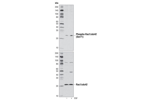
| Phospho-Rac1/cdc42 (Ser71) Antibody 2461 | 20 µl |
|
H | 28 | Rabbit |
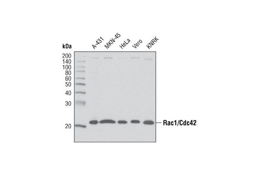
| Rac1/Cdc42 Antibody 4651 | 20 µl |
|
H M R Mk | 21 | Rabbit |
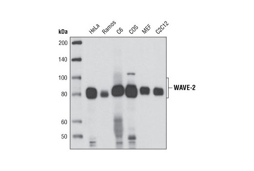
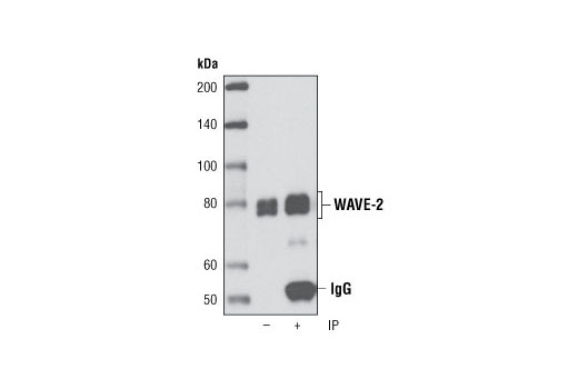
| WAVE-2 (D2C8) XP® Rabbit mAb 3659 | 20 µl |
|
H M R Mk | 80 | Rabbit IgG |
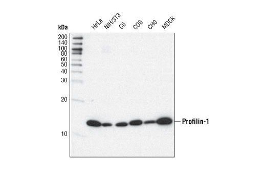
| Profilin-1 (C56B8) Rabbit mAb 3246 | 20 µl |
|
H M R Hm Mk Dg | 15 | Rabbit IgG |
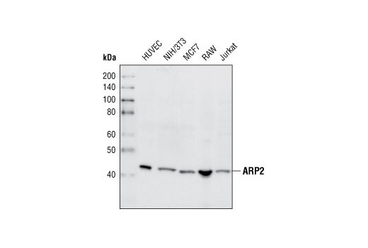
| ARP2 Antibody 3128 | 20 µl |
|
H M R Hm Mk Dm | 44 | Rabbit |
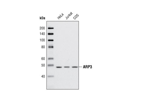
| ARP3 Antibody 4738 | 20 µl |
|
H Mk | 47 | Rabbit |
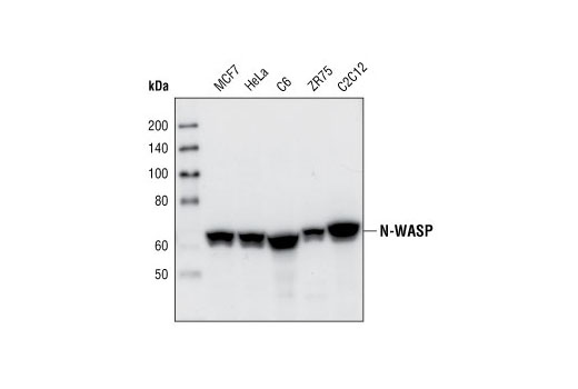
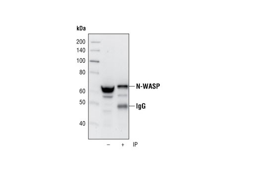
| N-WASP (30D10) Rabbit mAb 4848 | 20 µl |
|
H M R Hm Mk B | 65 | Rabbit IgG |
| Anti-rabbit IgG, HRP-linked Antibody 7074 | 100 µl |
|
Rab | Goat |
Product Information
Monoclonal antibodies are produced by immunizing animals with a synthetic peptide corresponding to residues near the amino terminus of human profilin-1 protein, the sequence of human N-WASP protein, or central residues of human WAVE-2 protein.
Activation state specific polyclonal antibodies are produced by immunizing animals with a synthetic phosphopeptide corresponding to residues surrounding Ser71 of human Rac1/cdc42 protein. Polyclonal antibodies are produced by immunizing animals with a synthetic peptide corresponding to residues near the amino termini of human rac1 and cdc42 proteins, the sequence of human ARP2 protein, or the amino terminus of human ARP3 protein. Polyclonal antibodies are purified by protein A and peptide affinity chromatography.
Actin nucleation is the process of forming new actin filaments and is necessary to stimulate actin polymerization. Actin polymerization is vital for cell motility, cell division, and cell adhesion. Rac and Cdc42, members of the Rho-GTPase family, play key roles in actin dynamics, membrane trafficking, transcriptional regulation, cell growth, and development (1). GTP binding stimulates the activity of Rac/Cdc42, and the hydrolysis of GTP to GDP through the intrinsic GTPase activity or Rac/Cdc42, rendering it inactive. GTP hydrolysis is aided by GTPase activating proteins (GAPs), while exchange of GDP for GTP is facilitated by guanine nucleotide exchange factors (GEFs). Another level of regulation is achieved through binding of RhoGDI, a guanine dissociation inhibitor, which retains Rho family GTPases, including Rac and Cdc42, in their inactive GDP-bound state (2,3). Hematopoietic WASP and ubiquitously expressed N-WASP are autoinhibited in unstimulated cells. Upon stimulation they are activated by Cdc42, which relieves the autoinhibition in conjunction with phosphatidyl inositol 4,5-bisphosphate (4). Three WAVE (Wasf, SCAR) family proteins are similar in sequence to WASP and N-WASP, but lack the WASP/N-WASP autoinhibition domains and are indirectly activated by Rac (4). WAVE-2 is widely distributed, while WAVE-1 and WAVE-3 are strongly expressed in the brain (5). The highly conserved ARP2/3 complex is an important actin nucleation protein complex consisting of ARP2, ARP3, and ARPC1-5. The ARP2/3 complex promotes branching of existing actin filaments and formation of daughter filaments following activation by nucleation-promoting factors, such as WASP/WAVE or cortactin (6). Profilins are conserved actin binding proteins that affect the rate of actin polymerization by binding actin monomers and promoting exchange of ADP for ATP (reviewed in 7). Profilins bind to proteins involved in the regulation of actin dynamics including paladin (8), dynamin-I (9), VASP (10), and N-WASP (11).
Explore pathways related to this product.
STRING - Known and Predicted Protein-Protein Interactions.
Except as otherwise expressly agreed in a writing signed by a legally authorized representative of CST, the following terms apply to Products provided by CST, its affiliates or its distributors. Any Customer's terms and conditions that are in addition to, or different from, those contained herein, unless separately accepted in writing by a legally authorized representative of CST, are rejected and are of no force or effect.
Products are labeled with For Research Use Only or a similar labeling statement and have not been approved, cleared, or licensed by the FDA or other regulatory foreign or domestic entity, for any purpose. Customer shall not use any Product for any diagnostic or therapeutic purpose, or otherwise in any manner that conflicts with its labeling statement. Products sold or licensed by CST are provided for Customer as the end-user and solely for research and development uses. Any use of Product for diagnostic, prophylactic or therapeutic purposes, or any purchase of Product for resale (alone or as a component) or other commercial purpose, requires a separate license from CST. Customer shall (a) not sell, license, loan, donate or otherwise transfer or make available any Product to any third party, whether alone or in combination with other materials, or use the Products to manufacture any commercial products, (b) not copy, modify, reverse engineer, decompile, disassemble or otherwise attempt to discover the underlying structure or technology of the Products, or use the Products for the purpose of developing any products or services that would compete with CST products or services, (c) not alter or remove from the Products any trademarks, trade names, logos, patent or copyright notices or markings, (d) use the Products solely in accordance with CST Product Terms of Sale and any applicable documentation, and (e) comply with any license, terms of service or similar agreement with respect to any third party products or services used by Customer in connection with the Products.