| Cat. # | Size | Qty. | Price |
|---|---|---|---|
| 9406S | 100 µl |
|
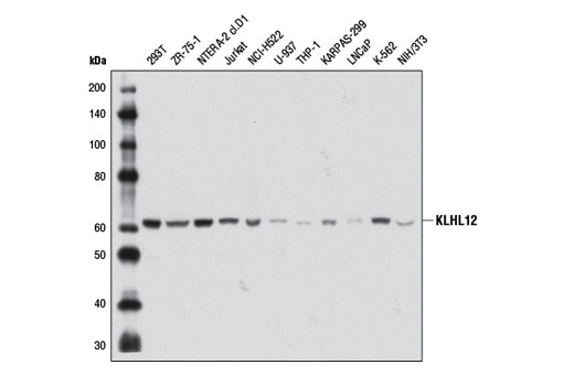
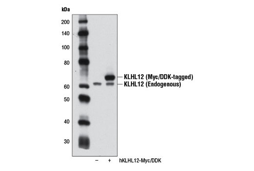
| REACTIVITY | H M Mk |
| SENSITIVITY | Endogenous |
| MW (kDa) | 62 |
| Source/Isotype | Mouse IgG1 |
Product Information
| Application | Dilution |
|---|---|
| Western Blotting | 1:1000 |
For western blots, incubate membrane with diluted primary antibody in 5% w/v nonfat dry milk, 1X TBS, 0.1% Tween® 20 at 4°C with gentle shaking, overnight.
NOTE: Please refer to primary antibody product webpage for recommended antibody dilution.
From sample preparation to detection, the reagents you need for your Western Blot are now in one convenient kit: #12957 Western Blotting Application Solutions Kit
NOTE: Prepare solutions with reverse osmosis deionized (RODI) or equivalent grade water.
Load 20 µl onto SDS-PAGE gel (10 cm x 10 cm).
NOTE: Loading of prestained molecular weight markers (#59329, 10 µl/lane) to verify electrotransfer and biotinylated protein ladder (#7727, 10 µl/lane) to determine molecular weights are recommended.
NOTE: Volumes are for 10 cm x 10 cm (100 cm2) of membrane; for different sized membranes, adjust volumes accordingly.
* Avoid repeated exposure to skin.
posted June 2005
revised June 2020
Protocol Id: 19
Human, Mouse, Monkey
Monoclonal antibody is produced by immunizing animals with a recombinant protein specific to the carboxy terminus of human KLHL12 protein.
Cullins are proteins that function as molecular scaffolds for modular ubiquitin ligases typified by the SCF (Skp1-CUL1-F-box) complex (1-3). The substrate selectivity of these E3 ligases is dictated by a specificity module that binds cullins. In the SCF complex, this module is composed of Skp1, which binds directly to CUL1, and a member of the F-box family of proteins such as Skp2 (1-4). CUL3 has been shown to be required for embryonic development in mammals and Caenorhabditis elegans (5-7) but until recently, its substrate specificity adaptor had yet to be elucidated. It is now recognized that substrate adaptors for CUL3-based ubiquitin ligase complexes contain a conserved BTB/POZ (Pox virus and Zinc finger) domain. This domain, which was initially identified in the Drosophila transcriptional repressors broad complex, tramtrack, and bric-a-brac is present in more than 190 human proteins. BTB proteins contain a variety of putative protein-protein interaction domains, including MATH domains, zinc finger repeats, and kelch repeats (8).
There are several lines of evidence suggesting that Kelch-like 12 protein (KLHL12) is a substrate-specific adaptor for the CUL3-based ubiquitin ligase complex. Analysis of the amino acid sequence of KLHL12 reveals an amino-terminal BTB motif, a central linker region, and a carboxy-terminal kelch domain composed of kelch repeats. Furthermore, KLHL12 has been shown to negatively regulate Wnt signaling by binding Disheveled and targeting it for ubiquitin-dependent proteasomal degradation (9). More recently, KLHL12 was shown to drive the assembly of large COPII vesicles by promoting the monoubiquitination of the COPII component Sec31. As a result, CUL3-KLHL12-dependent ubiquitination is essential for collagen export, a step that is required for integrin-dependent mouse embryonic stem cell division (10).
Except as otherwise expressly agreed in a writing signed by a legally authorized representative of CST, the following terms apply to Products provided by CST, its affiliates or its distributors. Any Customer's terms and conditions that are in addition to, or different from, those contained herein, unless separately accepted in writing by a legally authorized representative of CST, are rejected and are of no force or effect.
Products are labeled with For Research Use Only or a similar labeling statement and have not been approved, cleared, or licensed by the FDA or other regulatory foreign or domestic entity, for any purpose. Customer shall not use any Product for any diagnostic or therapeutic purpose, or otherwise in any manner that conflicts with its labeling statement. Products sold or licensed by CST are provided for Customer as the end-user and solely for research and development uses. Any use of Product for diagnostic, prophylactic or therapeutic purposes, or any purchase of Product for resale (alone or as a component) or other commercial purpose, requires a separate license from CST. Customer shall (a) not sell, license, loan, donate or otherwise transfer or make available any Product to any third party, whether alone or in combination with other materials, or use the Products to manufacture any commercial products, (b) not copy, modify, reverse engineer, decompile, disassemble or otherwise attempt to discover the underlying structure or technology of the Products, or use the Products for the purpose of developing any products or services that would compete with CST products or services, (c) not alter or remove from the Products any trademarks, trade names, logos, patent or copyright notices or markings, (d) use the Products solely in accordance with CST Product Terms of Sale and any applicable documentation, and (e) comply with any license, terms of service or similar agreement with respect to any third party products or services used by Customer in connection with the Products.