Revision 1
#14702
Store at -20C
877-616-CELL (2355)
877-678-TECH (8324)
3 Trask Lane | Danvers | Massachusetts | 01923 | USA
For Research Use Only. Not for Use in Diagnostic Procedures.
Applications:
W, IP, IF-IC
Reactivity:
H
Sensitivity:
Transfected Only
MW (kDa):
87 (Akt-GFP)
Source/Isotype:
Rabbit IgG
UniProt ID:
#P31751, #Q9Y243, #P31749
Entrez-Gene Id:
208, 10000, 207
Product Usage Information
| Application | Dilution |
|---|---|
| Western Blotting | 1:1000 |
| Immunoprecipitation | 1:50 |
| Immunofluorescence (Immunocytochemistry) | 1:800 |
Storage
Specificity/Sensitivity
Species predicted to react based on 100% sequence homology
Source / Purification
Background
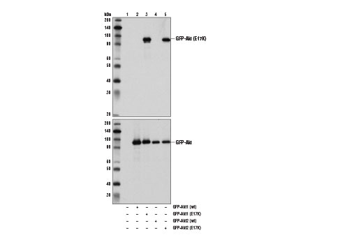
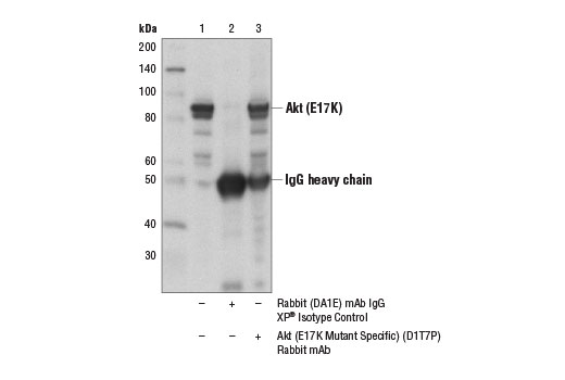
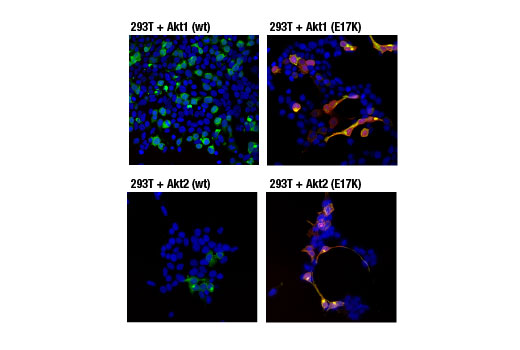
Mutation of the glutamic acid at residue 17 to lysine (E17K) of Akt was initially identified in human breast, colorectal, and ovarian cancers (20). This conserved glutamic acid residue is located at the lipid-binding pocket of the Akt1 plextrin homology domain. The E17K mutation increases the affinity between Akt1 and phospholipids at the plasma membrane, leading to increased Akt1 recruitment, super-activation of the Akt pathway, cellular transformation, and tumor formation (20,21). Additional studies detect the presence of the Akt1 (E17K) mutation in multiple cancers, including lung cancer, prostate cancer, and endometrial carcinoma (22,23). The presence of mutant Akt3 (E17K) protein has also been seen in cases of melanoma (24).
Background References
- Franke, T.F. et al. (1997) Cell 88, 435-7.
- Burgering, B.M. and Coffer, P.J. (1995) Nature 376, 599-602.
- Franke, T.F. et al. (1995) Cell 81, 727-36.
- Alessi, D.R. et al. (1996) EMBO J 15, 6541-51.
- Sarbassov, D.D. et al. (2005) Science 307, 1098-101.
- Jacinto, E. et al. (2006) Cell 127, 125-37.
- Cardone, M.H. et al. (1998) Science 282, 1318-21.
- Brunet, A. et al. (1999) Cell 96, 857-68.
- Zimmermann, S. and Moelling, K. (1999) Science 286, 1741-4.
- Cantley, L.C. and Neel, B.G. (1999) Proc Natl Acad Sci USA 96, 4240-5.
- Vlahos, C.J. et al. (1994) J Biol Chem 269, 5241-8.
- Hajduch, E. et al. (2001) FEBS Lett 492, 199-203.
- Cross, D.A. et al. (1995) Nature 378, 785-9.
- Diehl, J.A. et al. (1998) Genes Dev 12, 3499-511.
- Gesbert, F. et al. (2000) J Biol Chem 275, 39223-30.
- Zhou, B.P. et al. (2001) Nat Cell Biol 3, 245-52.
- Navé, B.T. et al. (1999) Biochem J 344 Pt 2, 427-31.
- Inoki, K. et al. (2002) Nat Cell Biol 4, 648-57.
- Manning, B.D. et al. (2002) Mol Cell 10, 151-62.
- Carpten, J.D. et al. (2007) Nature 448, 439-44.
- Landgraf, K.E. et al. (2008) Biochemistry 47, 12260-9.
- Malanga, D. et al. (2008) Cell Cycle 7, 665-9.
- Cohen, Y. et al. (2010) Gynecol Oncol 116, 88-91.
- Davies, M.A. et al. (2008) Br J Cancer 99, 1265-8.
Species Reactivity
Species reactivity is determined by testing in at least one approved application (e.g., western blot).
Western Blot Buffer
IMPORTANT: For western blots, incubate membrane with diluted primary antibody in 5% w/v BSA, 1X TBS, 0.1% Tween® 20 at 4°C with gentle shaking, overnight.
Applications Key
W: Western Blotting IP: Immunoprecipitation IF-IC: Immunofluorescence (Immunocytochemistry)
Cross-Reactivity Key
H: Human
Trademarks and Patents
Cell Signaling Technology is a trademark of Cell Signaling Technology, Inc.
All other trademarks are the property of their respective owners. Visit cellsignal.com/trademarks for more information.
Limited Uses
Except as otherwise expressly agreed in a writing signed by a legally authorized representative of CST, the following terms apply to Products provided by CST, its affiliates or its distributors. Any Customer's terms and conditions that are in addition to, or different from, those contained herein, unless separately accepted in writing by a legally authorized representative of CST, are rejected and are of no force or effect.
Products are labeled with For Research Use Only or a similar labeling statement and have not been approved, cleared, or licensed by the FDA or other regulatory foreign or domestic entity, for any purpose. Customer shall not use any Product for any diagnostic or therapeutic purpose, or otherwise in any manner that conflicts with its labeling statement. Products sold or licensed by CST are provided for Customer as the end-user and solely for research and development uses. Any use of Product for diagnostic, prophylactic or therapeutic purposes, or any purchase of Product for resale (alone or as a component) or other commercial purpose, requires a separate license from CST. Customer shall (a) not sell, license, loan, donate or otherwise transfer or make available any Product to any third party, whether alone or in combination with other materials, or use the Products to manufacture any commercial products, (b) not copy, modify, reverse engineer, decompile, disassemble or otherwise attempt to discover the underlying structure or technology of the Products, or use the Products for the purpose of developing any products or services that would compete with CST products or services, (c) not alter or remove from the Products any trademarks, trade names, logos, patent or copyright notices or markings, (d) use the Products solely in accordance with CST Product Terms of Sale and any applicable documentation, and (e) comply with any license, terms of service or similar agreement with respect to any third party products or services used by Customer in connection with the Products.
Revision 1