Revision 10
#3633
Store at -20C
877-616-CELL (2355)
877-678-TECH (8324)
3 Trask Lane | Danvers | Massachusetts | 01923 | USA
For Research Use Only. Not for Use in Diagnostic Procedures.
Applications:
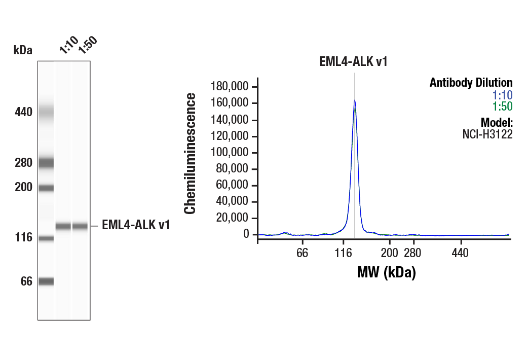
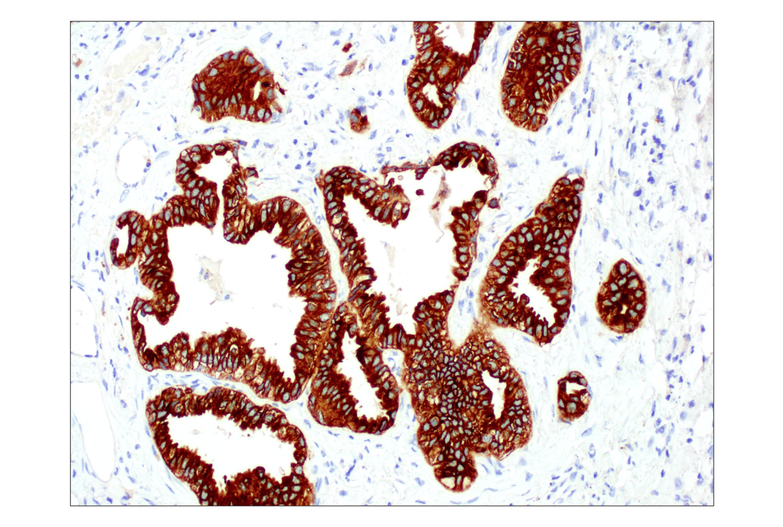
W, W-S, IP, IHC-P, FC-FP
Reactivity:
H
Sensitivity:
Endogenous
MW (kDa):
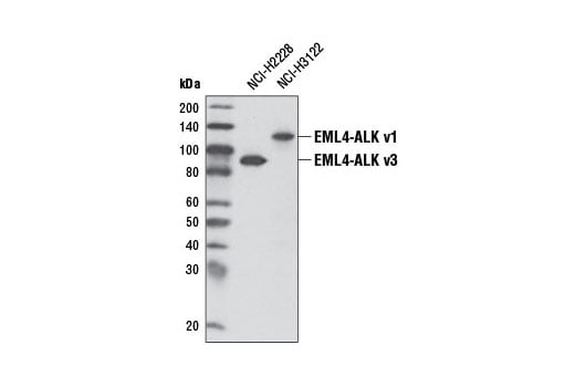
220 (ALK), 80 (NPM-ALK), 117 (EML4-ALK v1), 86 (EML4-ALK v3)
Source/Isotype:
Rabbit IgG
UniProt ID:
#Q9UM73
Entrez-Gene Id:
238
Product Usage Information
| Application | Dilution |
|---|---|
| Western Blotting | 1:2000 |
| Simple Western™ | 1:10 - 1:50 |
| Immunoprecipitation | 1:100 |
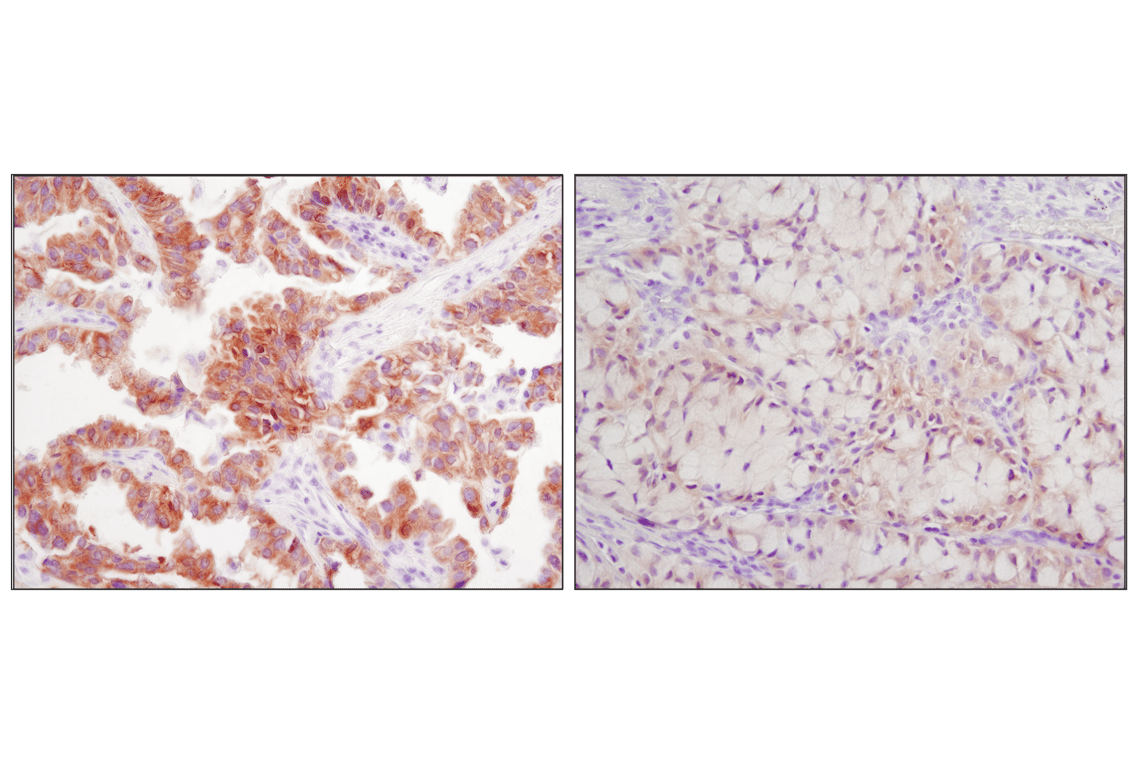
| Immunohistochemistry (Paraffin) | 1:100 - 1:400 |
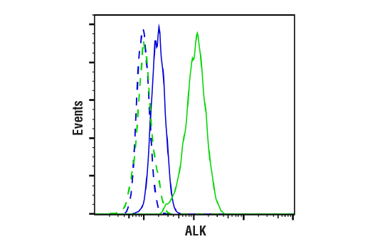
| Flow Cytometry (Fixed/Permeabilized) | 1:400 - 1:800 |
Storage
For a carrier free (BSA and azide free) version of this product see product #91392.
Specificity/Sensitivity
Source / Purification
Background
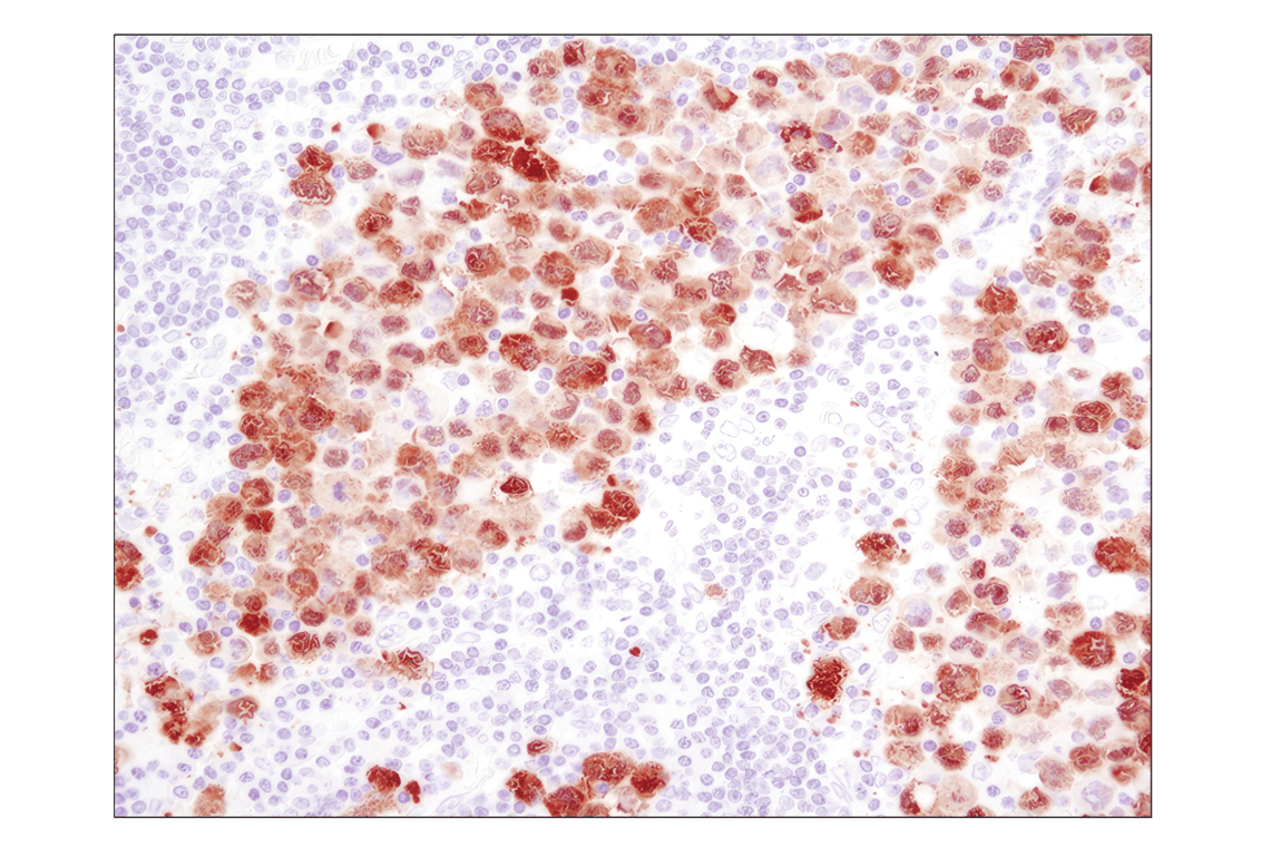
A distinct ALK oncogenic fusion protein involving ALK and echinoderm microtubule-associated protein like 4 (EML4) has been described in the research literature from a non-small cell lung cancer (NSCLC) cell line, with corresponding fusion transcripts present in some cases of lung adenocarcinoma. The short, amino-terminal region of the microtubule-associated protein EML4 is fused to the kinase domain of ALK (6-8).
Investigators have identified ALK translocations with other fusion partners, such as TRK-fused gene (TFG) and KIF5B, which have also been associated with NSCLC (6,7). In particular, the EML4-ALK fusion protein has been found in 3-7% of NSCLC samples (6-14).
Background References
- Stoica, G.E. et al. (2001) J Biol Chem 276, 16772-9.
- Iwahara, T. et al. (1997) Oncogene 14, 439-49.
- Morris, S.W. et al. (1997) Oncogene 14, 2175-88.
- Morris, S.W. et al. (1994) Science 263, 1281-4.
- Bai, R.Y. et al. (1998) Mol Cell Biol 18, 6951-61.
- Rikova, K. et al. (2007) Cell 131, 1190-203.
- Takeuchi, K. et al. (2008) Clin Cancer Res 14, 6618-24.
- Soda, M. et al. (2007) Nature 448, 561-6.
- Takeuchi, K. et al. (2009) Clin Cancer Res 15, 3143-9.
- Palmer, R.H. et al. (2009) Biochem J 420, 345-61.
- Horn, L. and Pao, W. (2009) J Clin Oncol 27, 4232-5.
- Rodig, S.J. et al. (2009) Clin Cancer Res 15, 5216-23.
- Mino-Kenudson, M. et al. (2010) Clin Cancer Res 16, 1561-71.
- Kwak, E.L. et al. (2010) N Engl J Med 363, 1693-703.
Species Reactivity
Species reactivity is determined by testing in at least one approved application (e.g., western blot).
Western Blot Buffer
IMPORTANT: For western blots, incubate membrane with diluted primary antibody in 5% w/v nonfat dry milk, 1X TBS, 0.1% Tween® 20 at 4°C with gentle shaking, overnight.
Applications Key
W: Western Blotting W-S: Simple Western™ IP: Immunoprecipitation IHC-P: Immunohistochemistry (Paraffin) FC-FP: Flow Cytometry (Fixed/Permeabilized)
Cross-Reactivity Key
H: Human
Trademarks and Patents
Cell Signaling Technology is a trademark of Cell Signaling Technology, Inc.
D5F3 is a registered trademark of Cell Signaling Technology, Inc.
All other trademarks are the property of their respective owners. Visit cellsignal.com/trademarks for more information.
Limited Uses
Except as otherwise expressly agreed in a writing signed by a legally authorized representative of CST, the following terms apply to Products provided by CST, its affiliates or its distributors. Any Customer's terms and conditions that are in addition to, or different from, those contained herein, unless separately accepted in writing by a legally authorized representative of CST, are rejected and are of no force or effect.
Products are labeled with For Research Use Only or a similar labeling statement and have not been approved, cleared, or licensed by the FDA or other regulatory foreign or domestic entity, for any purpose. Customer shall not use any Product for any diagnostic or therapeutic purpose, or otherwise in any manner that conflicts with its labeling statement. Products sold or licensed by CST are provided for Customer as the end-user and solely for research and development uses. Any use of Product for diagnostic, prophylactic or therapeutic purposes, or any purchase of Product for resale (alone or as a component) or other commercial purpose, requires a separate license from CST. Customer shall (a) not sell, license, loan, donate or otherwise transfer or make available any Product to any third party, whether alone or in combination with other materials, or use the Products to manufacture any commercial products, (b) not copy, modify, reverse engineer, decompile, disassemble or otherwise attempt to discover the underlying structure or technology of the Products, or use the Products for the purpose of developing any products or services that would compete with CST products or services, (c) not alter or remove from the Products any trademarks, trade names, logos, patent or copyright notices or markings, (d) use the Products solely in accordance with CST Product Terms of Sale and any applicable documentation, and (e) comply with any license, terms of service or similar agreement with respect to any third party products or services used by Customer in connection with the Products.
Revision 10
Revision 10