Revision 1
#35277
Store at -20C
AMPK Substrate Antibody Sampler Kit
1 Kit
(8 x 20 microliters)
877-616-CELL (2355)
877-678-TECH (8324)
3 Trask Lane | Danvers | Massachusetts | 01923 | USA
For Research Use Only. Not for Use in Diagnostic Procedures.
| Product Includes | Product # | Quantity | Mol. Wt | Isotype/Source |
|---|---|---|---|---|
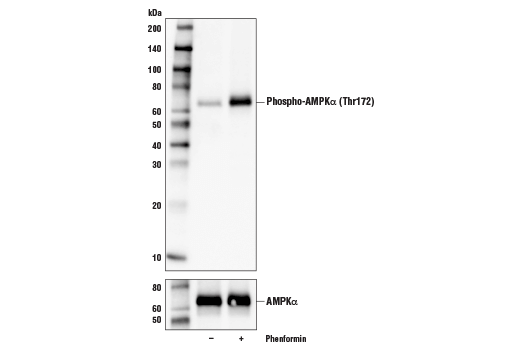
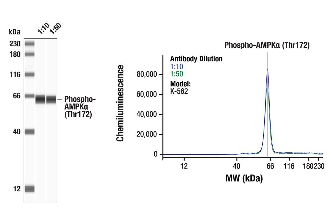
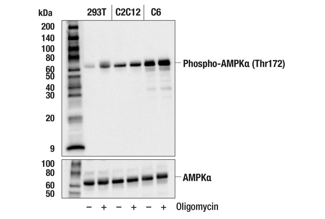
| Phospho-AMPK alpha (Thr172) (D4D6D) Rabbit Monoclonal Antibody | 50081 | 20 µl | 62 kDa | Rabbit IgG |
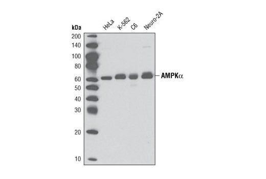
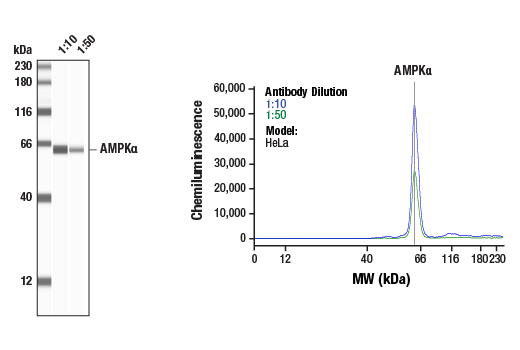
| AMPK alpha (D5A2) Rabbit Monoclonal Antibody | 5831 | 20 µl | 62 kDa | Rabbit IgG |
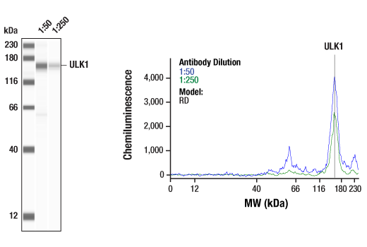
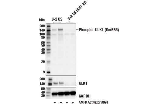
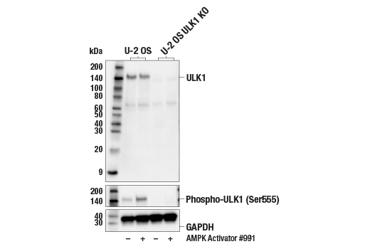
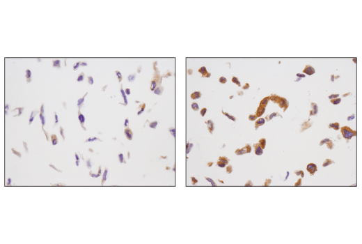
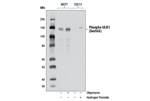
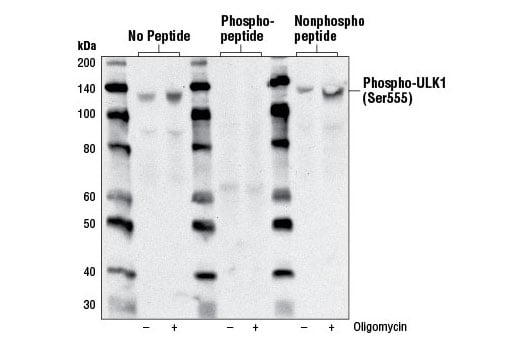
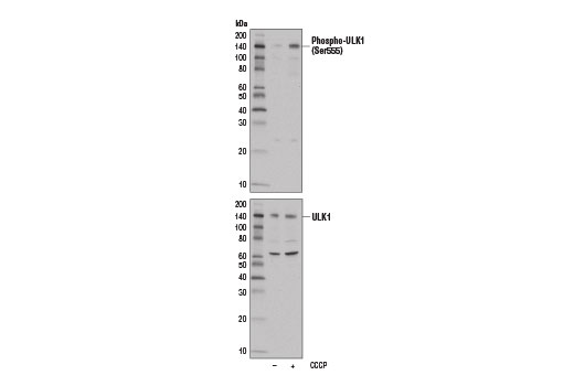
| Phospho-ULK1 (Ser555) (D1H4) Rabbit Monoclonal Antibody | 5869 | 20 µl | 140-150 kDa | Rabbit IgG |
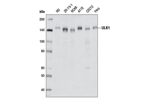
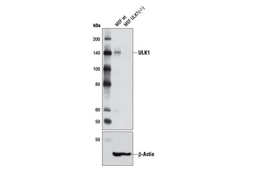
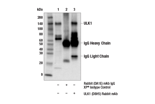
| ULK1 (D8H5) Rabbit Monoclonal Antibody | 8054 | 20 µl | 150 kDa | Rabbit IgG |
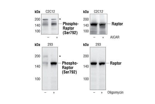
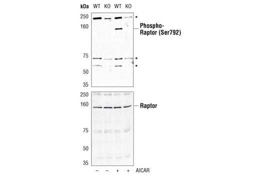
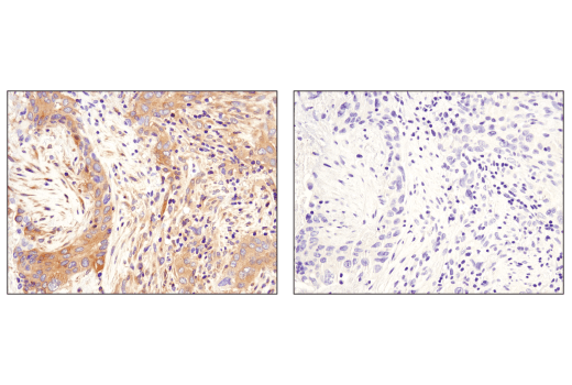
| Phospho-Raptor (Ser792) Antibody | 2083 | 20 µl | 150 kDa | Rabbit |
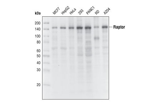
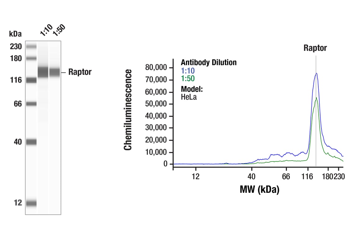
| Raptor (24C12) Rabbit Monoclonal Antibody | 2280 | 20 µl | 150 kDa | Rabbit |
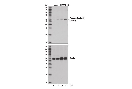
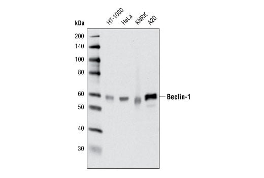
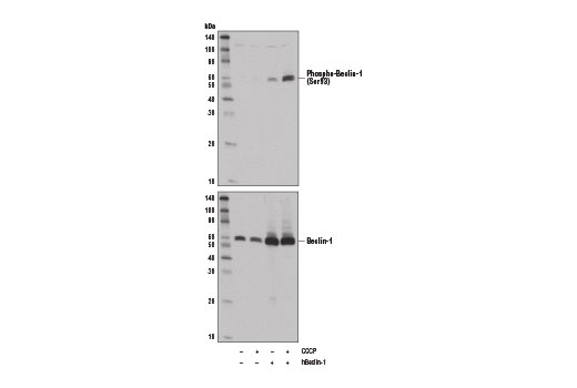
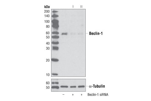
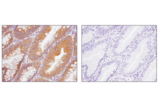
| Beclin-1 (D40C5) Rabbit Monoclonal Antibody | 3495 | 20 µl | 60 kDa | Rabbit IgG |
| Phospho-Beclin-1 (Ser93) (D9A5G) Rabbit Monoclonal Antibody | 14717 | 20 µl | 60 kDa | Rabbit IgG |
| Anti-rabbit IgG, HRP-linked Antibody | 7074 | 100 µl | Goat |
Please visit cellsignal.com for individual component applications, species cross-reactivity, dilutions, protocols, and additional product information.
Description
Storage
Background
AMPK phosphorylates a number of targets controlling cellular processes such as metabolism, cell growth, and autophagy (6). It suppresses the activity of the mammalian target of rapamycin (mTOR), that plays a key role in promoting cell growth. The regulatory associated protein of mTOR (Raptor) was identified as an mTOR binding partner that mediates mTOR signaling to downstream targets (7,8). Raptor binds to mTOR substrates, including 4E-BP1 and p70 S6 kinase, through their TOR signaling (TOS) motifs and is required for mTOR-mediated phosphorylation of these substrates (9,10). AMPK directly phosphorylates Raptor at Ser722/Ser792, and this phosphorylation is essential for inhibition of the raptor-containing mTOR complex 1 (mTORC1) and induces cell cycle arrest when cells are stressed for energy (11). AMPK also promotes autophagy by directly phosphorylating ULK1 (11,12). ULK1 is a Ser/Thr kinase required for the Initiation and formation of the autophagosome. AMPK, activated during low nutrient conditions, directly phosphorylates ULK1 at multiple sites including Ser317, Ser555, and Ser777 (11,12). Conversely, mTOR, which is a regulator of cell growth and an inhibitor of autophagy, phosphorylates ULK1 at Ser757 and disrupts the interaction between ULK1 and AMPK (11). AMPK can also directly phosphorylate Beclin-1, a component of the complex downstream of ULK1 in autophagosome formation that activates the class III phosphatidylinositol 3-kinase VPS34. AMPK phosphorylates Beclin-1 at Ser93 and Ser96 residues in human, which correspond to murine Ser91 and Ser94 (14).
Background References
- Hardie, D.G. (2004) J Cell Sci 117, 5479-87.
- Carling, D. (2004) Trends Biochem Sci 29, 18-24.
- Hawley, S.A. et al. (1996) J Biol Chem 271, 27879-87.
- Lizcano, J.M. et al. (2004) EMBO J 23, 833-43.
- Shaw, R.J. et al. (2004) Proc Natl Acad Sci U S A 101, 3329-35.
- Mihaylova, M.M. and Shaw, R.J. (2011) Nat Cell Biol 13, 1016-23.
- Hara, K. et al. (2002) Cell 110, 177-89.
- Kim, D.H. et al. (2002) Cell 110, 163-75.
- Beugnet, A. et al. (2003) J Biol Chem 278, 40717-22.
- Nojima, H. et al. (2003) J Biol Chem 278, 15461-4.
- Gwinn, D.M. et al. (2008) Mol Cell 30, 214-26.
- Kim, J. et al. (2011) Nat Cell Biol 13, 132-41.
- Egan, D.F. et al. (2011) Science 331, 456-61.
- Kim, J. et al. (2013) Cell 152, 290-303.
Trademarks and Patents
Cell Signaling Technology is a trademark of Cell Signaling Technology, Inc.
All other trademarks are the property of their respective owners. Visit cellsignal.com/trademarks for more information.
Limited Uses
Except as otherwise expressly agreed in a writing signed by a legally authorized representative of CST, the following terms apply to Products provided by CST, its affiliates or its distributors. Any Customer's terms and conditions that are in addition to, or different from, those contained herein, unless separately accepted in writing by a legally authorized representative of CST, are rejected and are of no force or effect.
Products are labeled with For Research Use Only or a similar labeling statement and have not been approved, cleared, or licensed by the FDA or other regulatory foreign or domestic entity, for any purpose. Customer shall not use any Product for any diagnostic or therapeutic purpose, or otherwise in any manner that conflicts with its labeling statement. Products sold or licensed by CST are provided for Customer as the end-user and solely for research and development uses. Any use of Product for diagnostic, prophylactic or therapeutic purposes, or any purchase of Product for resale (alone or as a component) or other commercial purpose, requires a separate license from CST. Customer shall (a) not sell, license, loan, donate or otherwise transfer or make available any Product to any third party, whether alone or in combination with other materials, or use the Products to manufacture any commercial products, (b) not copy, modify, reverse engineer, decompile, disassemble or otherwise attempt to discover the underlying structure or technology of the Products, or use the Products for the purpose of developing any products or services that would compete with CST products or services, (c) not alter or remove from the Products any trademarks, trade names, logos, patent or copyright notices or markings, (d) use the Products solely in accordance with CST Product Terms of Sale and any applicable documentation, and (e) comply with any license, terms of service or similar agreement with respect to any third party products or services used by Customer in connection with the Products.
Revision 1
*Cross-reacting bands at 200 kDa.
Revision 1
Revision 1

Revision 1
*Cross-reacting bands at 60, 70 and 240 kDa
Revision 1
Revision 1

Revision 1
Revision 1
Revision 1
Revision 1