Revision 5
#61949
Store at -20C
Androgen Receptor Antibody Sampler Kit
1 Kit
(3 x 20 microliters)
877-616-CELL (2355)
877-678-TECH (8324)
3 Trask Lane | Danvers | Massachusetts | 01923 | USA
For Research Use Only. Not for Use in Diagnostic Procedures.
| Product Includes | Product # | Quantity | Mol. Wt | Isotype/Source |
|---|---|---|---|---|
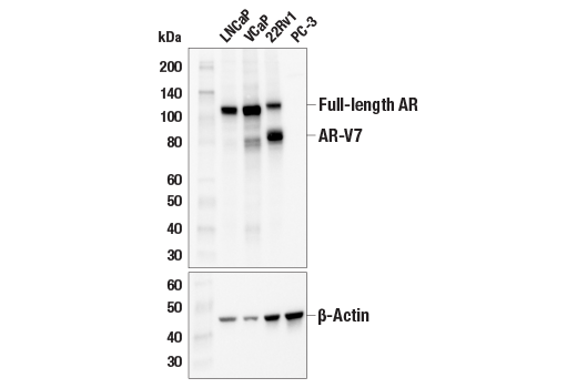
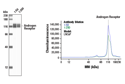
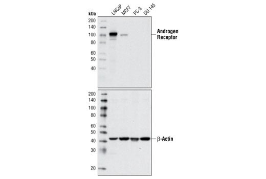
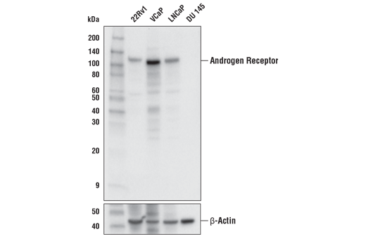
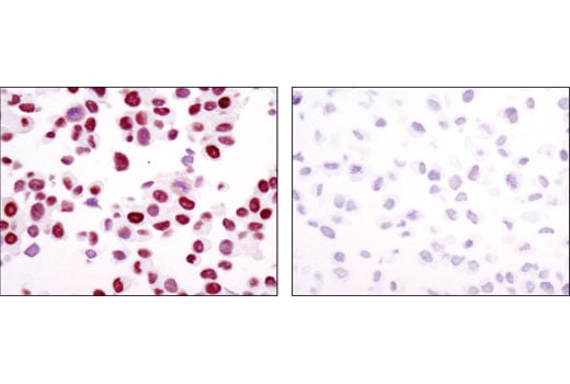
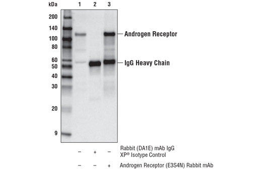
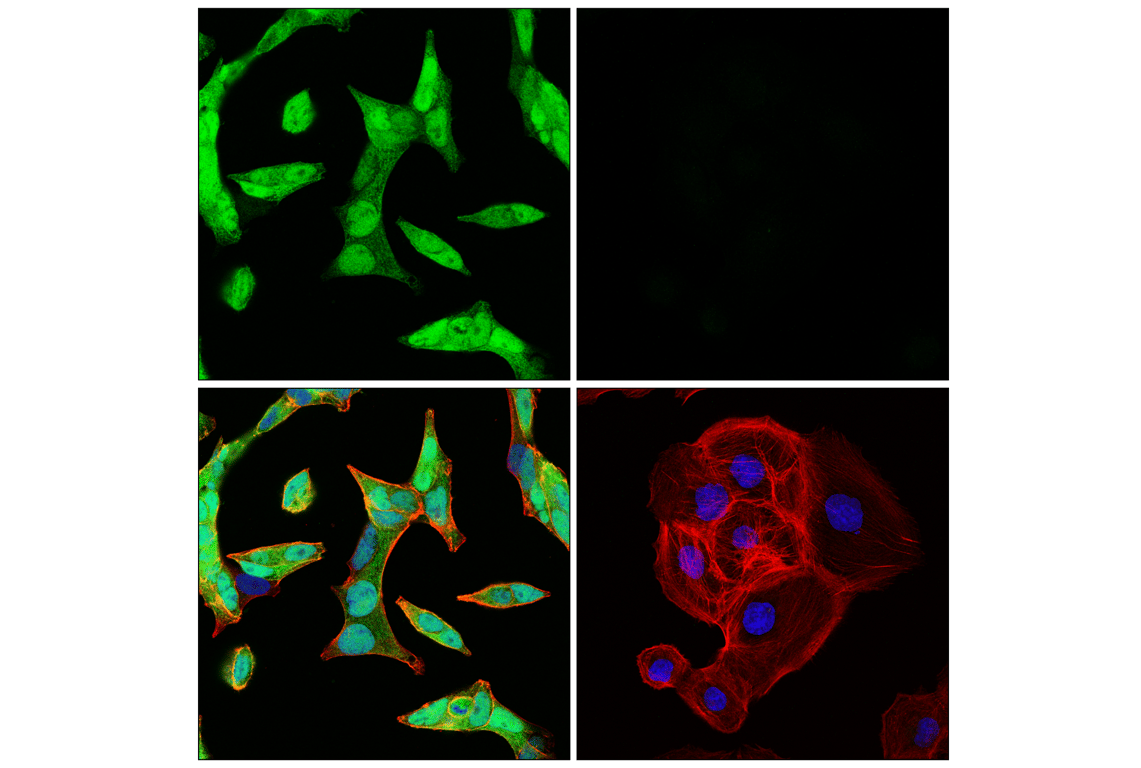
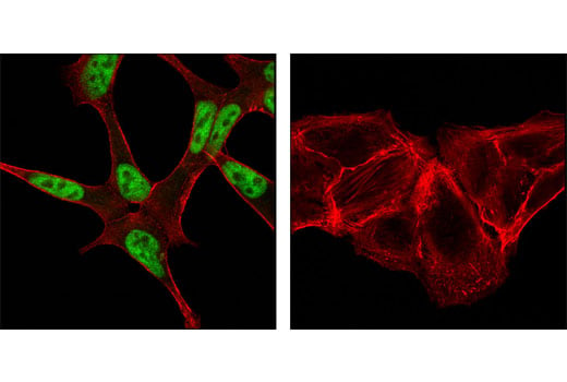
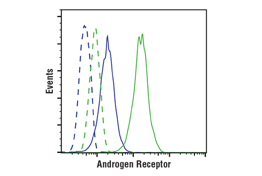
| Androgen Receptor (E3S4N) Rabbit Monoclonal Antibody (Carboxy-terminal Antigen) | 70317 | 20 µl | 110 kDa | Rabbit IgG |
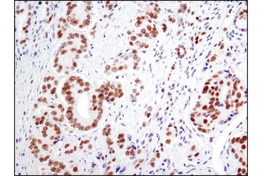
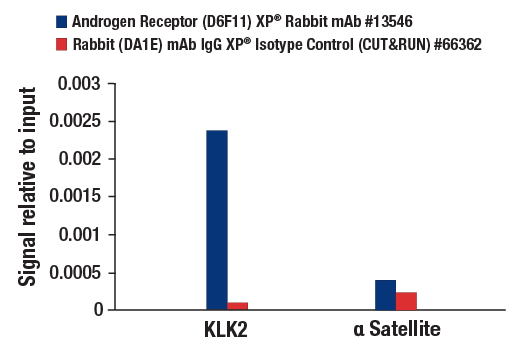
| Androgen Receptor (D6F11) Rabbit Monoclonal Antibody | 5153 | 20 µl | 110 kDa | Rabbit IgG |
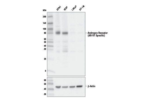
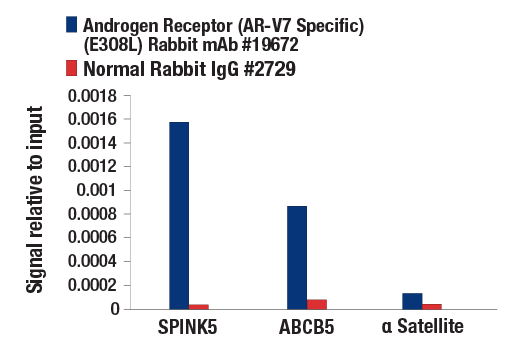
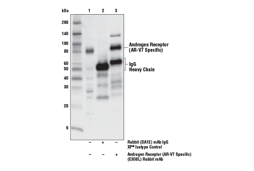
| Androgen Receptor (AR-V7 Specific) (E3O8L) Rabbit Monoclonal Antibody | 19672 | 20 µl | 80 kDa | Rabbit IgG |
| Anti-rabbit IgG, HRP-linked Antibody | 7074 | 100 µl | Goat |
Please visit cellsignal.com for individual component applications, species cross-reactivity, dilutions, protocols, and additional product information.
Description
Storage
Background
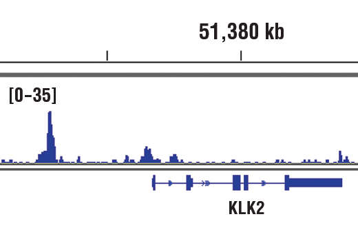
The AR3 or AR-V7 isoform, which lacks the typical ligand binding domain, is created through the alternative splicing of cryptic exons (4-5). AR-V7 is frequently expressed in castration-resistant prostate cancer (CRPC) and while dependent on the activity of the full-length androgen receptor (AR-FL), AR-V7 can activate a completely distinct transcriptional program (6-8). While enzalutamide and abiraterone have been beneficial in treating CRPC through the ligand binding domain of AR-FL, resistance in patients has been shown to be associated with AR-V7 detection in circulating tumor cells (9-12). Studies probing into mechanisms of overcoming this resistance are currently being explored and may help in stratifying patient populations for more personalized therapies (13-15).
Background References
- Li, J. and Al-Azzawi, F. (2009) Maturitas 63, 142-8.
- Avila, D.M. et al. J Steroid Biochem Mol Biol 76, 135-42.
- Montgomery, J.S. et al. (2001) J Pathol 195, 138-46.
- Hu, R. et al. (2009) Cancer Res 69, 16-22.
- Guo, Z. et al. (2009) Cancer Res 69, 2305-13.
- Watson, P.A. et al. (2010) Proc Natl Acad Sci U S A 107, 16759-65.
- Sun, S. et al. (2010) J Clin Invest 120, 2715-30.
- Hu, R. et al. (2012) Cancer Res 72, 3457-62.
- Scher, H.I. et al. (2012) N Engl J Med 367, 1187-97.
- de Bono, J.S. et al. (2011) N Engl J Med 364, 1995-2005.
- Ryan, C.J. et al. (2013) N Engl J Med 368, 138-48.
- Antonarakis, E.S. et al. (2014) N Engl J Med 371, 1028-38.
- Liu, C. et al. (2014) Clin Cancer Res 20, 3198-3210.
- Sarwar, M. et al. (2016) Oncotarget 7, 63065-63081.
- Ku, S.Y. et al. (2017) Science 355, 78-83.
Trademarks and Patents
Cell Signaling Technology is a trademark of Cell Signaling Technology, Inc.
All other trademarks are the property of their respective owners. Visit cellsignal.com/trademarks for more information.
Limited Uses
Except as otherwise expressly agreed in a writing signed by a legally authorized representative of CST, the following terms apply to Products provided by CST, its affiliates or its distributors. Any Customer's terms and conditions that are in addition to, or different from, those contained herein, unless separately accepted in writing by a legally authorized representative of CST, are rejected and are of no force or effect.
Products are labeled with For Research Use Only or a similar labeling statement and have not been approved, cleared, or licensed by the FDA or other regulatory foreign or domestic entity, for any purpose. Customer shall not use any Product for any diagnostic or therapeutic purpose, or otherwise in any manner that conflicts with its labeling statement. Products sold or licensed by CST are provided for Customer as the end-user and solely for research and development uses. Any use of Product for diagnostic, prophylactic or therapeutic purposes, or any purchase of Product for resale (alone or as a component) or other commercial purpose, requires a separate license from CST. Customer shall (a) not sell, license, loan, donate or otherwise transfer or make available any Product to any third party, whether alone or in combination with other materials, or use the Products to manufacture any commercial products, (b) not copy, modify, reverse engineer, decompile, disassemble or otherwise attempt to discover the underlying structure or technology of the Products, or use the Products for the purpose of developing any products or services that would compete with CST products or services, (c) not alter or remove from the Products any trademarks, trade names, logos, patent or copyright notices or markings, (d) use the Products solely in accordance with CST Product Terms of Sale and any applicable documentation, and (e) comply with any license, terms of service or similar agreement with respect to any third party products or services used by Customer in connection with the Products.
Revision 5
Revision 5

Revision 5

Revision 5
Revision 5
Revision 5
Revision 5