Revision 5
#32934
Store at -20C
877-616-CELL (2355)
877-678-TECH (8324)
3 Trask Lane | Danvers | Massachusetts | 01923 | USA
For Research Use Only. Not for Use in Diagnostic Procedures.
Applications:
W, IP, IHC-P, IF-IC, FC-FP
Reactivity:
H
Sensitivity:
Endogenous
MW (kDa):
38
Source/Isotype:
Rabbit IgG
UniProt ID:
#P04083
Entrez-Gene Id:
301
Product Usage Information
| Application | Dilution |
|---|---|
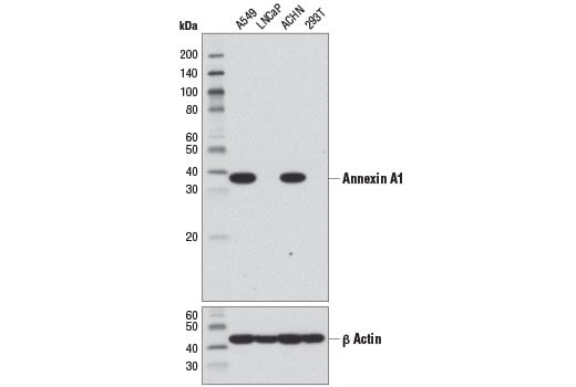
| Western Blotting | 1:1000 |
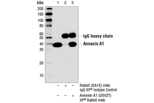
| Immunoprecipitation | 1:50 |
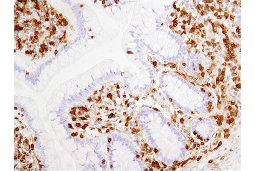
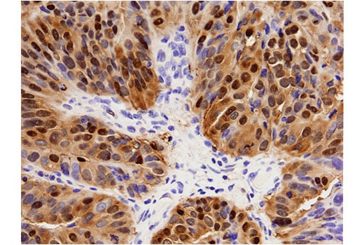
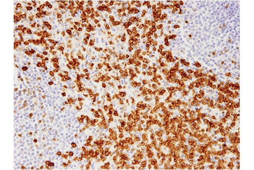
| Immunohistochemistry (Paraffin) | 1:400 |
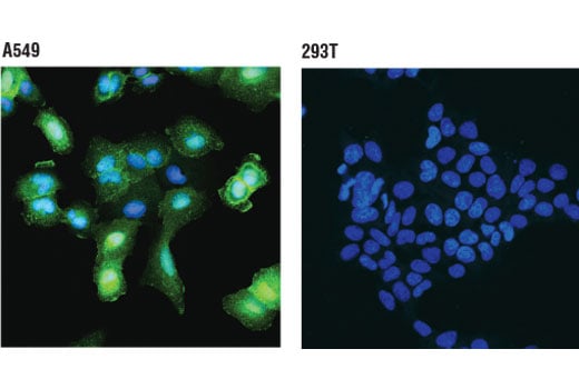
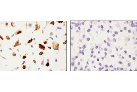
| Immunofluorescence (Immunocytochemistry) | 1:400 |
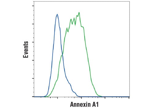
| Flow Cytometry (Fixed/Permeabilized) | 1:200 |
Storage
For a carrier free (BSA and azide free) version of this product see product #30797.
Specificity/Sensitivity
Source / Purification
Background
Background References
- Raynal, P. and Pollard, H.B. (1994) Biochim Biophys Acta 1197, 63-93.
- Blackwell, G.J. et al. (1980) Nature 287, 147-9.
- Rothhut, B. et al. (1983) Biochem Biophys Res Commun 117, 878-84.
- Hirata, F. et al. (1981) Proc Natl Acad Sci USA 78, 3190-4.
- Kim, K.M. et al. (1994) FEBS Lett 343, 251-5.
- Kim, S.W. et al. (2001) J Biol Chem 276, 15712-9.
- White, I.J. et al. (2006) EMBO J 25, 1-12.
- Varticovski, L. et al. (1988) Biochemistry 27, 3682-90.
- Dorovkov, M.V. and Ryazanov, A.G. (2004) J Biol Chem 279, 50643-6.
- Wang, W. and Creutz, C.E. (1994) Biochemistry 33, 275-82.
- Arur, S. et al. (2003) Dev Cell 4, 587-98.
- Perretti, M. and Gavins, F.N. (2003) News Physiol Sci 18, 60-4.
- Parente, L. and Solito, E. (2004) Inflamm Res 53, 125-32.
- Lim, L.H. and Pervaiz, S. (2007) FASEB J 21, 968-75.
Species Reactivity
Species reactivity is determined by testing in at least one approved application (e.g., western blot).
Western Blot Buffer
IMPORTANT: For western blots, incubate membrane with diluted primary antibody in 5% w/v BSA, 1X TBS, 0.1% Tween® 20 at 4°C with gentle shaking, overnight.
Applications Key
W: Western Blotting IP: Immunoprecipitation IHC-P: Immunohistochemistry (Paraffin) IF-IC: Immunofluorescence (Immunocytochemistry) FC-FP: Flow Cytometry (Fixed/Permeabilized)
Cross-Reactivity Key
H: Human
Trademarks and Patents
Cell Signaling Technology is a trademark of Cell Signaling Technology, Inc.
Alexa Fluor is a registered trademark of Life Technologies Corporation.
This product is provided under an intellectual property license from Life Technologies Corporation. The transfer of this product is conditioned on the buyer using the purchased product solely in research conducted by the buyer, excluding contract research or any fee for service research, and the buyer must not (1) use this product or its components for (a) diagnostic, therapeutic or prophylactic purposes; (b) testing, analysis or screening services, or information in return for compensation on a per-test basis; or (c) manufacturing or quality assurance or quality control, and/or (2) sell or transfer this product or its components for resale, whether or not resold for use in research. For information on purchasing a license to this product for purposes other than as described above, contact Life Technologies Corporation, 5791 Van Allen Way, Carlsbad, CA 92008 USA or [email protected].
All other trademarks are the property of their respective owners. Visit cellsignal.com/trademarks for more information.
Limited Uses
Except as otherwise expressly agreed in a writing signed by a legally authorized representative of CST, the following terms apply to Products provided by CST, its affiliates or its distributors. Any Customer's terms and conditions that are in addition to, or different from, those contained herein, unless separately accepted in writing by a legally authorized representative of CST, are rejected and are of no force or effect.
Products are labeled with For Research Use Only or a similar labeling statement and have not been approved, cleared, or licensed by the FDA or other regulatory foreign or domestic entity, for any purpose. Customer shall not use any Product for any diagnostic or therapeutic purpose, or otherwise in any manner that conflicts with its labeling statement. Products sold or licensed by CST are provided for Customer as the end-user and solely for research and development uses. Any use of Product for diagnostic, prophylactic or therapeutic purposes, or any purchase of Product for resale (alone or as a component) or other commercial purpose, requires a separate license from CST. Customer shall (a) not sell, license, loan, donate or otherwise transfer or make available any Product to any third party, whether alone or in combination with other materials, or use the Products to manufacture any commercial products, (b) not copy, modify, reverse engineer, decompile, disassemble or otherwise attempt to discover the underlying structure or technology of the Products, or use the Products for the purpose of developing any products or services that would compete with CST products or services, (c) not alter or remove from the Products any trademarks, trade names, logos, patent or copyright notices or markings, (d) use the Products solely in accordance with CST Product Terms of Sale and any applicable documentation, and (e) comply with any license, terms of service or similar agreement with respect to any third party products or services used by Customer in connection with the Products.
Revision 5
Revision 5
Revision 5