Revision 6
#39327
Store at -20C
877-616-CELL (2355)
877-678-TECH (8324)
3 Trask Lane | Danvers | Massachusetts | 01923 | USA
For Research Use Only. Not for Use in Diagnostic Procedures.
Applications:
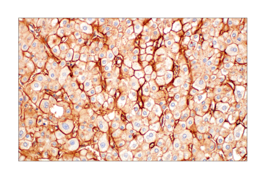
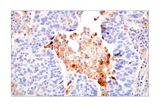
W, W-S, IHC-P
Reactivity:
H
Sensitivity:
Endogenous
MW (kDa):
35
Source/Isotype:
Rabbit IgG
UniProt ID:
#P02649
Entrez-Gene Id:
348
Product Usage Information
| Application | Dilution |
|---|---|
| Western Blotting | 1:1000 |
| Simple Western™ | 1:10 - 1:50 |
| Immunohistochemistry (Paraffin) | 1:400 - 1:1600 |
Storage
For a carrier free (BSA and azide free) version of this product see product #34392.
Specificity/Sensitivity
Source / Purification
Background
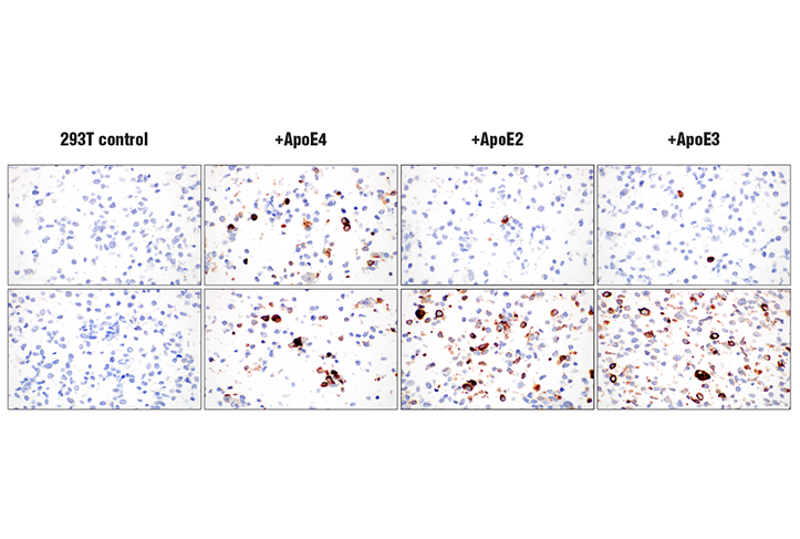
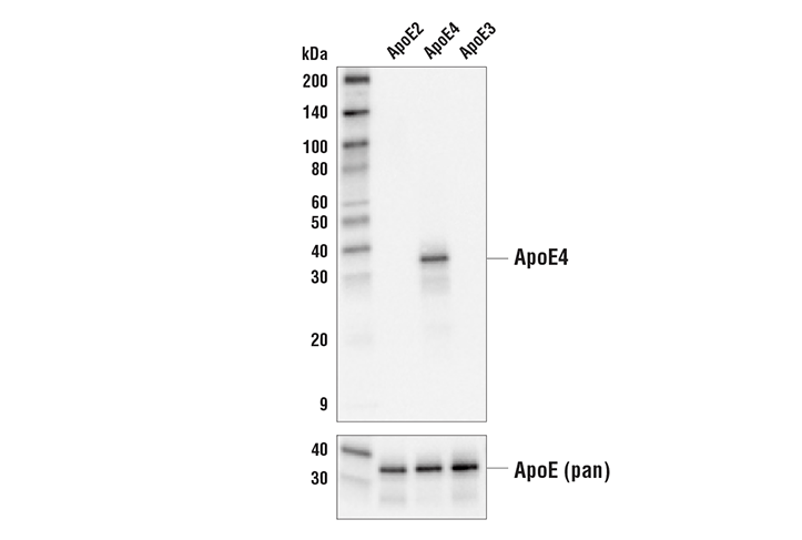
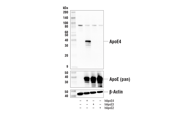
Human ApoE has three isoforms: ApoE2, ApoE3, and ApoE4. These three isoforms differ in the combination of cysteine and arginine residues located at positions 130 and 176. The ApoE4 isoform contains arginine at both locations. Research studies have linked ApoE4 function to neuronal plasticity, synaptogenesis, and neurodegenerative diseases (3). ApoE4 is produced in the liver and brain, although it is widely expressed in other tissues, such as the lung, spleen, and ovary. Investigators have established the ApoE4 allele as a genetic risk factor for Alzheimer’s disease (AD), accounting for 50-60% of the genetic variation in the disease (4). Research studies indicate that patients expressing ApoE4 have a reduced capacity for synaptic plasticity, an earlier age of onset of AD, and an increase in amyloid-beta (Aβ) deposition. The increase in Aβ suggests a role for ApoE4 in the impairment of amyloid clearance (5).
Species Reactivity
Species reactivity is determined by testing in at least one approved application (e.g., western blot).
Western Blot Buffer
IMPORTANT: For western blots, incubate membrane with diluted primary antibody in 5% w/v BSA, 1X TBS, 0.1% Tween® 20 at 4°C with gentle shaking, overnight.
Applications Key
W: Western Blotting W-S: Simple Western™ IHC-P: Immunohistochemistry (Paraffin)
Cross-Reactivity Key
H: Human
Trademarks and Patents
Cell Signaling Technology is a trademark of Cell Signaling Technology, Inc.
SignalStain is a registered trademark of Cell Signaling Technology, Inc.
All other trademarks are the property of their respective owners. Visit cellsignal.com/trademarks for more information.
Limited Uses
Except as otherwise expressly agreed in a writing signed by a legally authorized representative of CST, the following terms apply to Products provided by CST, its affiliates or its distributors. Any Customer's terms and conditions that are in addition to, or different from, those contained herein, unless separately accepted in writing by a legally authorized representative of CST, are rejected and are of no force or effect.
Products are labeled with For Research Use Only or a similar labeling statement and have not been approved, cleared, or licensed by the FDA or other regulatory foreign or domestic entity, for any purpose. Customer shall not use any Product for any diagnostic or therapeutic purpose, or otherwise in any manner that conflicts with its labeling statement. Products sold or licensed by CST are provided for Customer as the end-user and solely for research and development uses. Any use of Product for diagnostic, prophylactic or therapeutic purposes, or any purchase of Product for resale (alone or as a component) or other commercial purpose, requires a separate license from CST. Customer shall (a) not sell, license, loan, donate or otherwise transfer or make available any Product to any third party, whether alone or in combination with other materials, or use the Products to manufacture any commercial products, (b) not copy, modify, reverse engineer, decompile, disassemble or otherwise attempt to discover the underlying structure or technology of the Products, or use the Products for the purpose of developing any products or services that would compete with CST products or services, (c) not alter or remove from the Products any trademarks, trade names, logos, patent or copyright notices or markings, (d) use the Products solely in accordance with CST Product Terms of Sale and any applicable documentation, and (e) comply with any license, terms of service or similar agreement with respect to any third party products or services used by Customer in connection with the Products.
Revision 6
Revision 6
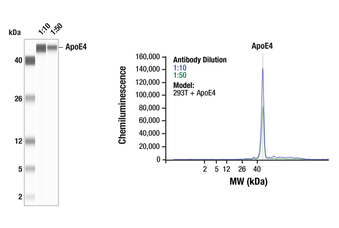
Simple Western™ analysis of lysates (0.1 mg/mL) from 293T cells transfected with constructs expressing full-length human ApoE4 protein using ApoE4 (E5M4L) Rabbit mAb #39327. The virtual lane view (left) shows the target band (as indicated) at 1:10 and 1:50 dilutions of primary antibody. The corresponding electropherogram view (right) plots chemiluminescence by molecular weight along the capillary at 1:10 (blue line) and 1:50 (green line) dilutions of primary antibody. This experiment was performed under reducing conditions on the Jess™ Simple Western instrument from ProteinSimple, a BioTechne brand, using the 2-40 kDa separation module.
Revision 6
Revision 6