| Cat. # | Size | Qty. | Price |
|---|---|---|---|
| 93589T | 1 Kit (8 x 20 microliters) |
|
| Product Includes | Quantity | Applications | Reactivity | MW(kDa) | Isotype |
|---|---|---|---|---|---|
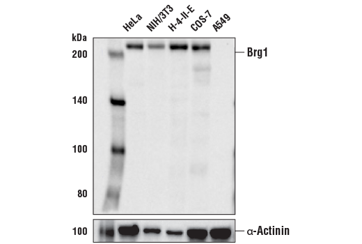
| Brg1 (E8V5B) Mouse mAb 72182 | 20 µl |
|
H M R Mk | 220 | Mouse IgG1 |
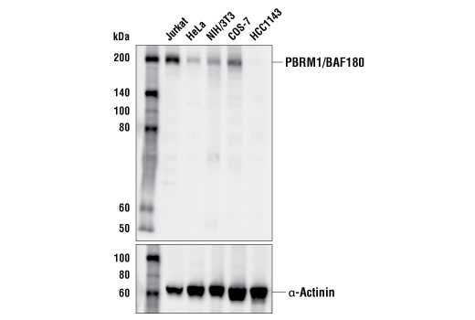
| PBRM1/BAF180 (D4L9X) Rabbit mAb 38439 | 20 µl |
|
H M R Mk | 205 | Rabbit IgG |
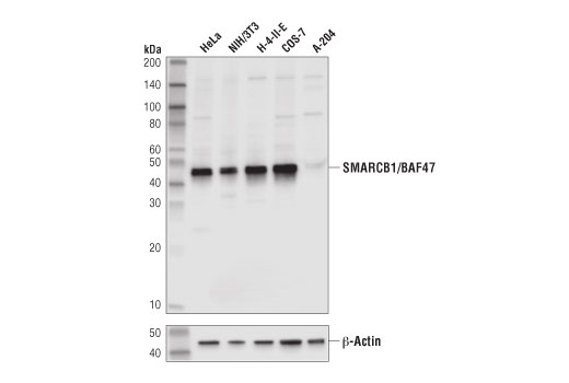
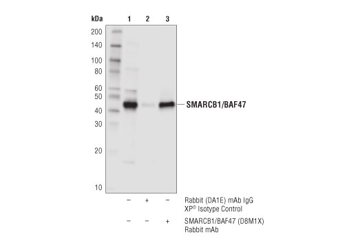
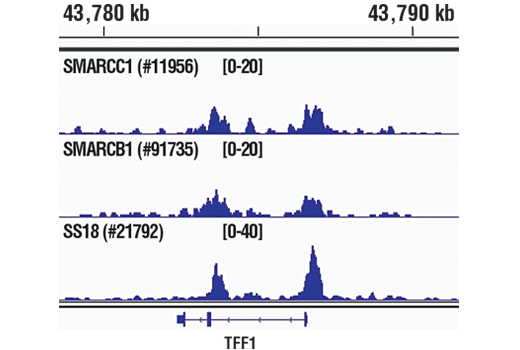
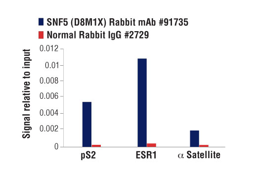
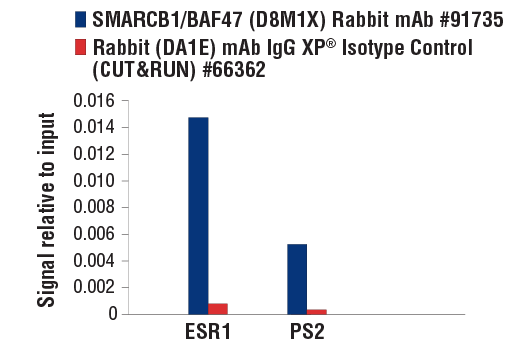
| SMARCB1/BAF47 (D8M1X) Rabbit mAb 91735 | 20 µl |
|
H M R Mk | 44 | Rabbit IgG |
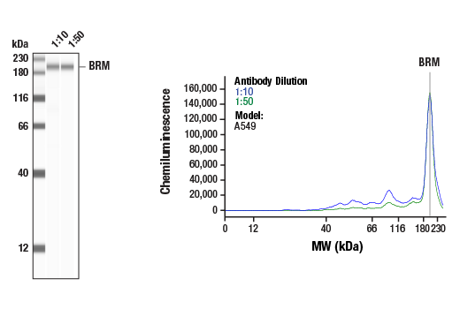
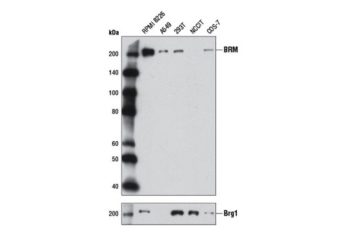
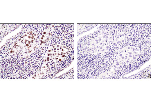
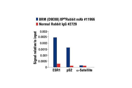
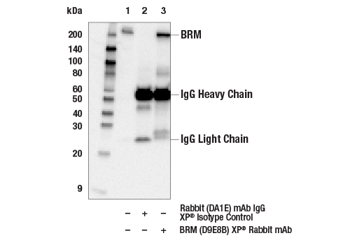
| BRM (D9E8B) XP® Rabbit mAb 11966 | 20 µl |
|
H Mk | 200 | Rabbit IgG |
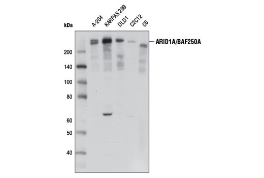
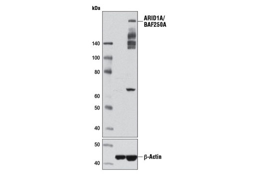
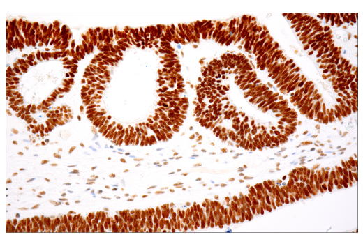
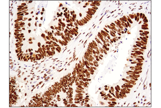
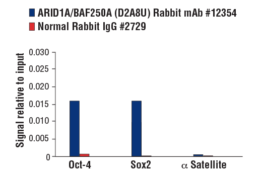
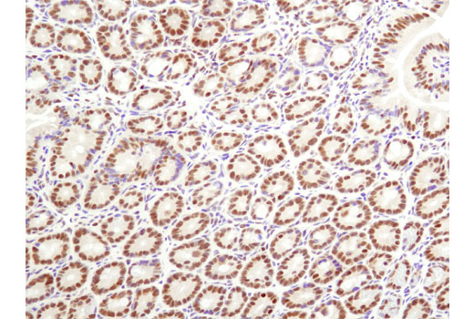
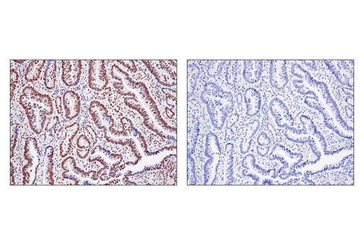
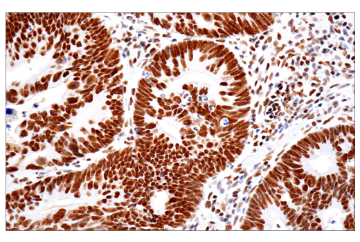
| ARID1A/BAF250A (D2A8U) Rabbit mAb 12354 | 20 µl |
|
H M R Mk | 270 | Rabbit IgG |
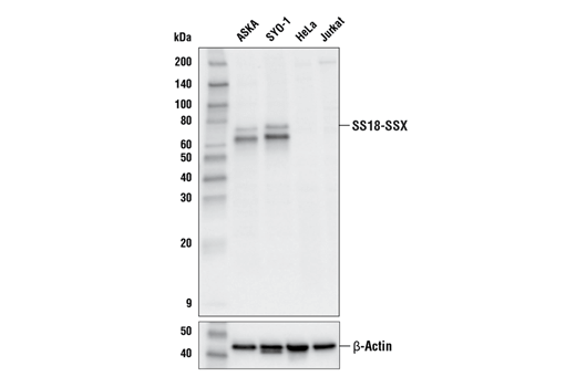
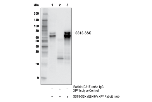
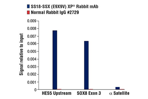
| SS18-SSX (E9X9V) XP® Rabbit mAb 72364 | 20 µl |
|
H | 65, 75 | Rabbit IgG |
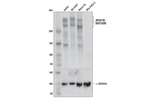
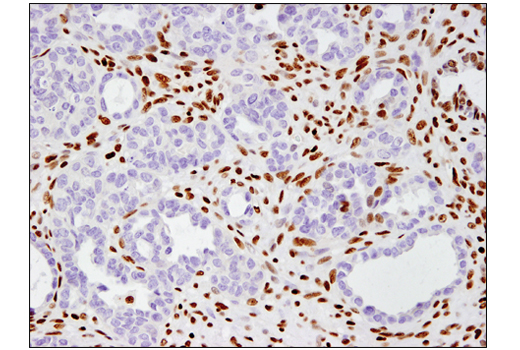
| ARID1B/BAF250B (E1U7D) Rabbit mAb 65747 | 20 µl |
|
H M | 250, 280 | Rabbit IgG |
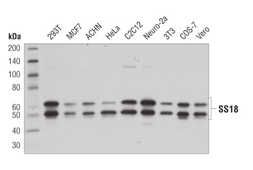
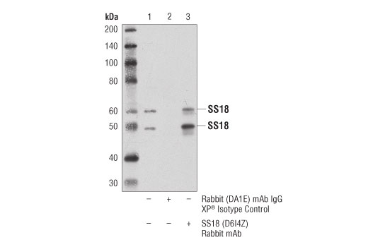
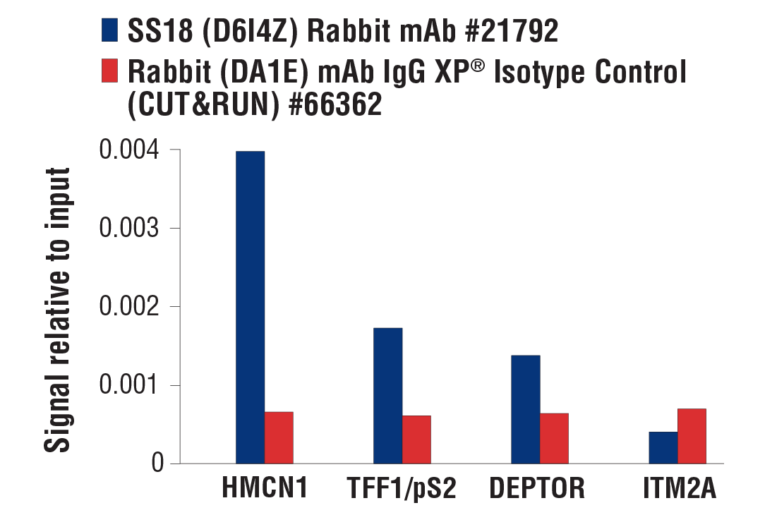
| SS18 (D6I4Z) Rabbit mAb 21792 | 20 µl |
|
H M R Mk | Iso1 60, Iso2 50 | Rabbit IgG |
| Anti-rabbit IgG, HRP-linked Antibody 7074 | 100 µl |
|
Goat | ||
| Anti-mouse IgG, HRP-linked Antibody 7076 | 100 µl |
|
Horse |
Product Information
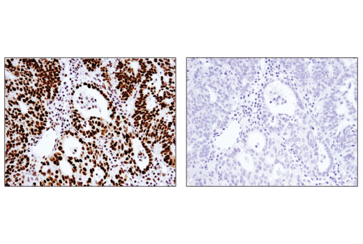
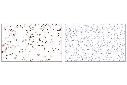
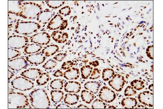
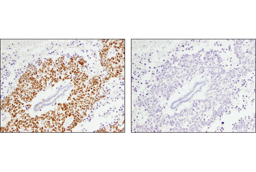
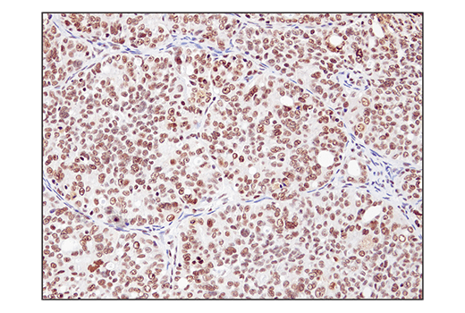
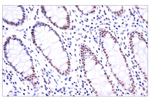
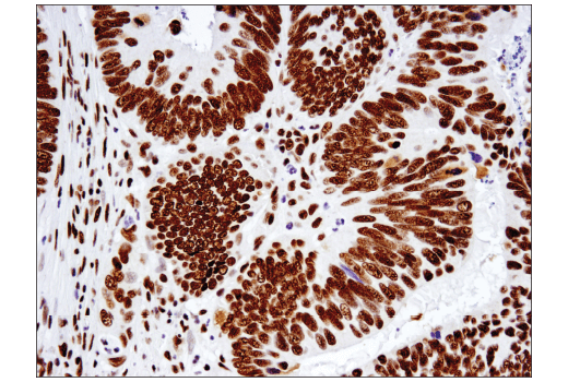
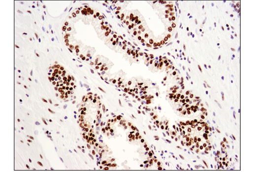
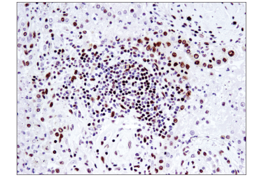
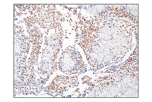
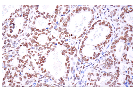
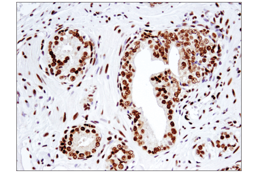
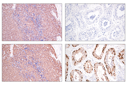
Monoclonal antibodies are produced by immunizing animals with synthetic peptides corresponding to residues surrounding Gly1293 of human ARID1A/BAF250A protein, Ala1320 of human ARID1B/BAF250B protein, Pro57 of human Brg1 protein, Gly264 of human BRM protein, Leu120 of human SMARCB1/BAF47 protein, Gln394 of human SS18 protein, and surrounding the fusion site of human SS18-SSX protein. Monoclonal antibody is produced by immunizing animals with recombinant protein specific to the amino terminus of human PBRM1/BAF180 protein.
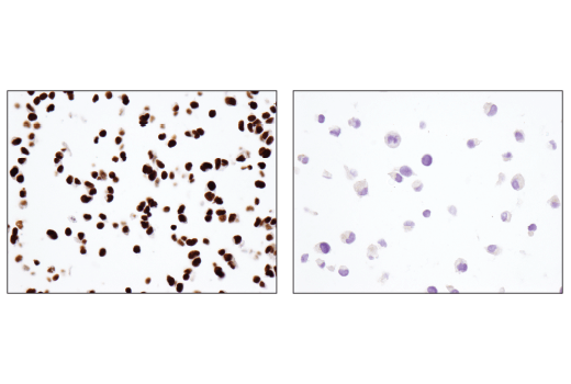
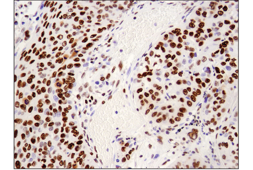
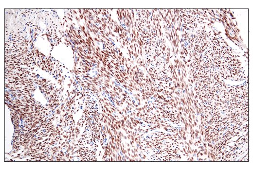
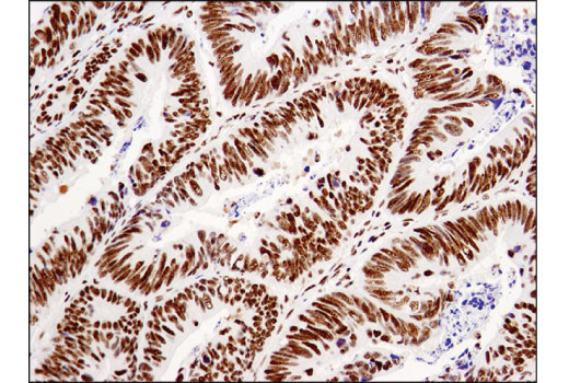
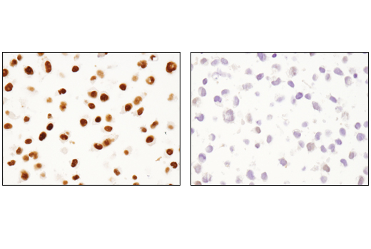
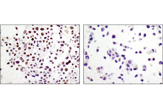
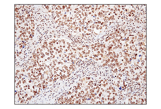
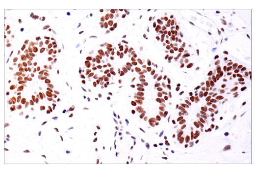
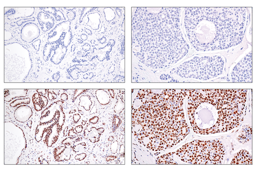
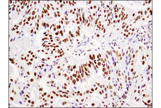
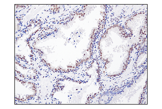
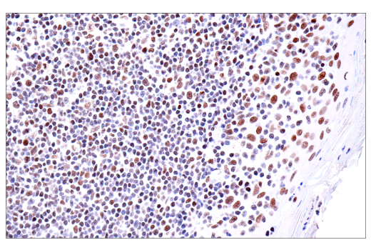
The modulation of chromatin structure is an essential component in the regulation of transcriptional activation and repression. Modifications can be made by at least two evolutionarily conserved strategies, through the disruption of histone-DNA contacts by ATP-dependent chromatin remodelers, or by histone tail modifications including methylation and acetylation. One of the four classes of ATP-dependent histone remodelers is the SWI/SNF complex, the central catalytic subunit of which is Brg1 or the highly related protein hBRM (1). This SWI/SNF complex contains varying subunits but its association with either Brg1 or hBRM remains constant (1). SWI/SNF complexes have been shown to regulate gene activation, cell growth, the cell cycle, and differentiation (1). Brg1/hBRM have been shown to regulate transcription through enhancing transcriptional activation of glucocorticoid receptors (2). Although usually associated with transcriptional activation, Brg1/hBRM have also been found in complexes associated with transcriptional repression, including HDACs, Rb, and Tif1β (3-5). Brg1/hBRM plays a vital role in the regulation of gene transcription during early mammalian embryogenesis. In addition, Brg1/hBRM also plays a role as a tumor suppressor and Brg1 is mutated in several tumor cell lines (6-8).
Except as otherwise expressly agreed in a writing signed by a legally authorized representative of CST, the following terms apply to Products provided by CST, its affiliates or its distributors. Any Customer's terms and conditions that are in addition to, or different from, those contained herein, unless separately accepted in writing by a legally authorized representative of CST, are rejected and are of no force or effect.
Products are labeled with For Research Use Only or a similar labeling statement and have not been approved, cleared, or licensed by the FDA or other regulatory foreign or domestic entity, for any purpose. Customer shall not use any Product for any diagnostic or therapeutic purpose, or otherwise in any manner that conflicts with its labeling statement. Products sold or licensed by CST are provided for Customer as the end-user and solely for research and development uses. Any use of Product for diagnostic, prophylactic or therapeutic purposes, or any purchase of Product for resale (alone or as a component) or other commercial purpose, requires a separate license from CST. Customer shall (a) not sell, license, loan, donate or otherwise transfer or make available any Product to any third party, whether alone or in combination with other materials, or use the Products to manufacture any commercial products, (b) not copy, modify, reverse engineer, decompile, disassemble or otherwise attempt to discover the underlying structure or technology of the Products, or use the Products for the purpose of developing any products or services that would compete with CST products or services, (c) not alter or remove from the Products any trademarks, trade names, logos, patent or copyright notices or markings, (d) use the Products solely in accordance with CST Product Terms of Sale and any applicable documentation, and (e) comply with any license, terms of service or similar agreement with respect to any third party products or services used by Customer in connection with the Products.