Revision 1
#12109
Store at -20C
877-616-CELL (2355)
877-678-TECH (8324)
3 Trask Lane | Danvers | Massachusetts | 01923 | USA
For Research Use Only. Not for Use in Diagnostic Procedures.
Applications:
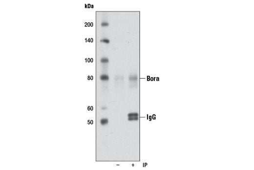
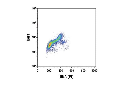
W, IP, FC-FP
Reactivity:
H
Sensitivity:
Endogenous
MW (kDa):
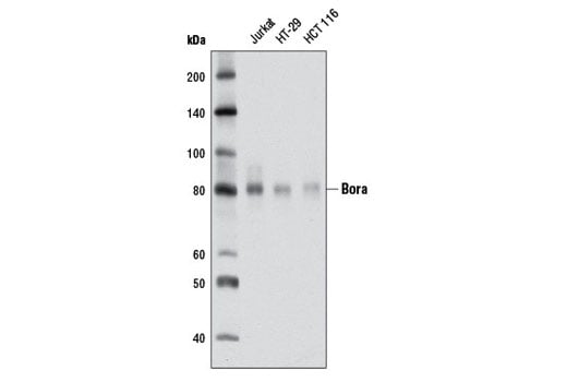
80
Source/Isotype:
Rabbit IgG
UniProt ID:
#Q6PGQ7
Entrez-Gene Id:
79866
Product Usage Information
| Application | Dilution |
|---|---|
| Western Blotting | 1:1000 |
| Immunoprecipitation | 1:50 |
| Flow Cytometry (Fixed/Permeabilized) | 1:200 |
Storage
Specificity/Sensitivity
Source / Purification
Background
Found to be conserved from C. elegans to humans, Bora is translocated from the nucleus to the cytoplasm upon activation of cdc2 at the onset of mitosis. Once present in the cytoplasm, Bora binds to and activates Aurora-A and PLK1 (3-5). It has been proposed that the binding of human Bora to PLK1 may lead to a conformational change in the protein that disrupts the autoinhibition by the Polo-Box Domain (PBD). This would allow for Thr210 on PLK1 to become more accessible for phosphorylation by Aurora-A (reviewed in 6). Active PLK1 then initiates the PLK1-cdc25-cdc2 positive feedback loop, leading to mitotic entry and the phosphorylation of Bora. Once phosphorylated in prophase, Bora is degraded allowing for normal mitotic progression (7).
Background References
- Nigg, E.A. (2001) Nat Rev Mol Cell Biol 2, 21-32.
- Archambault, V. and Carmena, M. (2012) Cell Cycle 11, 1490-5.
- Hutterer, A. et al. (2006) Dev Cell 11, 147-57.
- Seki, A. et al. (2008) Science 320, 1655-8.
- Chan, E.H. et al. (2008) Chromosoma 117, 457-69.
- Macurek, L. et al. (2009) Cancer Res 69, 4555-8.
- Seki, A. et al. (2008) J Cell Biol 181, 65-78.
Species Reactivity
Species reactivity is determined by testing in at least one approved application (e.g., western blot).
Western Blot Buffer
IMPORTANT: For western blots, incubate membrane with diluted primary antibody in 5% w/v BSA, 1X TBS, 0.1% Tween® 20 at 4°C with gentle shaking, overnight.
Applications Key
W: Western Blotting IP: Immunoprecipitation FC-FP: Flow Cytometry (Fixed/Permeabilized)
Cross-Reactivity Key
H: Human
Trademarks and Patents
Cell Signaling Technology is a trademark of Cell Signaling Technology, Inc.
Alexa Fluor is a registered trademark of Life Technologies Corporation.
All other trademarks are the property of their respective owners. Visit cellsignal.com/trademarks for more information.
Limited Uses
Except as otherwise expressly agreed in a writing signed by a legally authorized representative of CST, the following terms apply to Products provided by CST, its affiliates or its distributors. Any Customer's terms and conditions that are in addition to, or different from, those contained herein, unless separately accepted in writing by a legally authorized representative of CST, are rejected and are of no force or effect.
Products are labeled with For Research Use Only or a similar labeling statement and have not been approved, cleared, or licensed by the FDA or other regulatory foreign or domestic entity, for any purpose. Customer shall not use any Product for any diagnostic or therapeutic purpose, or otherwise in any manner that conflicts with its labeling statement. Products sold or licensed by CST are provided for Customer as the end-user and solely for research and development uses. Any use of Product for diagnostic, prophylactic or therapeutic purposes, or any purchase of Product for resale (alone or as a component) or other commercial purpose, requires a separate license from CST. Customer shall (a) not sell, license, loan, donate or otherwise transfer or make available any Product to any third party, whether alone or in combination with other materials, or use the Products to manufacture any commercial products, (b) not copy, modify, reverse engineer, decompile, disassemble or otherwise attempt to discover the underlying structure or technology of the Products, or use the Products for the purpose of developing any products or services that would compete with CST products or services, (c) not alter or remove from the Products any trademarks, trade names, logos, patent or copyright notices or markings, (d) use the Products solely in accordance with CST Product Terms of Sale and any applicable documentation, and (e) comply with any license, terms of service or similar agreement with respect to any third party products or services used by Customer in connection with the Products.
Revision 1