Recombinant antibodies offer several key advantages compared to traditional antibodies. These include superior lot-to-lot consistency, continuous supply, and animal-free manufacturing. As such, recombinant antibodies are seeing increased use for scientific research, especially as a means of addressing the ongoing reproducibility crisis.

Traditional polyclonal and monoclonal antibodies are the product of normal B cell development and genetic recombination. They are generated by immunizing an animal with an antigen to elicit an immune response. While polyclonal antibodies are secreted by many different B cell clones and recognize multiple antigenic epitopes, monoclonals originate from a single B cell clone and are specific for just one epitope.
Recombinant antibodies are monoclonal, but their production involves in vitro genetic manipulation. After cloning the antibody genes into an expression vector, this is then transfected into an appropriate host cell line for antibody expression. Mammalian cell lines are most commonly used for recombinant antibody production, although cell lines of bacterial, yeast, or insect origin are also suitable.
Because recombinant antibody production involves sequencing the antibody light and heavy chains, it is a highly controlled and reliable process. In contrast, hybridoma-based systems for producing monoclonal antibodies are subject to genetic drift and instability, increasing the potential for lot-to-lot variability or loss of antibody expression. Recombinant antibodies are highly consistent from lot to lot, thereby ensuring reproducible experimental results.
In vitro methods for producing antibodies are amenable to large-scale production, meaning antibody availability is unlikely to become a limiting factor. Moreover, since the recombinant antibody sequence is known, continuity of supply is assured; in situations where an antibody will be used to support large, long-term studies, this can be an especially critical factor.
Unlike traditional methods for antibody production, recombinant approaches avoid the need to use animals. Where polyclonal antibodies are purified directly from the serum of the immunized host, and monoclonals are purified from either hybridoma-derived tissue culture supernatant or ascites, recombinant antibodies are instead purified from the tissue culture supernatants of transfected host cell lines. Regardless of whether an antibody is polyclonal, monoclonal or recombinant, it must always be properly validated in the intended application prior to experimental use. At CST, we adhere to the Hallmarks of Antibody Validation™, six complementary strategies for determining the specificity, sensitivity, and functionality of an antibody in any given assay. By carefully tailoring these strategies to each antibody product, we guarantee that CST antibodies will work as expected, to help you achieve results you can trust.

| Cat. # | Size | Qty. | Price |
|---|---|---|---|
| 37805T | 20 µl |
|
|
| 37805S | 100 µl |
|
| REACTIVITY | H |
| SENSITIVITY | Endogenous |
| MW (kDa) | 120, 145 |
| Source/Isotype | Rabbit IgG |
Product Information
| Application | Dilution |
|---|---|
| Western Blotting | 1:1000 |
| Immunoprecipitation | 1:200 |
| IHC Leica Bond | 1:100 |
| Immunohistochemistry (Paraffin) | 1:100 |
Supplied in 10 mM sodium HEPES (pH 7.5), 150 mM NaCl, 100 µg/ml BSA, 50% glycerol and less than 0.02% sodium azide. Store at –20°C. Do not aliquot the antibody.
For a carrier-free (BSA and azide free) version of this product see product #67295.
For western blots, incubate membrane with diluted primary antibody in 5% w/v nonfat dry milk, 1X TBS, 0.1% Tween® 20 at 4°C with gentle shaking, overnight.
NOTE: Please refer to primary antibody product webpage for recommended antibody dilution.
NOTE: Prepare solutions with reverse osmosis deionized (RODI) or equivalent grade water.
Load 20 µl onto SDS-PAGE gel (10 cm x 10 cm).
NOTE: Loading of prestained molecular weight markers (#59329, 10 µl/lane) to verify electrotransfer and biotinylated protein ladder (#7727, 10 µl/lane) to determine molecular weights are recommended.
NOTE: Volumes are for 10 cm x 10 cm (100 cm2) of membrane; for different sized membranes, adjust volumes accordingly.
* Avoid repeated exposure to skin.
posted June 2005
revised June 2020
Protocol Id: 263
This protocol is intended for immunoprecipitation of native proteins for analysis by western immunoblot or kinase activity utilizing Protein A magnetic separation.
NOTE: Prepare solutions with reverse osmosis deionized (RODI) or equivalent grade water.
10X Cell Lysis Buffer: (#9803) To prepare 10 ml of 1X cell lysis buffer, add 1 ml cell lysis buffer to 9 ml dH2O, mix.
NOTE: Add 1 mM PMSF (#8553) immediately prior to use.
A cell lysate pre-clearing step is highly recommended to reduce non-specific protein binding to the Protein A Magnetic beads. Pre-clear enough lysate for test samples and isotype controls.
IMPORTANT: Pre-wash #73778 magnetic beads just prior to use:
Carefully remove the buffer once the solution is clear. Add 500 μl of 1X cell lysis buffer to the magnetic bead pellet, briefly vortex to wash the beads. Place tube back in magnetic separation rack. Remove buffer once solution is clear. Repeat washing step once more.
IMPORTANT: The optimal lysate concentration will depend on the expression level of the protein of interest. A starting concentration between 250 μg/ml-1.0 mg/ml is recommended.
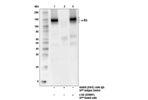
IMPORTANT: Appropriate isotype controls are highly recommended in order to show specific binding in your primary antibody immunoprecipitation. Use Normal Rabbit IgG #2729 for rabbit polyclonal primary antibodies, Rabbit (DA1E) mAb IgG XP® Isotype Control #3900 for rabbit monoclonal primary antibodies, Mouse (G3A1) mAb IgG1 Isotype Control #5415 for mouse monoclonal IgG1 primary antibodies, Mouse (E5Y6Q) mAb IgG2a Isotype Control #61656 for mouse monoclonal IgG2a primary antibodies, Mouse (E7Q5L) mAb IgG2b Isotype Control #53484 for mouse monoclonal IgG2b primary antibodies, and Mouse (E1D5H) mAb IgG3 Isotype Control #37988 for mouse monoclonal IgG3 primary antibodies. Isotype controls should be concentration matched and run alongside the primary antibody samples.
Proceed to one of the following specific set of steps.
NOTE: To minimize masking caused by denatured IgG heavy chains (~50 kDa), we recommend using Mouse Anti-Rabbit IgG (Light-Chain Specific) (D4W3E) mAb (#45262) or Mouse Anti-Rabbit IgG (Conformation Specific) (L27A9) mAb (#3678) (or HRP conjugate #5127). To minimize masking caused by denatured IgG light chains (~25 kDa), we recommend using Mouse Anti-Rabbit IgG (Conformation Specific) (L27A9) mAb (#3678) (or HRP conjugate #5127).
posted December 2008
revised April 2021
Protocol Id: 410
NOTE: Please see product datasheet or product webpage for appropriate antibody dilution^.
| Step | Reagents | Time/Temperature | |
|---|---|---|---|
| 1 | Dewax | BOND™ Dewax Solution, 100% Alcohol, BOND™ Wash Solution | Pre-programmed Leica® BOND™ |
| 2 | Antigen Retrieval | BOND™ Epitope Retrieval ER2 Solution | 20 min., 100˚C | Protocol: HIER 20 min with ER2 |
| 3 | Peroxide Block | Refine Detection Kit Peroxide Block* | 5 min. |
| WASH | BOND™ Wash Solution | 3x 0:00 min. | |
| 4 | Protein Block (optional) | #5425 NGS or #15019 Animal-Free Blocking Solution | 20 min. |
| 5 | Primary Antibody^ | Dilute in #8112 SignalStain® Antibody Diluent | 30 min. |
| WASH | BOND™ Wash Solution | 3x 2:00 min. | |
| NA | Post Primary Mouse Linker | Refine Detection Kit Post Primary* | Not Applied |
| 6 | Secondary Detection | Refine Detection Kit Polymer* | 10 min. |
| WASH | BOND™ Wash Solution/Deionized Water | Custom (see below) | |
| 7a | Visualization | Refine Detection Kit Mixed DAB Refine* | 0:00 min. |
| 7b | Visualization | Refine Detection Kit Mixed DAB Refine* | 10 min. |
| WASH | Deionized Water | 3x 0:00 min. | |
| 8 | Counterstain | Refine Detection Kit Hematoxylin* | 5 min. |
| WASH | Deionized Water | 0:00 min. | |
| WASH | BOND™ Wash Solution | 0:00 min. | |
| WASH | Deionized Water | 0:00 min. | |
| 9 | Dehydration (Offline): | ||
| Incubate sections in 95% ethanol two times for 10 seconds each. | |||
| Repeat in 100% ethanol, incubating sections two times for 10 seconds each. | |||
| Repeat in xylene, incubating sections two times for 10 seconds each. | |||
| 10 | Mount sections with coverslips and #14177 SignalStain® Mounting Medium | ||
| Optional Custom wash: | BOND™ Wash Solution | 2:00 | |
| BOND™ Wash Solution | Dispenser Type: OPEN 0:00 | ||
| BOND™ Wash Solution | 2:00 | ||
| BOND™ Wash Solution | Dispenser Type: OPEN 0:00 | ||
| BOND™ Wash Solution | 0:00 | ||
| Deionized Water | 0:00 | ||
*Reagent included in BOND™ Polymer Refine Detection Kit (Catalog No: DS9800)
LEICA® is a registered trademark of Leica Microsystems IR GmbH.
BOND™ is a trademark of Leica Biosystems Melbourne Pty. Ltd. No affiliation or sponsorship between CST and Leica Microsystems IR GmbH or Leica Biosystems Melbourne Pty. Ltd is implied.
posted August 2018
revised September 2018
Protocol Id: 1444
NOTE: Prepare solutions with reverse osmosis deionized (RODI) or equivalent grade water.
NOTE: Do not allow slides to dry at any time during this procedure.
For Citrate: Heat slides in a microwave submersed in 1X citrate unmasking solution until boiling is initiated; follow with 10 min at a sub-boiling temperature (95°-98°C). Cool slides on bench top for 30 min.
|
RECOMMENDED DETECTION REAGENTS |
SignalStain® Boost IHC Detection Reagent (HRP, Rabbit) #8114 | SignalStain® Boost IHC Detection Reagent (AP, Rabbit) #18653 |
|---|---|---|
|
COMPATIBLE CHROMOGEN |
SignalStain® DAB Substrate Kit #8059 | SignalStain® Vibrant Red Alkaline Phosphatase Substrate Kit #76713 |
| SignalStain® Vivid Purple Peroxidase Substrate Kit #96632 | SignalStain® Ultra Blue Alkaline Phosphatase Substrate Kit #12824 | |
| SignalStain® Deep Black Peroxidase Substrate Kit #72986 | ||
| SignalStain® Radiant Yellow Peroxidase Substrate Kit #69644 |
NOTE: Use of detection reagents other than those specified in this protocol may require further optimization of the primary antibody to account for the different sensitivities of the detection reagents.
posted February 2010
revised June 2020
Protocol Id: 283
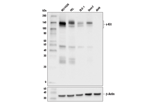
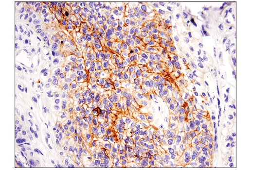
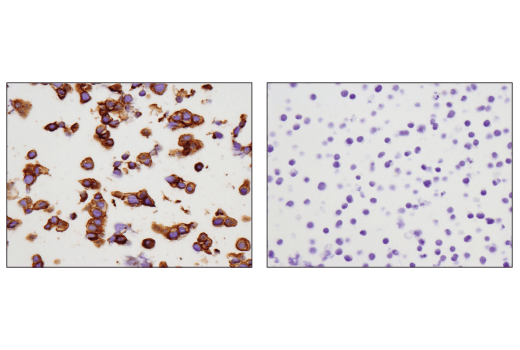
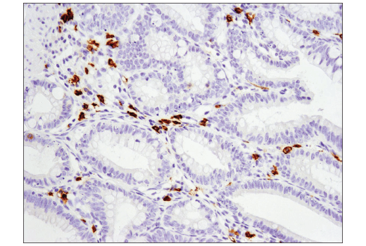
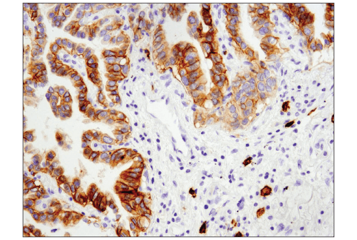
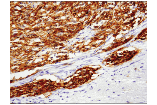
Human
Monoclonal antibody is produced by immunizing animals with a synthetic peptide corresponding to residues surrounding Val955 of human c-Kit protein.
c-Kit is a member of the subfamily of receptor tyrosine kinases that includes PDGF, CSF-1, and FLT3/flk-2 receptors (1,2). It plays a critical role in activation and growth in a number of cell types, including hematopoietic stem cells, mast cells, melanocytes, and germ cells (3). Upon binding with its stem cell factor (SCF) ligand, c-Kit undergoes dimerization/oligomerization and autophosphorylation. Activation of c-Kit results in the recruitment and tyrosine phosphorylation of downstream SH2-containing signaling components, including PLCγ, the p85 subunit of PI3 kinase, SHP2, and CrkL (4). Molecular lesions that impair the kinase activity of c-Kit are associated with a variety of developmental disorders (5), and mutations that constitutively activate c-Kit can lead to pathogenesis of mastocytosis and gastrointestinal stromal tumors (6). Tyr719 is located in the kinase insert region of the catalytic domain. c-Kit phosphorylated at Tyr719 binds to the p85 subunit of PI3 kinase in vitro and in vivo (7).
Explore pathways related to this product.
STRING - Known and Predicted Protein-Protein Interactions.
Except as otherwise expressly agreed in a writing signed by a legally authorized representative of CST, the following terms apply to Products provided by CST, its affiliates or its distributors. Any Customer's terms and conditions that are in addition to, or different from, those contained herein, unless separately accepted in writing by a legally authorized representative of CST, are rejected and are of no force or effect.
Products are labeled with For Research Use Only or a similar labeling statement and have not been approved, cleared, or licensed by the FDA or other regulatory foreign or domestic entity, for any purpose. Customer shall not use any Product for any diagnostic or therapeutic purpose, or otherwise in any manner that conflicts with its labeling statement. Products sold or licensed by CST are provided for Customer as the end-user and solely for research and development uses. Any use of Product for diagnostic, prophylactic or therapeutic purposes, or any purchase of Product for resale (alone or as a component) or other commercial purpose, requires a separate license from CST. Customer shall (a) not sell, license, loan, donate or otherwise transfer or make available any Product to any third party, whether alone or in combination with other materials, or use the Products to manufacture any commercial products, (b) not copy, modify, reverse engineer, decompile, disassemble or otherwise attempt to discover the underlying structure or technology of the Products, or use the Products for the purpose of developing any products or services that would compete with CST products or services, (c) not alter or remove from the Products any trademarks, trade names, logos, patent or copyright notices or markings, (d) use the Products solely in accordance with CST Product Terms of Sale and any applicable documentation, and (e) comply with any license, terms of service or similar agreement with respect to any third party products or services used by Customer in connection with the Products.