Revision 1
#9328
Store at -20C
c-Oncogene Antibody Sampler Kit
1 Kit
(9 x 20 microliters)
877-616-CELL (2355)
877-678-TECH (8324)
3 Trask Lane | Danvers | Massachusetts | 01923 | USA
For Research Use Only. Not for Use in Diagnostic Procedures.
| Product Includes | Product # | Quantity | Mol. Wt | Isotype/Source |
|---|---|---|---|---|
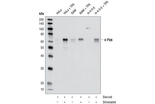
| c-Fos Antibody | 4384 | 20 µl | 62 kDa | Rabbit |
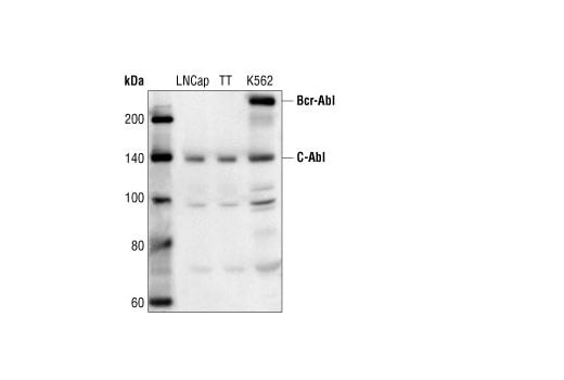
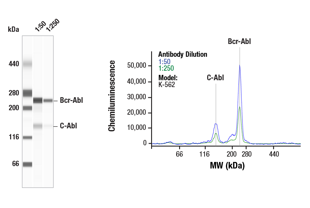
| c-Abl Antibody | 2862 | 20 µl | 135 (c-Abl); 210 (Bcr-Abl) kDa | Rabbit |
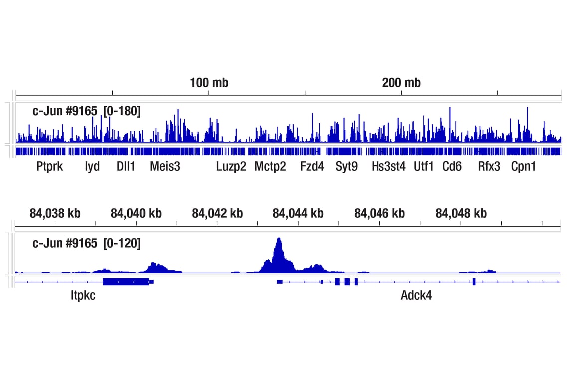
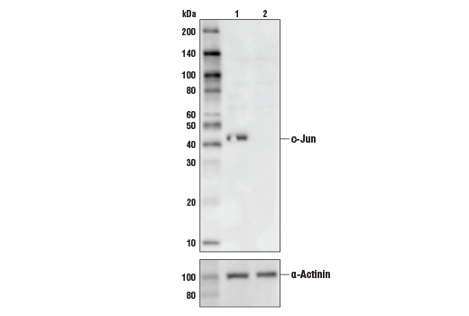
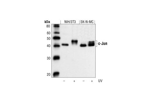
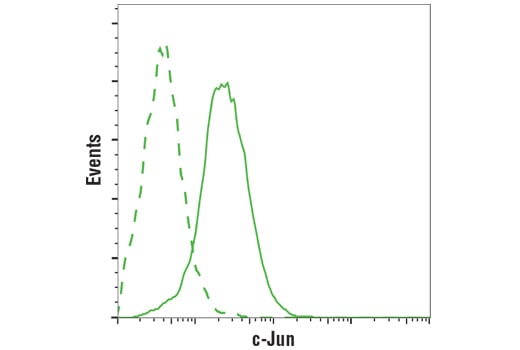
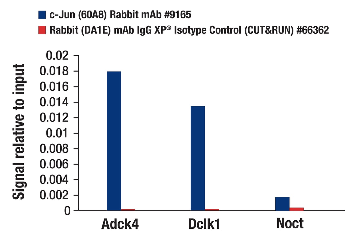
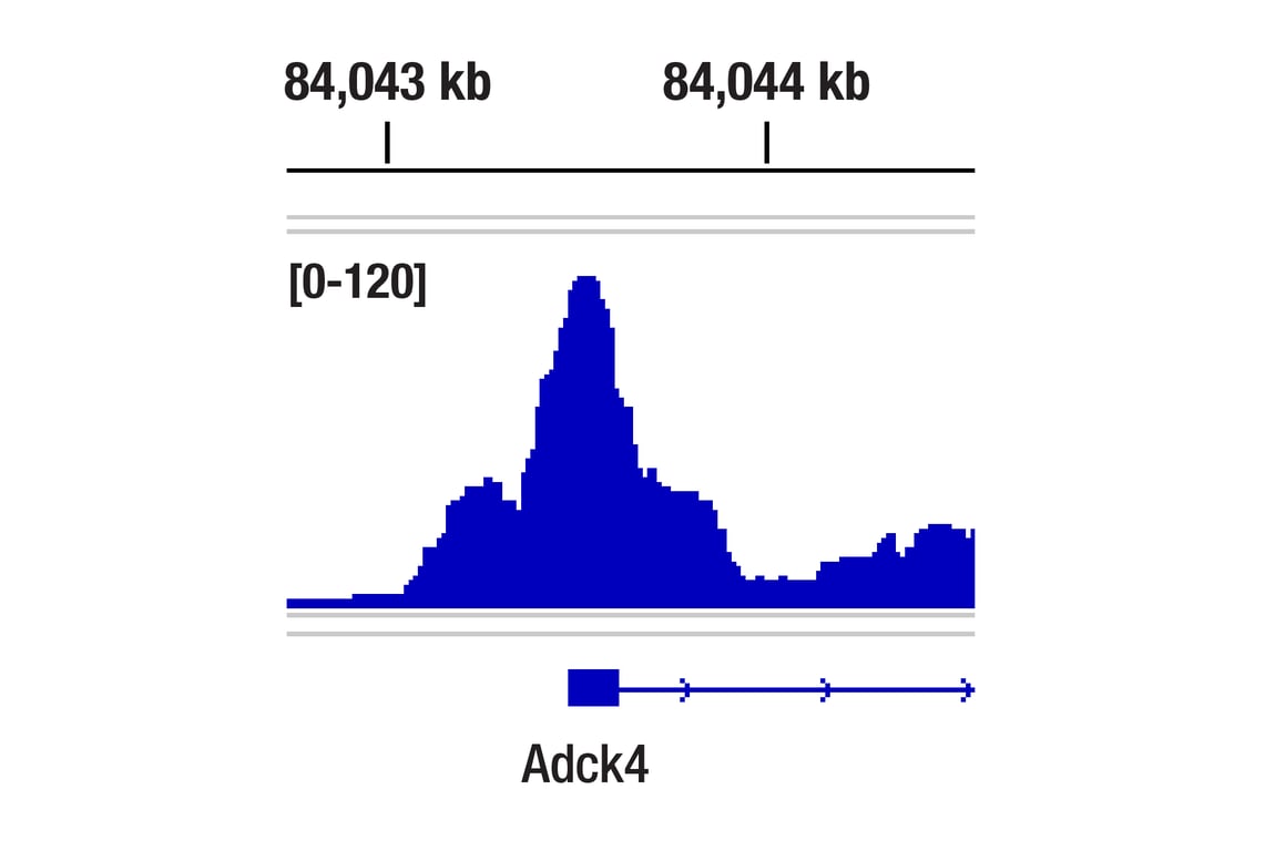
| c-Jun (60A8) Rabbit Monoclonal Antibody | 9165 | 20 µl | 43, 48 kDa | Rabbit IgG |
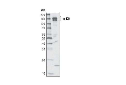
| c-Kit (D13A2) Rabbit Monoclonal Antibody | 3074 | 20 µl | 120 and 145 kDa | Rabbit IgG |
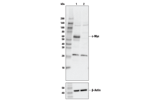
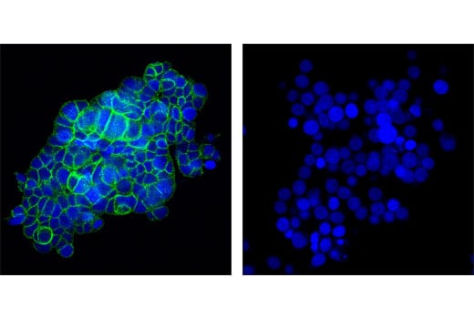
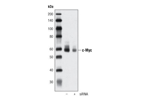
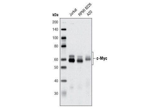
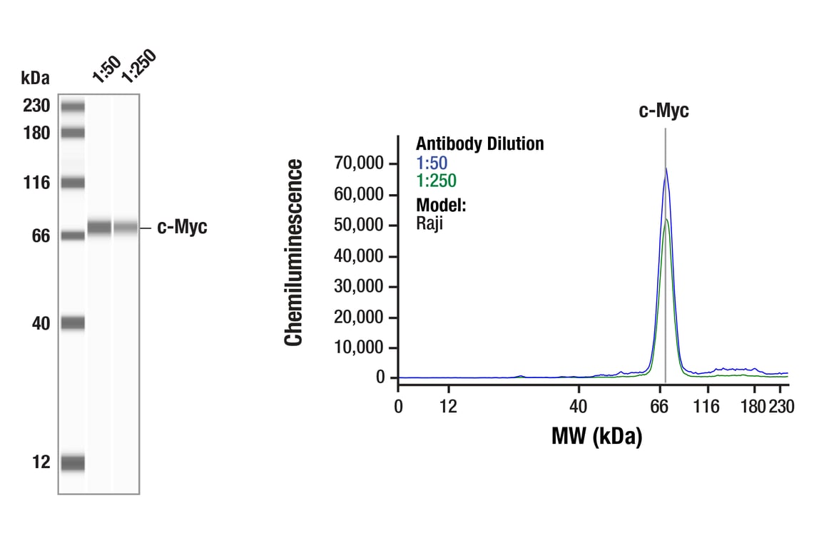
| c-Myc (D84C12) Rabbit Monoclonal Antibody | 5605 | 20 µl | 57-65 kDa | Rabbit IgG |
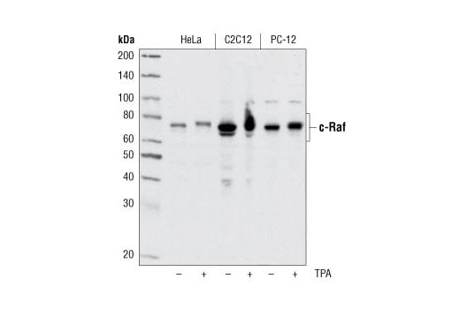
| c-Raf Antibody | 9422 | 20 µl | 65 to 75 kDa | Rabbit |
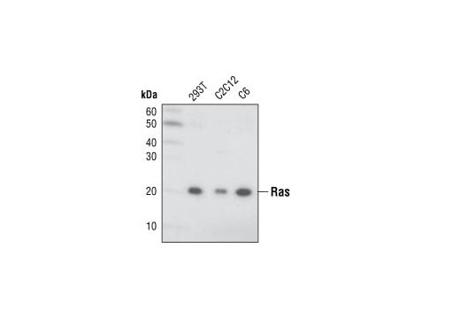
| Ras (27H5) Rabbit Monoclonal Antibody | 3339 | 20 µl | 21 kDa | Rabbit IgG |
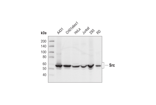
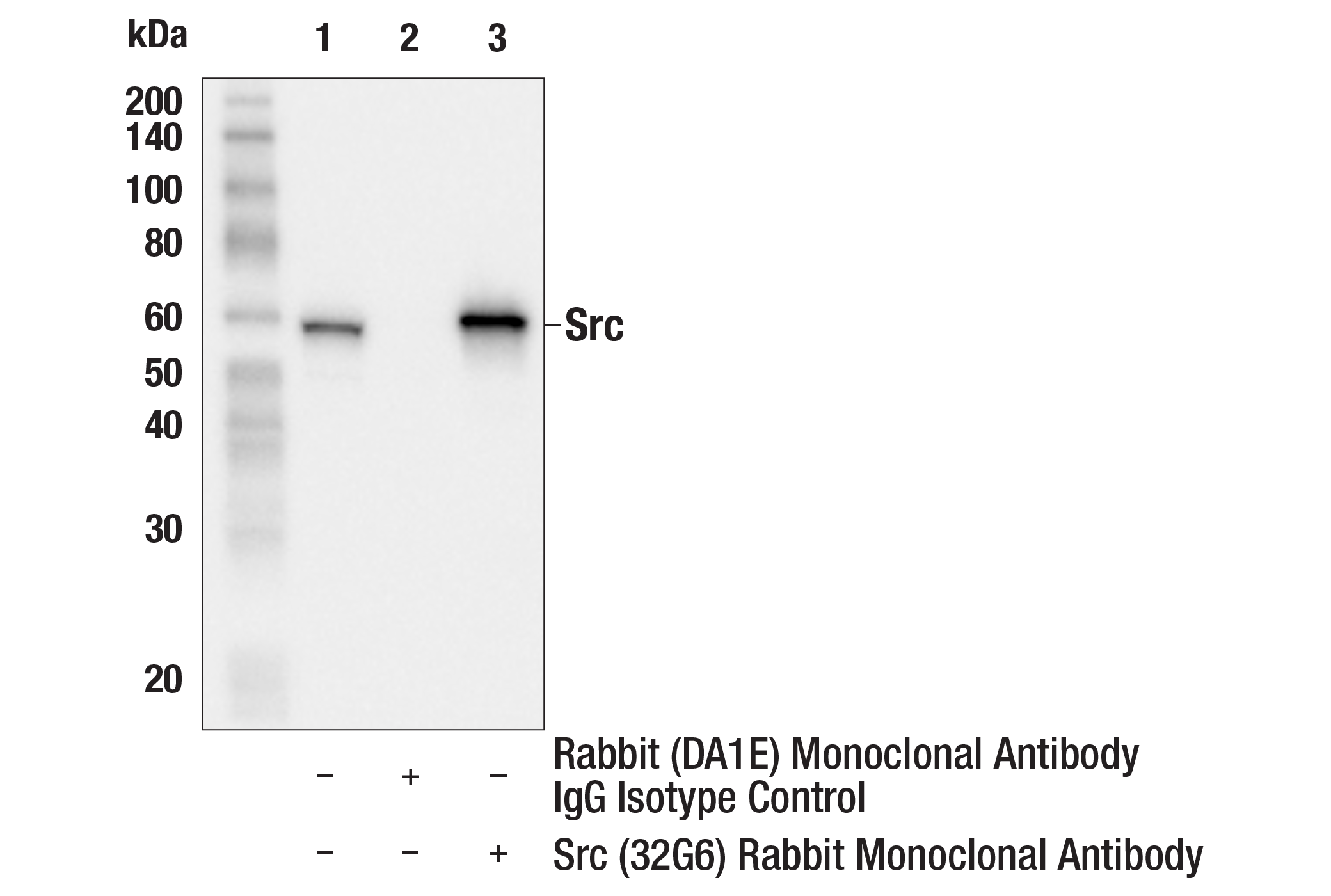
| Src (32G6) Rabbit Monoclonal Antibody | 2123 | 20 µl | 60 kDa | Rabbit IgG |
| Anti-rabbit IgG, HRP-linked Antibody | 7074 | 100 µl | Goat | |
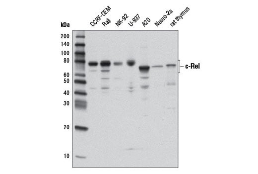
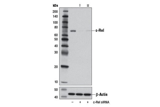
| c-Rel (D4Y6M) Rabbit Monoclonal Antibody | 12707 | 20 µl | 68-78 kDa | Rabbit IgG |
Please visit cellsignal.com for individual component applications, species cross-reactivity, dilutions, protocols, and additional product information.
Description
Storage
Background
Non-receptor (i.e. cytoplasmic, nuclear) tyrosine kinases such as c-Abl and Src play key roles in the regulation of cell proliferation, differentiation, apoptosis, cell adhesion and stress responses (2,3). Alteration of the corresponding c-Abl and Src proto-oncogenes is associated with oncogenesis; Abl1-BCR gene translocations result in chronic myelogenous leukemia (CML) while constitutively active Src is seen in some patients with colon cancer and altered Src expression is seen in a wide array of cancers (2,4). Regulation of Raf tyrosine kinase by Ras GTPase controls downstream kinases in the MEK/MAPK signaling pathway (5). Activation of the Ras and Raf proto-oncogenes are common in human cancers and both proteins are seen as potential therapeutic targets (6). The receptor tyrosine kinase c-Kit plays a critical role in activation and growth of hematopoietic stem cells (7); mutations that inhibit c-Kit kinase activity are associated with a variety of developmental disorders while mutations producing constitutively active c-Kit can result in mastocytosis and gastrointestinal stromal tumors (8). The alteration of key transcription factors such as c-Fos, c-Jun, c-Myc and c-Rel that are normally responsible for regulating cell and tissue growth, differentiation and the inflammation/immune response, can also result in unregulated, oncogenic cell growth (9-12).
Background References
- Croce, C.M. (2008) N Engl J Med 358, 502-11.
- Wang, J.Y. (2000) Oncogene 19, 5643-50.
- Thomas, S.M. and Brugge, J.S. (1997) Annu Rev Cell Dev Biol 13, 513-609.
- Dehm, S.M. and Bonham, K. (2004) Biochem Cell Biol 82, 263-74.
- Avruch, J. et al. (1994) Trends Biochem Sci 19, 279-83.
- Stites, E.C. et al. (2007) Science 318, 463-7.
- Gommerman, J.L. et al. (1997) J Biol Chem 272, 30519-25.
- Nocka, K. et al. (1990) EMBO J 9, 1805-13.
- Milde-Langosch, K. (2005) Eur J Cancer 41, 2449-61.
- Shaulian, E. and Karin, M. (2002) Nat Cell Biol 4, E131-6.
- Yokota, J. et al. (1986) Science 231, 261-5.
- Rayet, B. and Gélinas, C. (1999) Oncogene 18, 6938-47.
Trademarks and Patents
Cell Signaling Technology is a trademark of Cell Signaling Technology, Inc.
All other trademarks are the property of their respective owners. Visit cellsignal.com/trademarks for more information.
Limited Uses
Except as otherwise expressly agreed in a writing signed by a legally authorized representative of CST, the following terms apply to Products provided by CST, its affiliates or its distributors. Any Customer's terms and conditions that are in addition to, or different from, those contained herein, unless separately accepted in writing by a legally authorized representative of CST, are rejected and are of no force or effect.
Products are labeled with For Research Use Only or a similar labeling statement and have not been approved, cleared, or licensed by the FDA or other regulatory foreign or domestic entity, for any purpose. Customer shall not use any Product for any diagnostic or therapeutic purpose, or otherwise in any manner that conflicts with its labeling statement. Products sold or licensed by CST are provided for Customer as the end-user and solely for research and development uses. Any use of Product for diagnostic, prophylactic or therapeutic purposes, or any purchase of Product for resale (alone or as a component) or other commercial purpose, requires a separate license from CST. Customer shall (a) not sell, license, loan, donate or otherwise transfer or make available any Product to any third party, whether alone or in combination with other materials, or use the Products to manufacture any commercial products, (b) not copy, modify, reverse engineer, decompile, disassemble or otherwise attempt to discover the underlying structure or technology of the Products, or use the Products for the purpose of developing any products or services that would compete with CST products or services, (c) not alter or remove from the Products any trademarks, trade names, logos, patent or copyright notices or markings, (d) use the Products solely in accordance with CST Product Terms of Sale and any applicable documentation, and (e) comply with any license, terms of service or similar agreement with respect to any third party products or services used by Customer in connection with the Products.
Revision 1
Revision 1
Revision 1

Revision 1
Revision 1
Revision 1

Revision 1
Revision 1
Revision 1
Revision 1
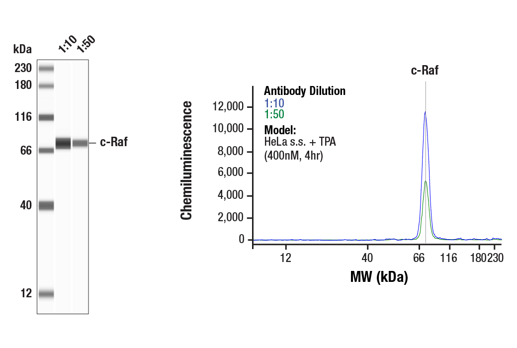
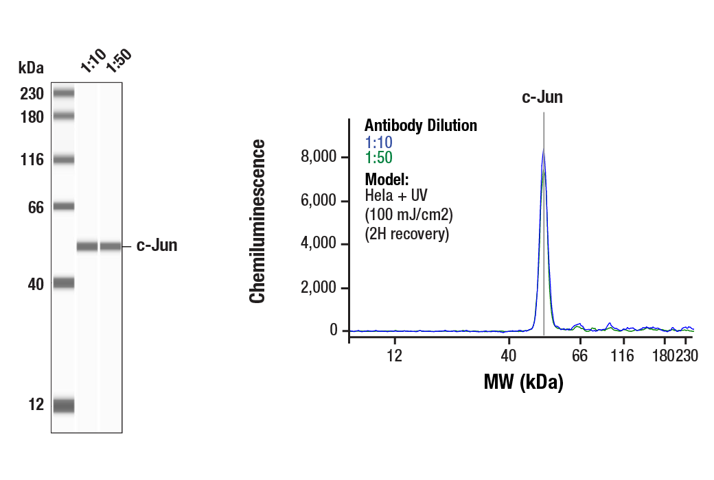
Simple Western™ analysis of lysates (0.1 mg/mL) from HeLa cells treated with UV (100 mJ/cm2; 2H recovery) using c-Jun (60A8) Rabbit mAb #9165. The virtual lane view (left) shows the target band (as indicated) at 1:10 and 1:50 dilutions of primary antibody. The corresponding electropherogram view (right) plots chemiluminescence by molecular weight along the capillary at 1:10 (blue line) and 1:50 (green line) dilutions of primary antibody. This experiment was performed under reducing conditions on the Jess™ Simple Western instrument from ProteinSimple, a BioTechne brand, using the 12-230 kDa separation module.
Revision 1