Revision 2

#9962

Store at -20C

Epitope Tag Antibody Sampler Kit

1 Kit

(5 x 20 microliters)

CST Logo
Orders:

877-616-CELL (2355)

[email protected]

Support:

877-678-TECH (8324)

3 Trask Lane | Danvers | Massachusetts | 01923 | USA

For Research Use Only. Not for Use in Diagnostic Procedures.

Product Includes Product # Quantity Mol. Wt Isotype/Source
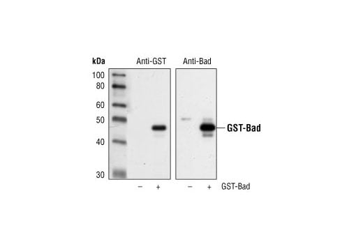
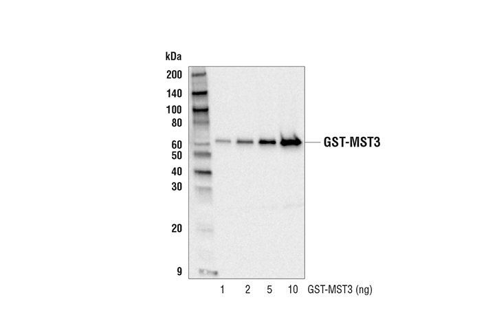
GST-Tag (91G1) Rabbit Monoclonal Antibody262520 µlRabbit IgG
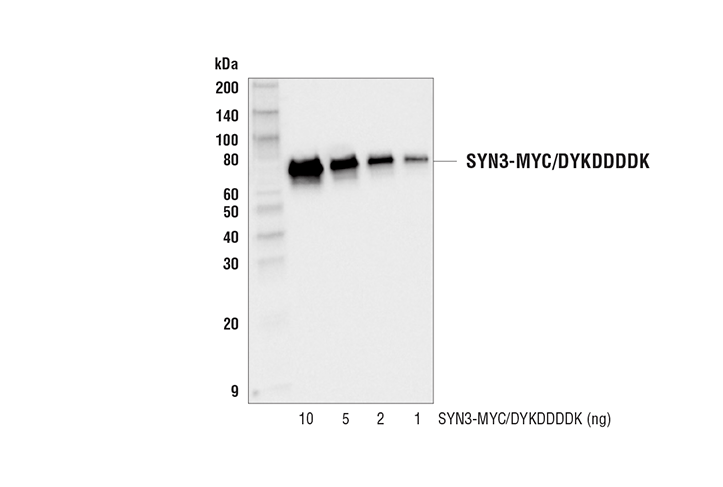
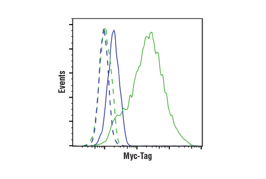
Myc-Tag (71D10) Rabbit Monoclonal Antibody227820 µlRabbit IgG
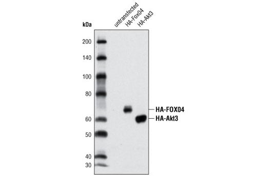
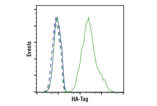
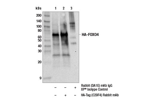
HA-Tag (C29F4) Rabbit Monoclonal Antibody372420 µlRabbit IgG
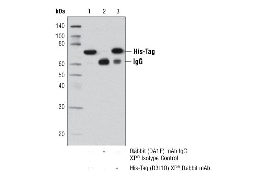
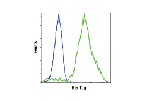
His-Tag (D3I1O) Rabbit Monoclonal Antibody1269820 µlRabbit IgG
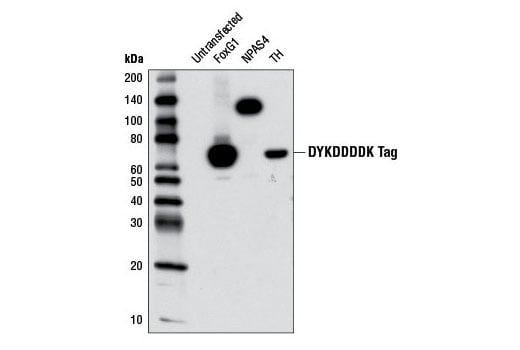
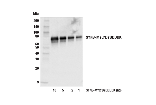
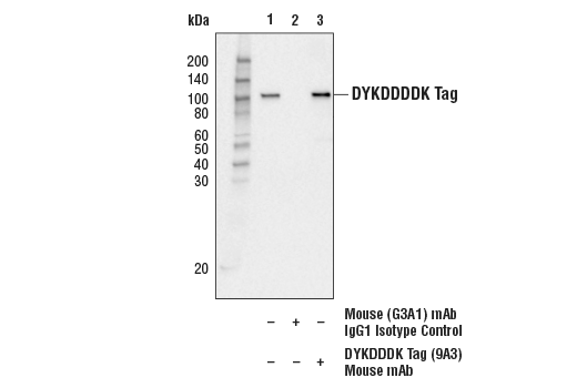
DYKDDDDK Tag (9A3) Mouse Monoclonal Antibody (Binds to same epitope as Sigma-Aldrich Anti-FLAG M2 antibody)814620 µlMouse IgG1
Anti-rabbit IgG, HRP-linked Antibody7074100 µlGoat
Anti-mouse IgG, HRP-linked Antibody7076100 µlHorse

Please visit cellsignal.com for individual component applications, species cross-reactivity, dilutions, protocols, and additional product information.

Description

The Epitope Tag Antibody Sampler Kit provides an economical means to analyze the expression of a variety of epitope tagged proteins. The kit contains enough primary and secondary antibodies to perform two Western blots per primary antibody.

Storage

Supplied in 10 mM sodium HEPES (pH 7.5), 150 mM NaCl, 100 µg/ml BSA, 50% glycerol and less than 0.02% sodium azide. Store at –20°C. Do not aliquot the antibody.

Background

Epitope tags are useful for the labeling and detection of proteins using immunoblotting, immunoprecipitation, and immunostaining techniques. Because of their small size, they are unlikely to affect the tagged protein’s biochemical properties.

Several different epitope tags are now commonly utilized and readily available. For instance, a variety of plasmids contain DNA that encodes an amino-terminal tag consisting of six histidine (6xHis) residues followed by an extended multiple cloning site. The 6xHis tag on the expressed recombinant proteins allows for efficient coupling to Ni2+ affinity resins and purification by single step chromatography (1). As is the case with other protein tag systems (2), this polyhistidine tag can often be cleaved at sites recognized by proteases such as thrombin and enterokinases to isolate the protein of interest (1). Glutathione S-transferase (GST) is another widely used fusion partner, since it provides both an easily detectable Tag and a simple purification process with little effect on the biological function of the protein of interest. Numerous vectors containing GST-Tag have been developed for both prokaryotic and eukaryotic systems over the past decade (3-5). The HA tag, derived from an epitope of the influenza hemagglutinin protein, has also been extensively used as a general epitope tag in expression vectors (6), while the Myc epitope tag is routinely used to detect expression of recombinant proteins in bacteria, yeast, insect and mammalian cell systems (7). Finally, the DYKDDDDK peptide has been used extensively as a general epitope tag in expression vectors and consists of only eight amino acids. This peptide can be expressed and detected with the protein of interested as an amino-terminal or carboxy-terminal fusion (8).

Trademarks and Patents

Cell Signaling Technology is a trademark of Cell Signaling Technology, Inc.

All other trademarks are the property of their respective owners. Visit cellsignal.com/trademarks for more information.

Limited Uses

Except as otherwise expressly agreed in a writing signed by a legally authorized representative of CST, the following terms apply to Products provided by CST, its affiliates or its distributors. Any Customer's terms and conditions that are in addition to, or different from, those contained herein, unless separately accepted in writing by a legally authorized representative of CST, are rejected and are of no force or effect.

Products are labeled with For Research Use Only or a similar labeling statement and have not been approved, cleared, or licensed by the FDA or other regulatory foreign or domestic entity, for any purpose. Customer shall not use any Product for any diagnostic or therapeutic purpose, or otherwise in any manner that conflicts with its labeling statement. Products sold or licensed by CST are provided for Customer as the end-user and solely for research and development uses. Any use of Product for diagnostic, prophylactic or therapeutic purposes, or any purchase of Product for resale (alone or as a component) or other commercial purpose, requires a separate license from CST. Customer shall (a) not sell, license, loan, donate or otherwise transfer or make available any Product to any third party, whether alone or in combination with other materials, or use the Products to manufacture any commercial products, (b) not copy, modify, reverse engineer, decompile, disassemble or otherwise attempt to discover the underlying structure or technology of the Products, or use the Products for the purpose of developing any products or services that would compete with CST products or services, (c) not alter or remove from the Products any trademarks, trade names, logos, patent or copyright notices or markings, (d) use the Products solely in accordance with CST Product Terms of Sale and any applicable documentation, and (e) comply with any license, terms of service or similar agreement with respect to any third party products or services used by Customer in connection with the Products.

Orders: 877-616-CELL (2355) [email protected] Support: 877-678-TECH (8324) [email protected] Web: cellsignal.com
For Research Use Only. Not for Use in Diagnostic Procedures.

Revision 2

Cell Signaling Technology Logo
Western blot analysis of extracts from 293T cells, untransfected or transfected with 6xHis-Tag fusion protein 1 or 6xHis-Tag fusion protein 2, using His-Tag (D3I1O) XP® Rabbit mAb.
undefined Image 1: Epitope Tag Antibody Sampler Kit
Western blot analysis of extracts from untransfected control cells (-) and transfected cells overexpressing Myc-Bcl-2 (+), using Bcl-2 Antibody (left) and Myc-Tag (71D10) Rabbit mAb (right).
undefined Image 2: Epitope Tag Antibody Sampler Kit
Western blot analysis of extracts from COS-7 cells untransfected (-) and transfected cells overexpressing GST-Bad (+), using GST-Tag (91G1) Rabbit mAb (left) and Bad Antibody #9292 (right).
undefined Image 3: Epitope Tag Antibody Sampler Kit
Orders: 877-616-CELL (2355) [email protected] Support: 877-678-TECH (8324) [email protected] Web: cellsignal.com
For Research Use Only. Not for Use in Diagnostic Procedures.

Revision 2

Cell Signaling Technology Logo
Western blot analysis of extracts from HeLa cells, untransfected or transfected with either HA-FoxO4 or HA-Akt3, using HA-Tag (C29F4) Rabbit mAb.
undefined Image 4: Epitope Tag Antibody Sampler Kit
After the primary antibody is bound to the target protein, a complex with HRP-linked secondary antibody is formed. The LumiGLO® is added and emits light during enzyme catalyzed decomposition.
undefined Image 5: Epitope Tag Antibody Sampler Kit
After the primary antibody is bound to the target protein, a complex with HRP-linked secondary antibody is formed. The LumiGLO* is added and emits light during enzyme catalyzed decomposition.
undefined Image 6: Epitope Tag Antibody Sampler Kit
Orders: 877-616-CELL (2355) [email protected] Support: 877-678-TECH (8324) [email protected] Web: cellsignal.com
For Research Use Only. Not for Use in Diagnostic Procedures.

Revision 2

Cell Signaling Technology Logo
Western blot analysis of extracts from 293T cells, untransfected or transfected with either DYKDDDDK-tagged FoxG1, NPAS4, or Tyrosine Hydroxylase (TH), using DYKDDDDK Tag (9A3) Mouse mAb.
undefined Image 7: Epitope Tag Antibody Sampler Kit
Immunoprecipitation of His-Tag protein from transfected 293T cells using Rabbit (DA1E) mAb IgG XP® Isotype Control #3900 (lane 2) or His-Tag (D3I1O) XP® Rabbit mAb (lane 3). Lane 1 is 10% input. Western blot analysis was performed using His-Tag (D3I1O) XP® Rabbit mAb.
undefined Image 8: Epitope Tag Antibody Sampler Kit
Western blot analysis of recombinant human SYN3-MYC/DYKDDDDK at various concentrations using, Myc-Tag (71D10) Rabbit mAb.
undefined Image 9: Epitope Tag Antibody Sampler Kit
Orders: 877-616-CELL (2355) [email protected] Support: 877-678-TECH (8324) [email protected] Web: cellsignal.com
For Research Use Only. Not for Use in Diagnostic Procedures.

Revision 2

Cell Signaling Technology Logo
Western blot analysis of recombinant human GST-MST3 at various concentrations, using GST-Tag (91G1) Rabbit mAb.
undefined Image 10: Epitope Tag Antibody Sampler Kit
Immunohistochemical analysis of paraffin-embedded COS cells, HA-Tag transfected (left) or untransfected (right), using HA-Tag (C29F4) Rabbit mAb.
undefined Image 11: Epitope Tag Antibody Sampler Kit
Western blot analysis of recombinant human SYN3-MYC/DYKDDDDK at various concentrations using, DYKDDDDK Tag (9A3) Mouse mAb.
undefined Image 12: Epitope Tag Antibody Sampler Kit
Orders: 877-616-CELL (2355) [email protected] Support: 877-678-TECH (8324) [email protected] Web: cellsignal.com
For Research Use Only. Not for Use in Diagnostic Procedures.

Revision 2

Cell Signaling Technology Logo
Confocal immunofluorescent analysis of 293T cells transfected with a His-Tagged protein using His-Tag (D3I1O) XP® Rabbit mAb (green). Actin filaments were labeled with DY-554 phalloidin (red). Blue pseudocolor= DRAQ5® #4084 (fluorescent DNA dye).
undefined Image 13: Epitope Tag Antibody Sampler Kit
Confocal immunofluorescent analysis of 293 cells stably expressing Myc-tagged ADORA2A (left) versus wild-type 293 cells (right) using Myc-Tag (71D10) Rabbit mAb (green). Blue = DAPI #4083 (fluorescent DNA dye).
undefined Image 14: Epitope Tag Antibody Sampler Kit
Simple Western™ analysis of lysates (0.1 mg/mL) from 293T cells, transfected with a construct expressing GST tagged BetaTrop1/trc using GST-Tag (91G1) Rabbit mAb #2625. The virtual lane view (left) shows the target band (as indicated) at 1:10 and 1:50 dilutions of primary antibody. The corresponding electropherogram view (right) plots chemiluminescence by molecular weight along the capillary at 1:10 (blue line) and 1:50 (green line) dilutions of primary antibody.  This experiment was performed under reducing conditions on the JessTM Simple Western instrument from ProteinSimple, a BioTechne brand, using the 12-230 kDa separation module.
undefined Image 15: Epitope Tag Antibody Sampler Kit
Orders: 877-616-CELL (2355) [email protected] Support: 877-678-TECH (8324) [email protected] Web: cellsignal.com
For Research Use Only. Not for Use in Diagnostic Procedures.

Revision 2

Cell Signaling Technology Logo
Confocal immunofluorescent analysis of COS cells, transfected with an HA-tagged protein (left) or mock-transfected (right), using HA-Tag (C29F4) Rabbit mAb (green). Actin filaments have been labeled using DY-554 phalloidin (red). Blue pseudocolor = DRAQ5® (fluorescent DNA dye).
undefined Image 16: Epitope Tag Antibody Sampler Kit
Immunoprecipitation of PI3K-DYKDDDDK tag protein from 293 transfected cell extracts. Lane 1 is 10% input, lane 2 is Mouse (G3A1) mAb IgG1 Isotype Control #5415, and lane 3 is DYKDDDDK Tag (9A3) Mouse mAb. Western blot analysis was performed using DYKDDDDK Tag (D6W5B) Rabbit mAb #14793. Anti-rabbit IgG, HRP-linked Antibody #7074 was used as a secondary antibody.
undefined Image 17: Epitope Tag Antibody Sampler Kit
Flow cytometric analysis of 293T cells, untransfected (blue) or transfected with a His-myc-Akt plasmid (green), using His-Tag (D3I1O) XP® Rabbit mAb. Anti-rabbit IgG (H+L), F(ab')2 Fragment (Alexa Fluor® 647 Conjugate) #4414 was used as a secondary antibody.
undefined Image 18: Epitope Tag Antibody Sampler Kit
Orders: 877-616-CELL (2355) [email protected] Support: 877-678-TECH (8324) [email protected] Web: cellsignal.com
For Research Use Only. Not for Use in Diagnostic Procedures.

Revision 2

Cell Signaling Technology Logo
Flow cytometric analysis of 293T cells, mock-transfected (blue) or transfected with a construct expressing Myc-Tag (green), using Myc-Tag (71D10) Rabbit mAb (solid lines) or concentration-matched Rabbit (DA1E) mAb IgG XP® Isotype Control #3900 (dashed lines). Anti-rabbit (Alexa Fluor® 488 Conjugate) #4412 was used as a secondary antibody.
undefined Image 19: Epitope Tag Antibody Sampler Kit
Flow cytometric analysis of 293T cells untransfected (blue) or transfected with HA-tagged Akt (green), using HA-Tag (C29F4) Rabbit mAb (solid lines) or concentration matched Rabbit (DA1E) mAb IgG XP® Isotype Control #3900 (dashed lines).Anti-rabbit IgG (H+L), F(ab')2 Fragment (Alexa Fluor® 488 Conjugate) #4412 was used as a secondary antibody.
undefined Image 20: Epitope Tag Antibody Sampler Kit
Immunohistochemical analysis of paraffin-embedded 293T cell pellets, control (left) or ROS-DYKDDDDK Tag transfected (right), using DYKDDDDK Tag (9A3) Mouse mAb.
undefined Image 21: Epitope Tag Antibody Sampler Kit
Orders: 877-616-CELL (2355) [email protected] Support: 877-678-TECH (8324) [email protected] Web: cellsignal.com
For Research Use Only. Not for Use in Diagnostic Procedures.

Revision 2

Cell Signaling Technology Logo
293T cells were either untransfected (left panel) or transfected with an His-tagged human Stat3 construct (right panel), then treated with Human Interleukin-6 (hIL-6) (100 ng/ml, 30 min). Chromatin immunoprecipitations were performed with cross-linked chromatin from cells and His-Tag (D3I1O) XP® Rabbit mAb, Stat3α (D1A5) XP® Rabbit mAb #8768, or Normal Rabbit IgG #2729 using SimpleChIP® Enzymatic Chromatin IP Kit (Magnetic Beads) #9003. The enriched DNA was quantified by real-time PCR using SimpleChIP® Human c-Fos Promoter Primers, human IRF-1 promoter primers, and SimpleChIP® Human α Satellite Repeat Primers #4486. The amount of immunoprecipitated DNA in each sample is represented as signal relative to the total amount of input chromatin, which is equivalent to one.
undefined Image 22: Epitope Tag Antibody Sampler Kit
Confocal immunofluorescent analysis of HeLa cells transfected with a DYKDDDDK-tagged IRF3 construct using DYKDDDDK Tag (9A3) Mouse mAb (green). Actin filaments were labeled with DY-554 phalloidin (red). Blue pseudocolor = DRAQ5® #4084 (fluorescent DNA dye).
undefined Image 23: Epitope Tag Antibody Sampler Kit
293T cells were either untransfected (left panel) or transfected with an HA-tagged human CBP construct (right panel), then treated with Forskolin #3828 (30 µM) . Chromatin immunoprecipitations were performed with cross-linked chromatin from cells and HA-Tag (C29F4) Rabbit mAb, CBP (D9B6) Rabbit mAb, or Normal Rabbit IgG #2729 using SimpleChIP® Enzymatic Chromatin IP Kit (Magnetic Beads) #9003. The enriched DNA was quantified by real-time PCR using human ALS2 exon 1 primers, SimpleChIP® Human NR4A3 Promoter Primers #4829, and SimpleChIP® Human α Satellite Repeat Primers #4486. The amount of immunoprecipitated DNA in each sample is represented as signal relative to the total amount of input chromatin, which is equivalent to one.
undefined Image 24: Epitope Tag Antibody Sampler Kit
Orders: 877-616-CELL (2355) [email protected] Support: 877-678-TECH (8324) [email protected] Web: cellsignal.com
For Research Use Only. Not for Use in Diagnostic Procedures.

Revision 2

Cell Signaling Technology Logo
Flow cytometric analysis of 293T cells, mock-transfected (blue, negative) or transfected with a construct expressing DYKDDDDK Tag (green, positive), using DYKDDDDK Tag (9A3) Mouse mAb (solid lines) or concentration-matched Mouse (G3A1) mAb IgG1 IsotypeControl #5415 (dashed lines). Anti-mouse IgG (H+L), F(ab')2 Fragment (Alexa Fluor® 647 Conjugate) #4410 was used as a secondary antibody.
undefined Image 25: Epitope Tag Antibody Sampler Kit
Immunoprecipitation of HA-FoxO4 tag protein from 293T transfected cell extracts. Lane 1 is 10% input, lane 2 is HA-Tag (C29F4) Rabbit mAb, and lane 3 is Rabbit (DA1E) mAb IgG XP® Isotype Control #3900. Western blot was performed using HA-Tag (C29F4) Rabbit mAb. A conformation specific secondary antibody was used to avoid reactivity with IgG.
undefined Image 26: Epitope Tag Antibody Sampler Kit

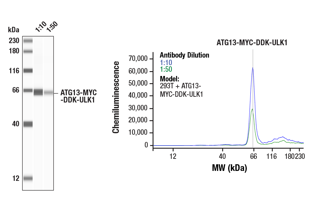
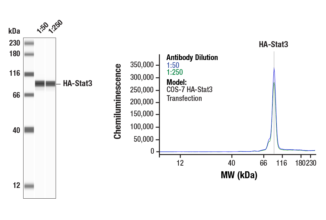
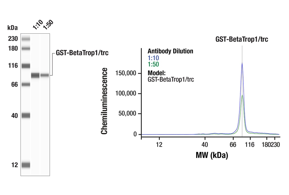
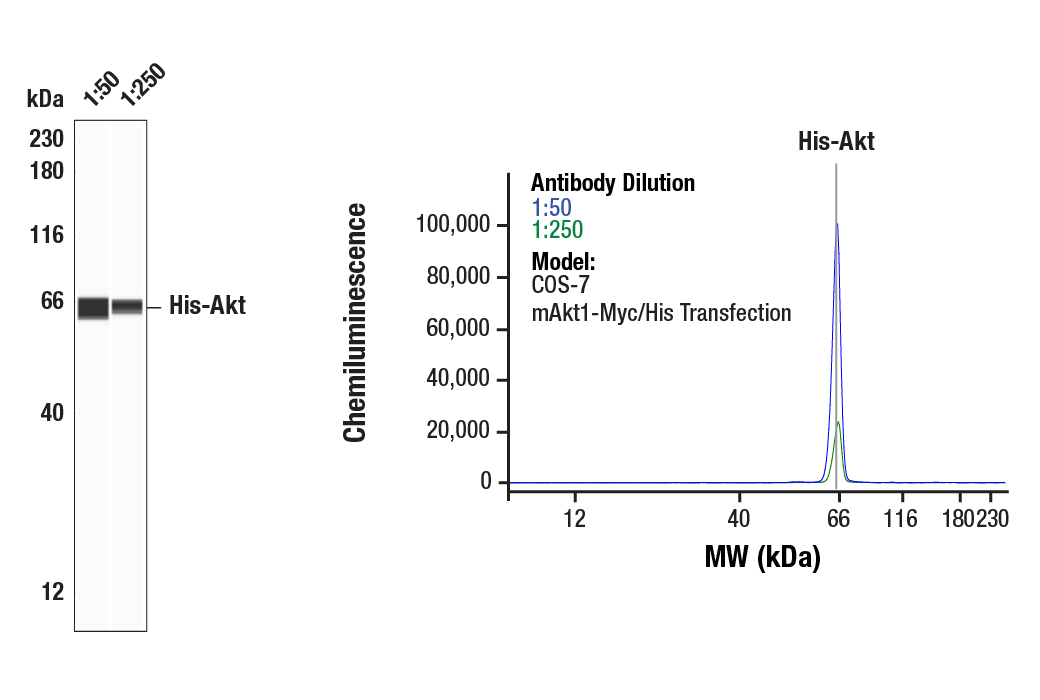
Simple Western™ analysis of lysates (0.1 mg/mL) from COS-7 mAkt1-Myc/His Transfection. cells using His-Tag (D3I1O) XP® Rabbit mAb #12698. The virtual lane view (left) shows the target band (as indicated) at 1:50 and 1:250 dilutions of primary antibody. The corresponding electropherogram view (right) plots chemiluminescence by molecular weight along the capillary at 1:50 (blue line) and 1:250 (green line) dilutions of primary antibody. This experiment was performed under reducing conditions on the Jess™ Simple Western instrument from ProteinSimple, a BioTechne brand, using the 12-230 kDa separation module.

undefined Image 27: Epitope Tag Antibody Sampler Kit
Orders: 877-616-CELL (2355) [email protected] Support: 877-678-TECH (8324) [email protected] Web: cellsignal.com
For Research Use Only. Not for Use in Diagnostic Procedures.

Revision 2

Cell Signaling Technology Logo
Simple Western™ analysis of lysates (1.0 mg/mL) from 293T cells transfected with ATG13-MYC-DDK-ULK1 using Myc-Tag (71D10) Rabbit mAb #2278. The virtual lane view (left) shows the target band (as indicated) at 1:10 and 1:50 dilutions of primary antibody. The corresponding electropherogram view (right) plots chemiluminescence by molecular weight along the capillary at 1:10 (blue line) and 1:50 (green line) dilutions of primary antibody. This experiment was performed under reducing conditions on the Jess™ Simple Western instrument from ProteinSimple, a BioTechne brand, using the 12-230 kDa separation module.
undefined Image 28: Epitope Tag Antibody Sampler Kit
Simple Western™ analysis of lysates (0.1 mg/mL) from COS-7 cells, transfected with a construct expressing HA-tagged Stat3 using HA-Tag (C29F4) Rabbit mAb #3724. The virtual lane view (left) shows a single target band (as indicated) at 1:50 and 1:250 dilutions of primary antibody. The corresponding electropherogram view (right) plots chemiluminescence by molecular weight along the capillary at 1:50 (blue line) and 1:250 (green line) dilutions of primary antibody. This experiment was performed under reducing conditions on the Jess™ Simple Western instrument from ProteinSimple, a BioTechne brand, using the 12-230 kDa separation module.
undefined Image 29: Epitope Tag Antibody Sampler Kit
Orders: 877-616-CELL (2355) [email protected] Support: 877-678-TECH (8324) [email protected] Web: cellsignal.com
For Research Use Only. Not for Use in Diagnostic Procedures.