See notes for additional testing details.
| Cat. # | Size | Qty. | Price |
|---|---|---|---|
| 48243T | 1 Kit (7 x 20 microliters) |
|
| Product Includes | Quantity | Applications | Reactivity | MW(kDa) | Isotype |
|---|---|---|---|---|---|
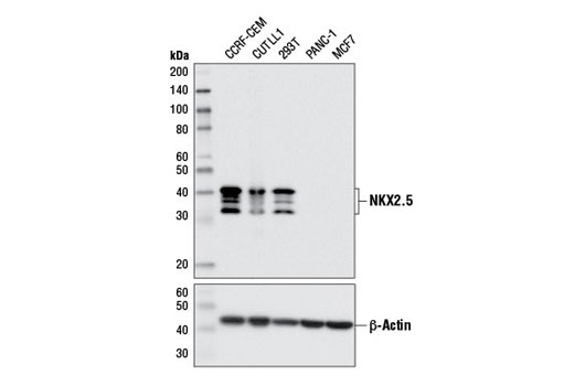
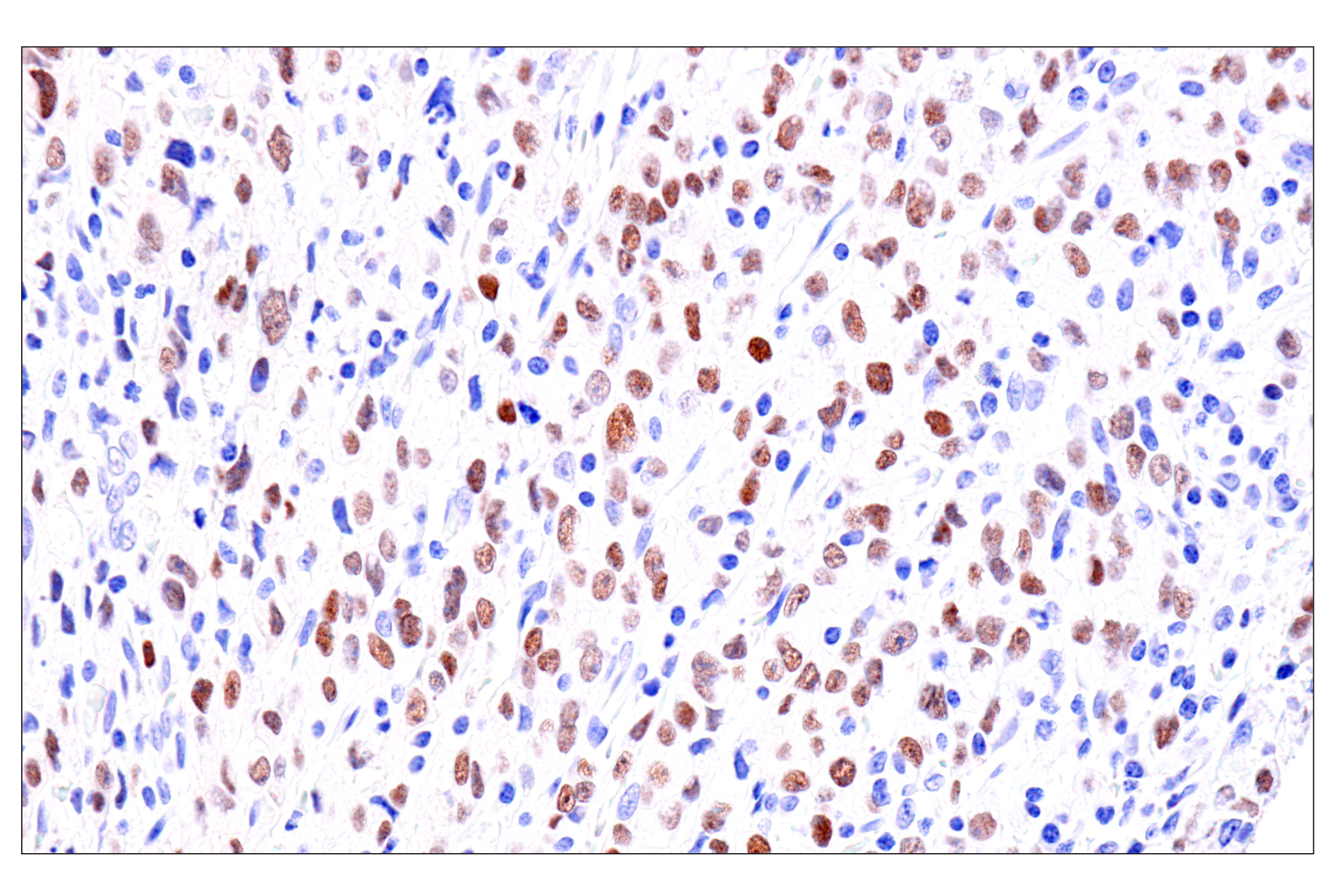
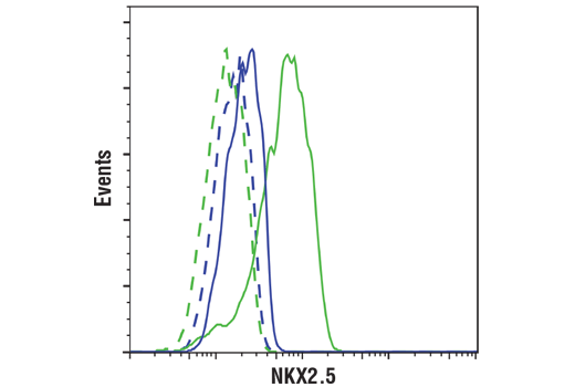
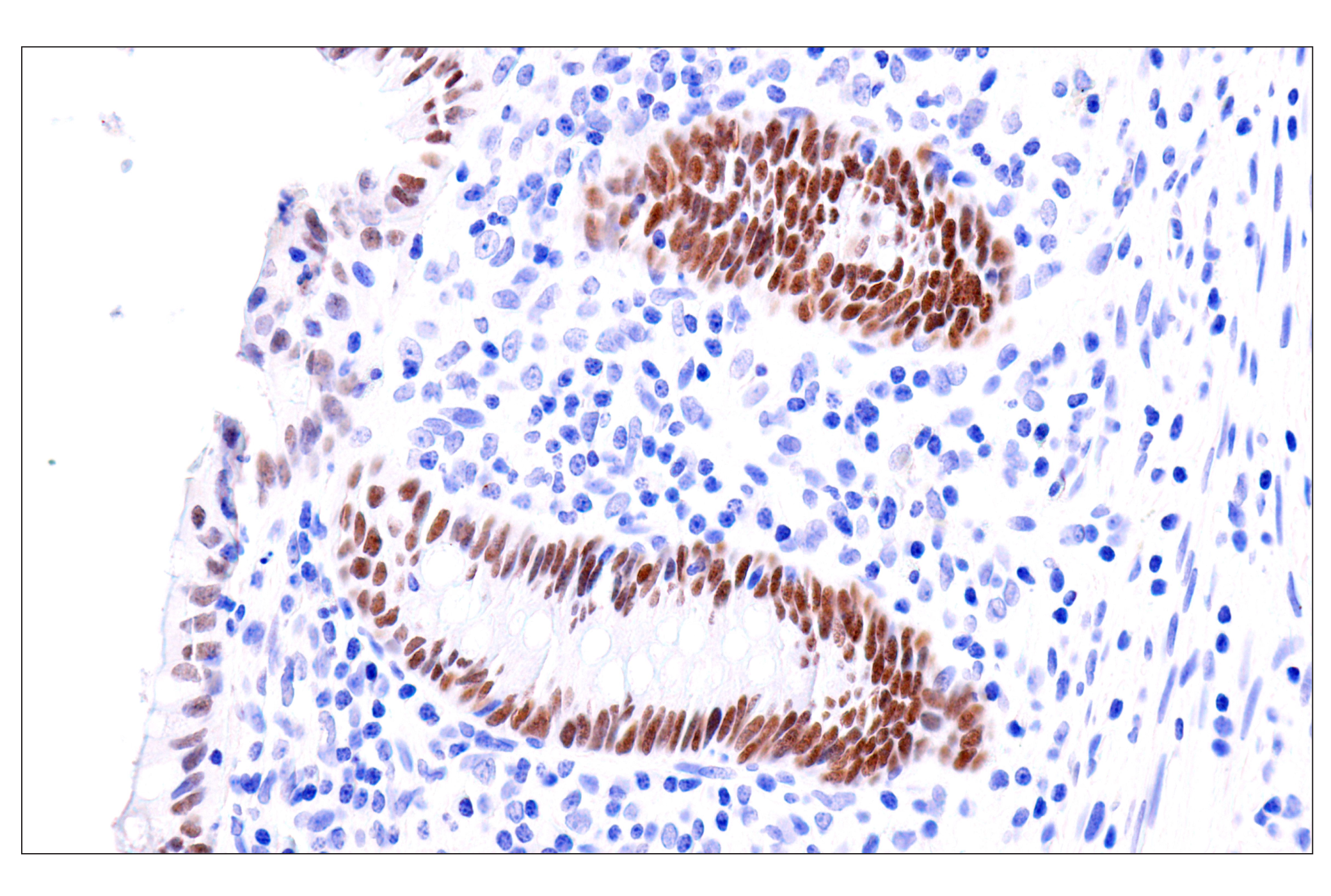
| NKX2.5 (E1Y8H) Rabbit mAb 8792 | 20 µl |
|
H | 30-42 | Rabbit IgG |
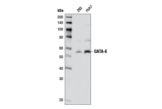
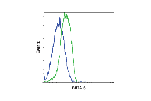
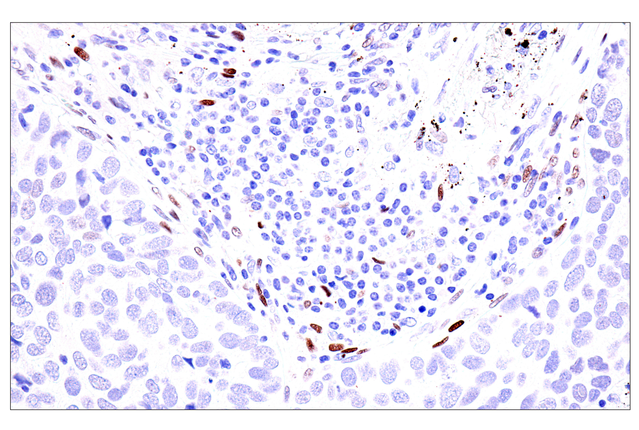
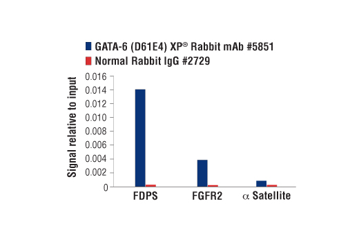
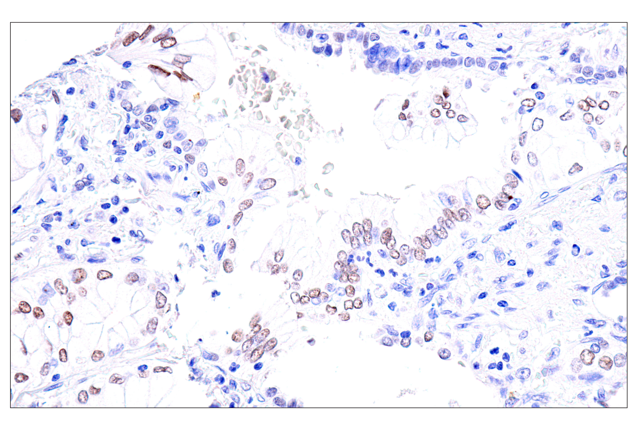
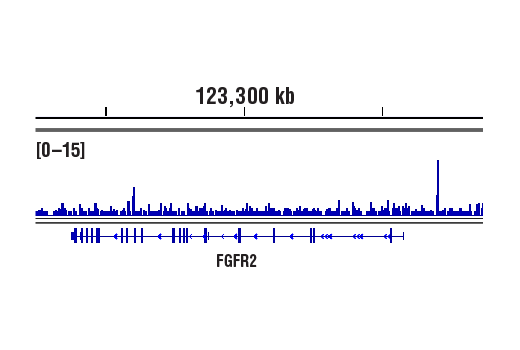
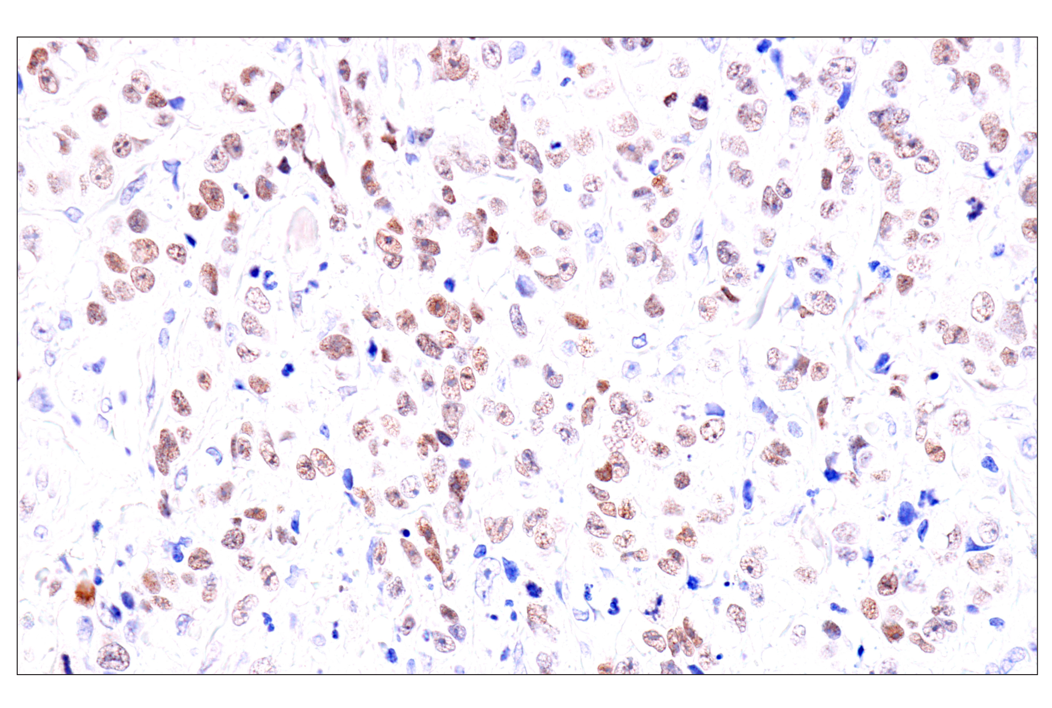
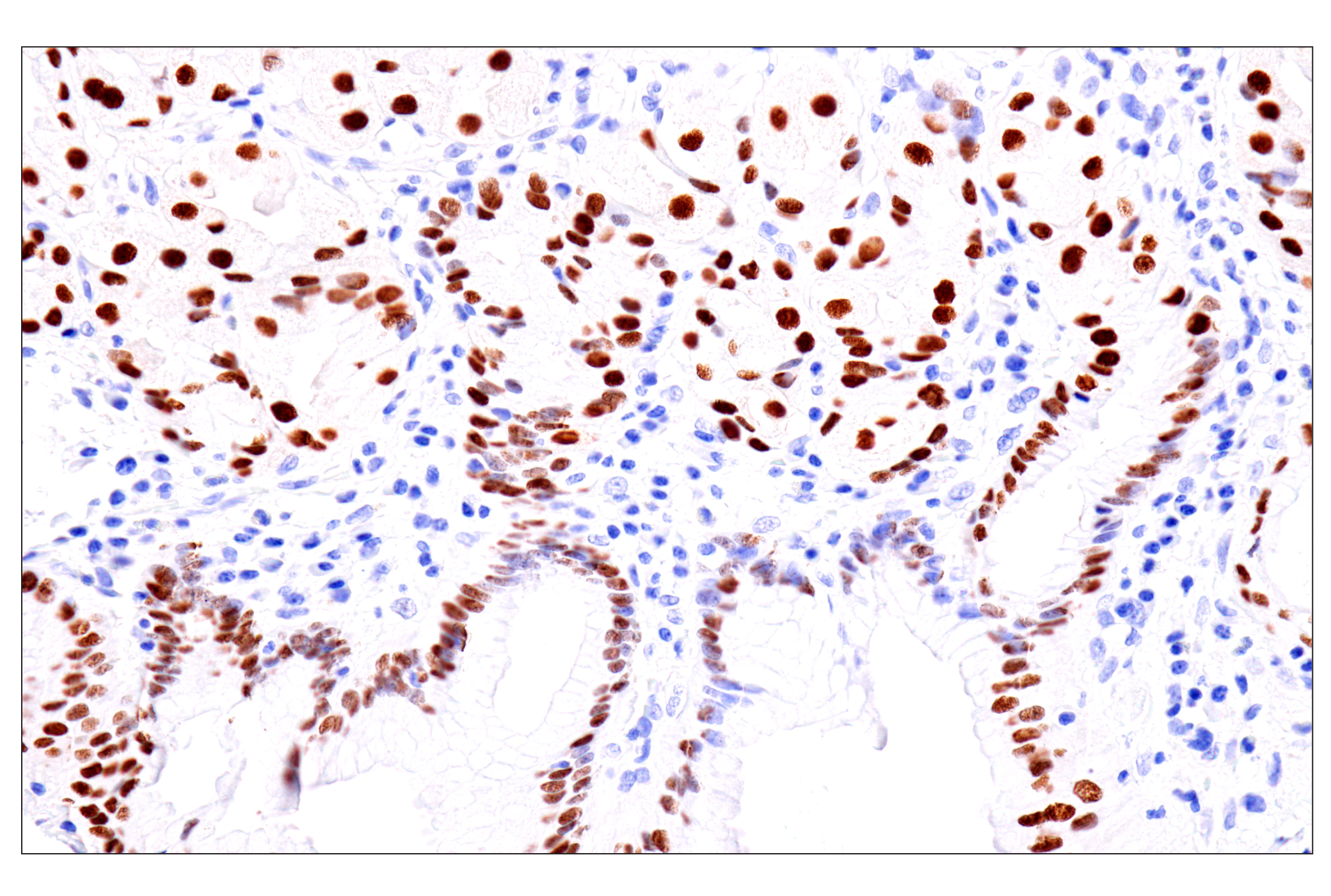
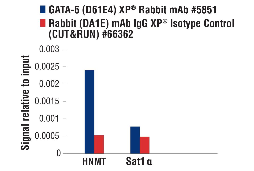
| GATA-6 (D61E4) XP® Rabbit mAb 5851 | 20 µl |
|
H M | 55 | Rabbit IgG |
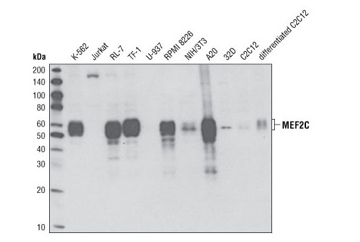
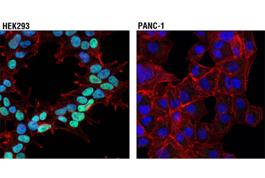
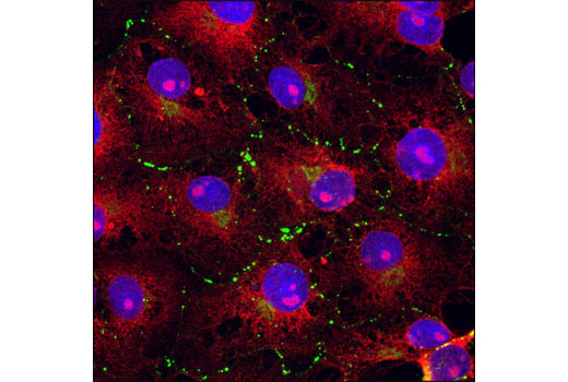
| MEF2C (D80C1) XP® Rabbit mAb 5030 | 20 µl |
|
H M | 50-60 | Rabbit IgG |
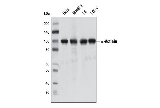
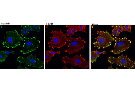
| α-Actinin (D6F6) XP® Rabbit mAb 6487 | 20 µl |
|
H M R Mk | 100 | Rabbit IgG |
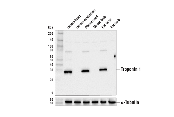
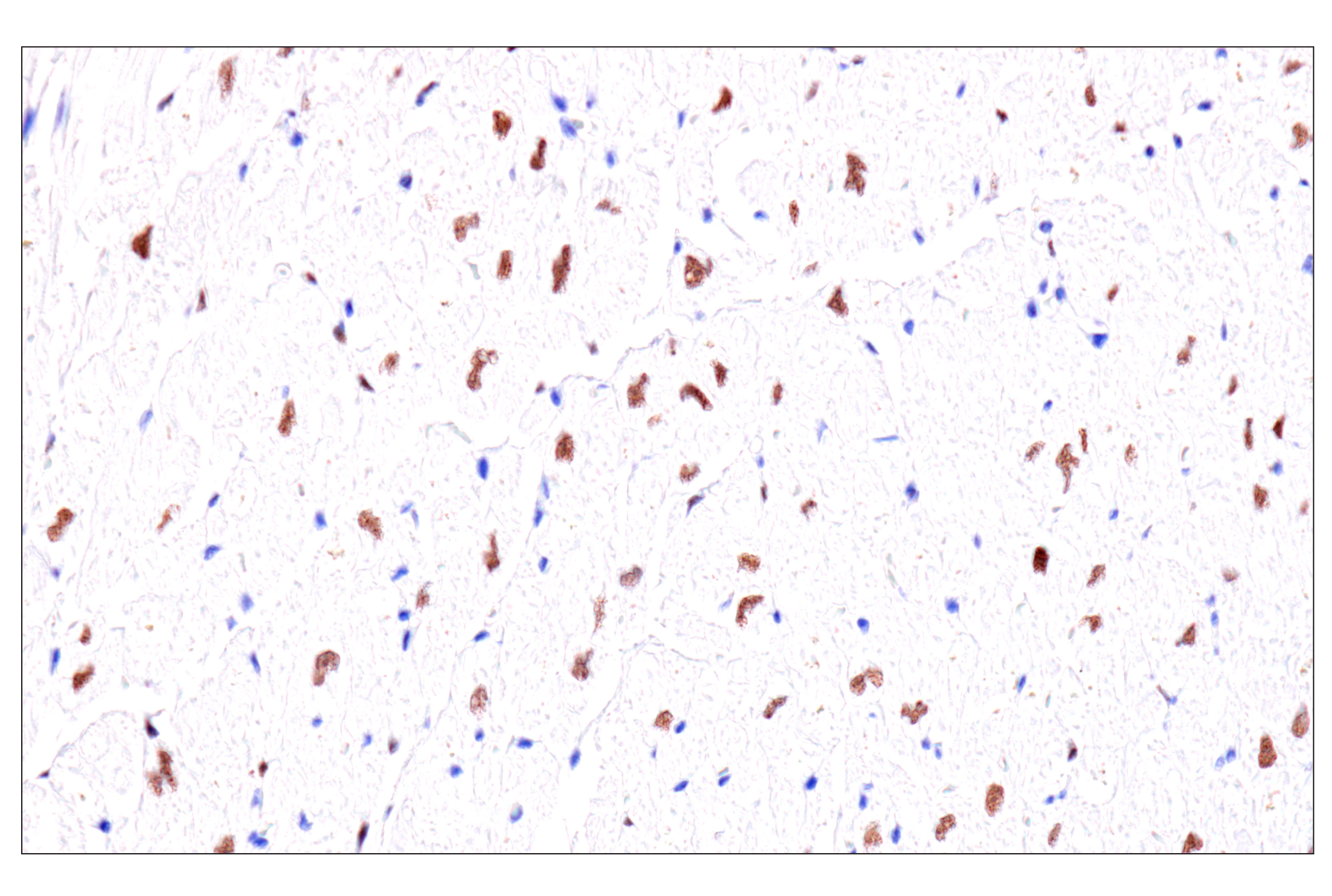
| Troponin I (D6F8) Rabbit mAb 13083 | 20 µl |
|
H M R | 28 | Rabbit IgG |
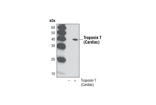
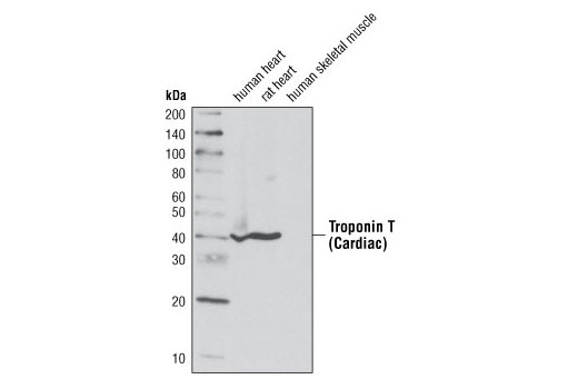
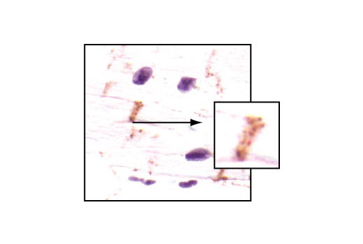
| Troponin T (Cardiac) Antibody 5593 | 20 µl |
|
H R | 40 | Rabbit |
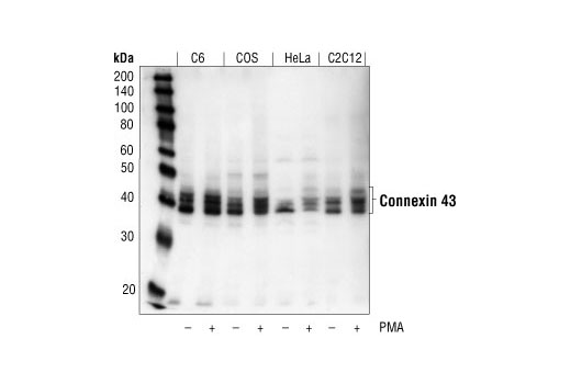
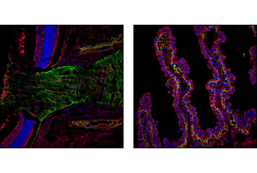
| Connexin 43 Antibody 3512 | 20 µl |
|
H M R Mk Z | 39, 41, 43, 44 | Rabbit |
| Anti-rabbit IgG, HRP-linked Antibody 7074 | 100 µl |
|
Goat |
Product Information
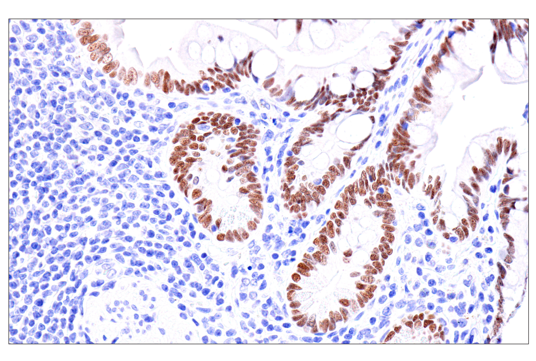
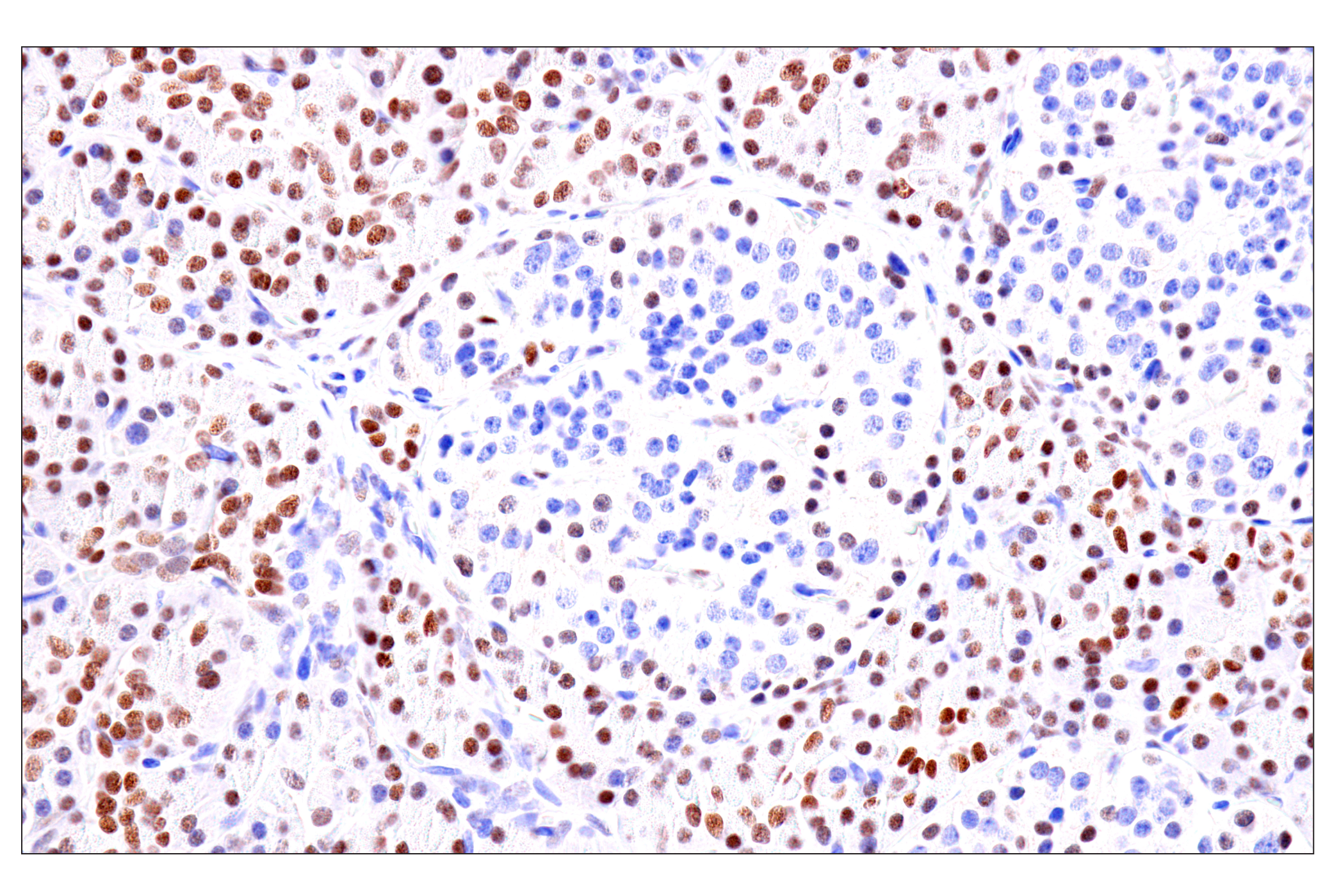
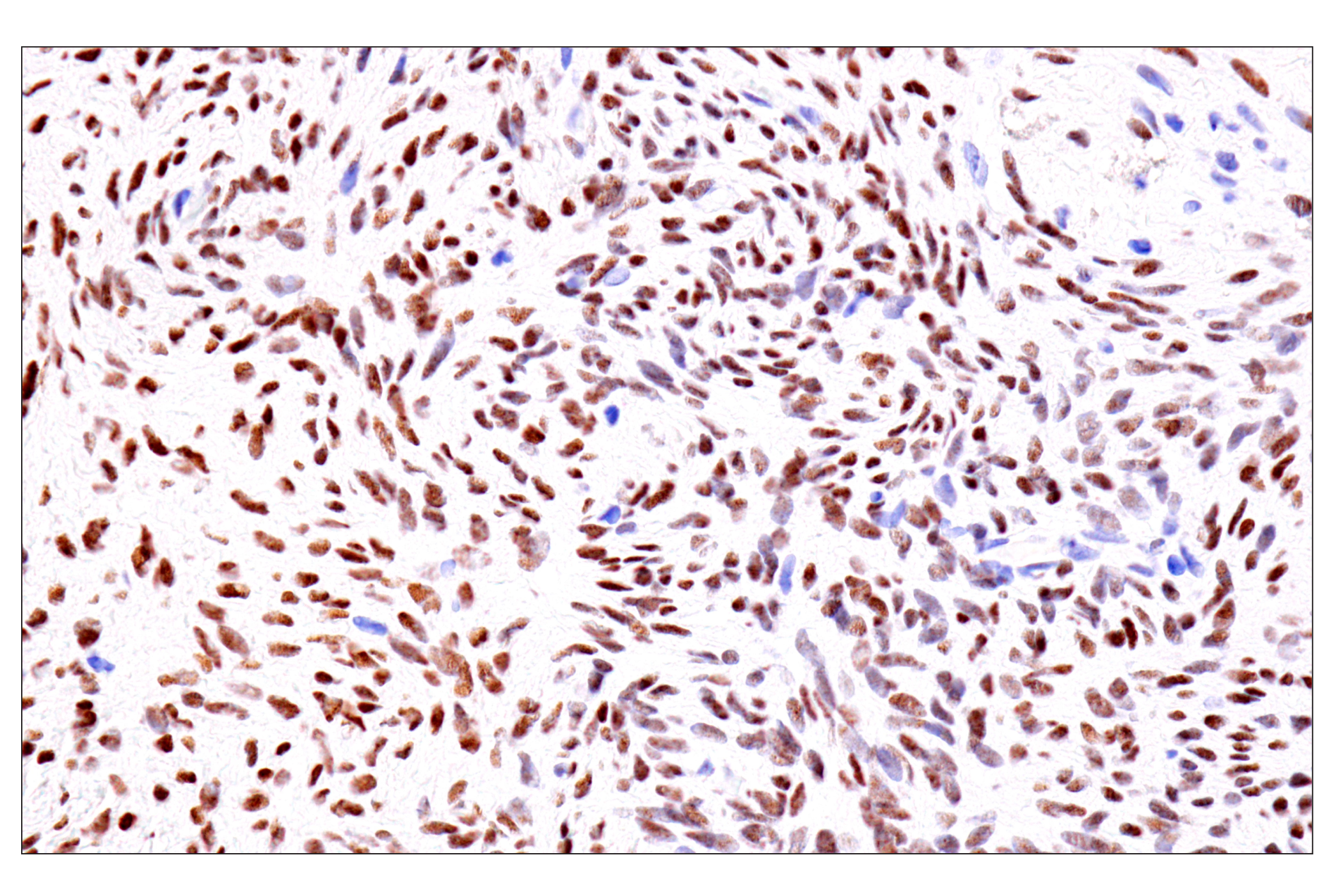
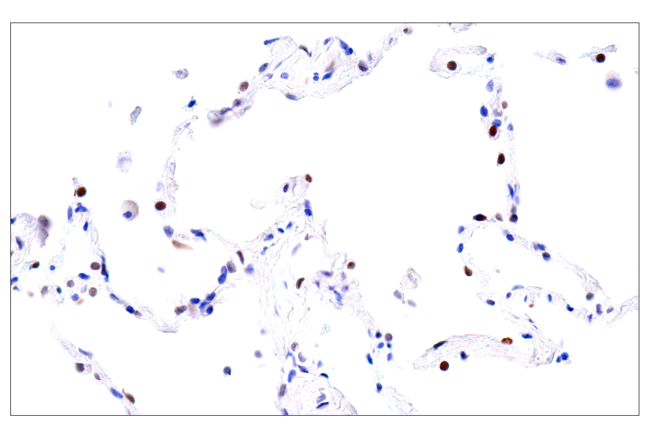
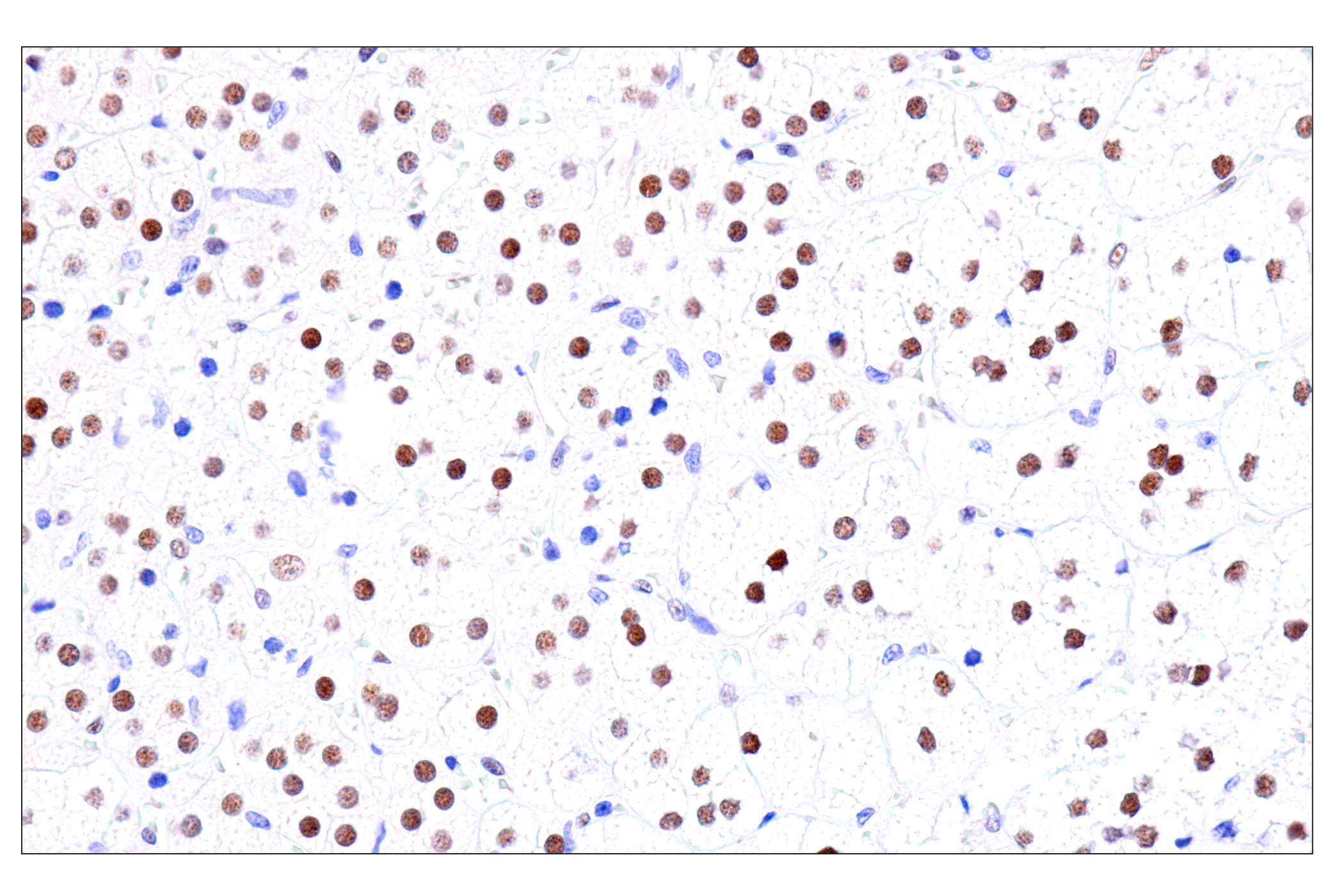
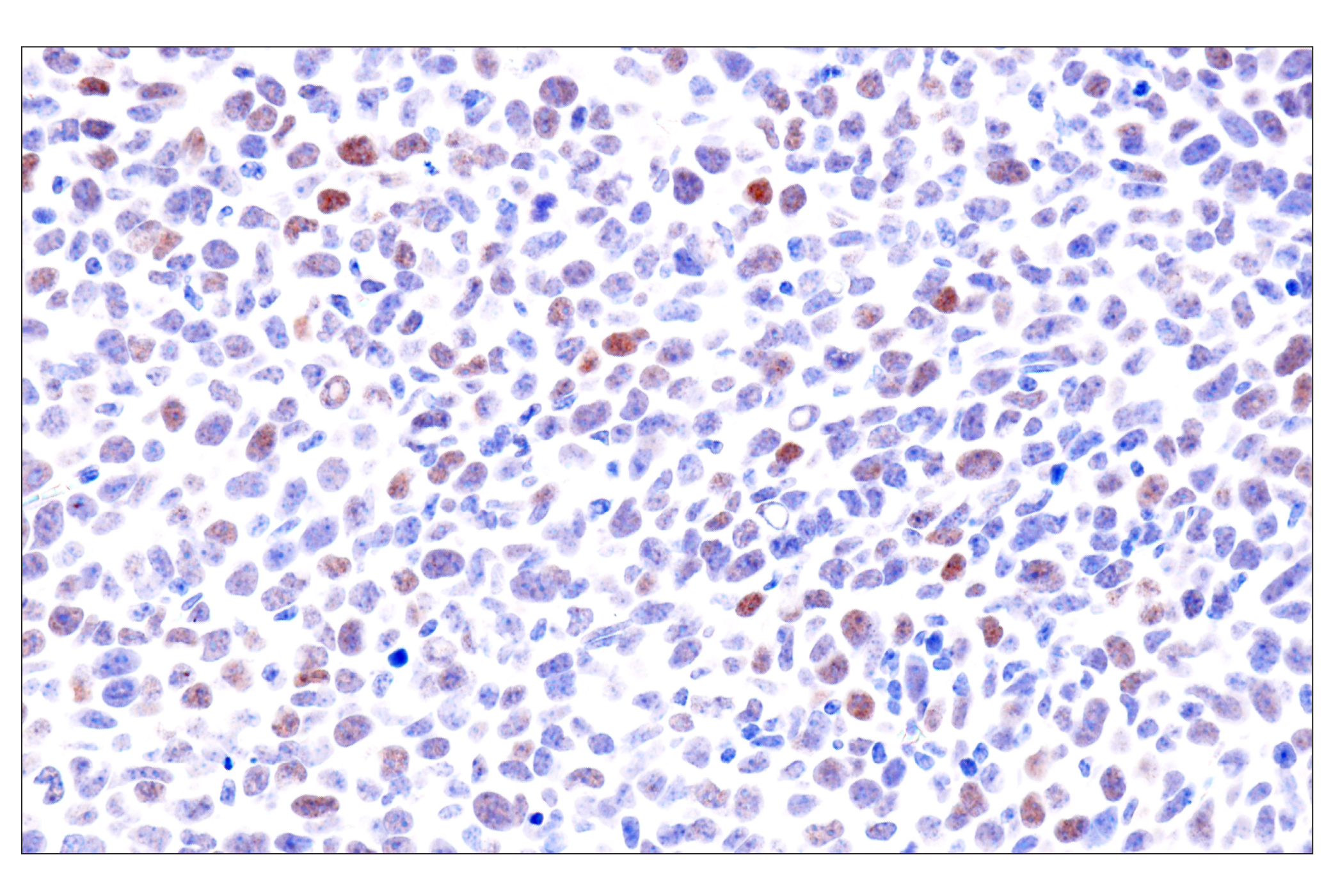
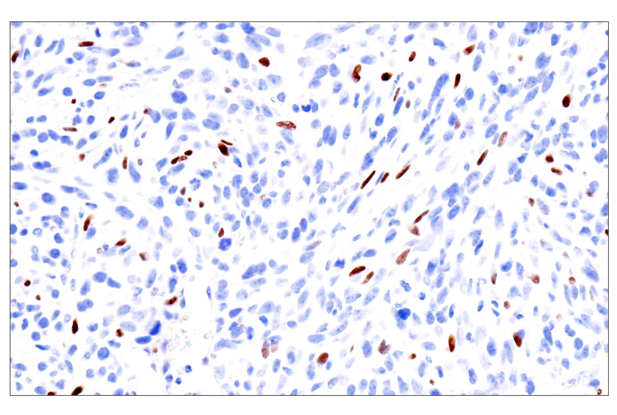
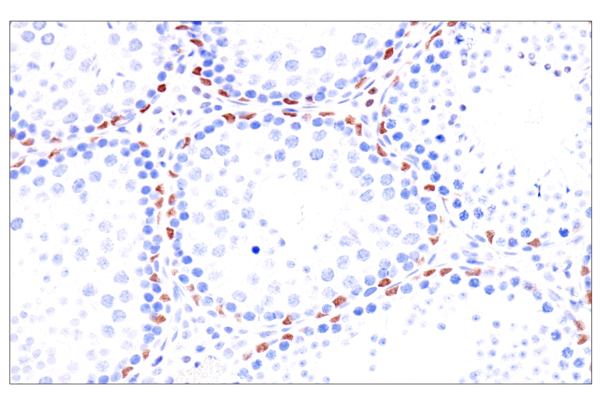
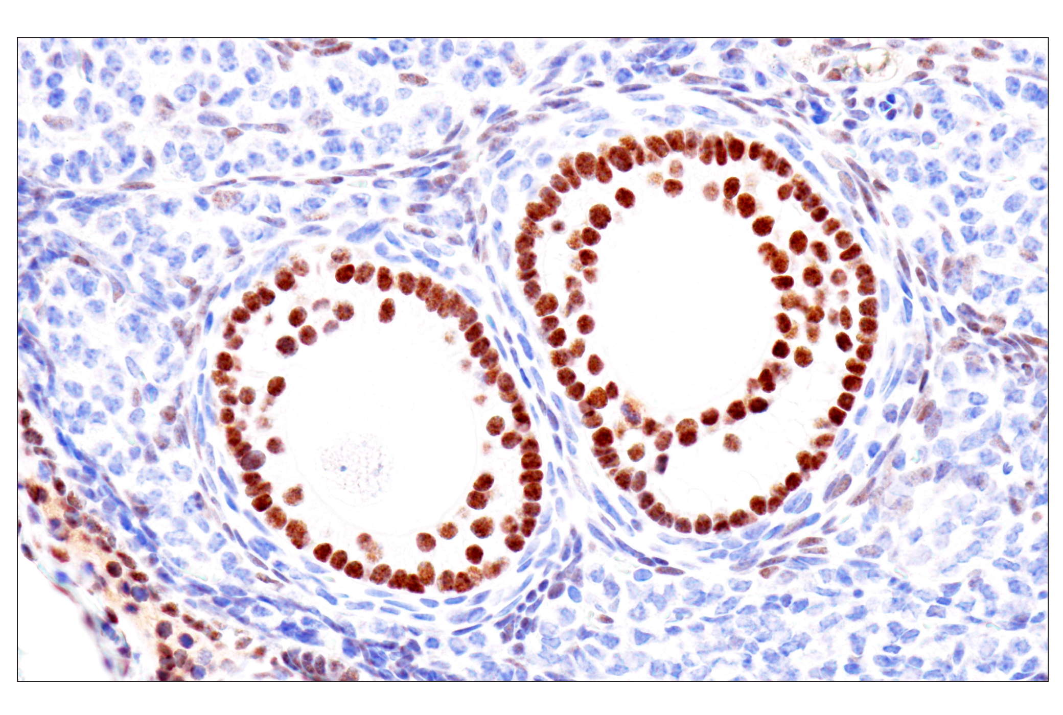
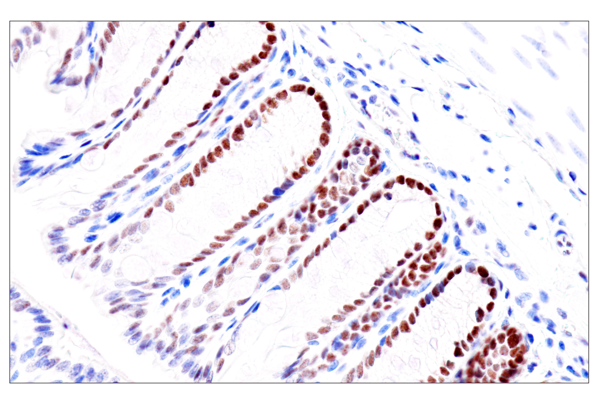
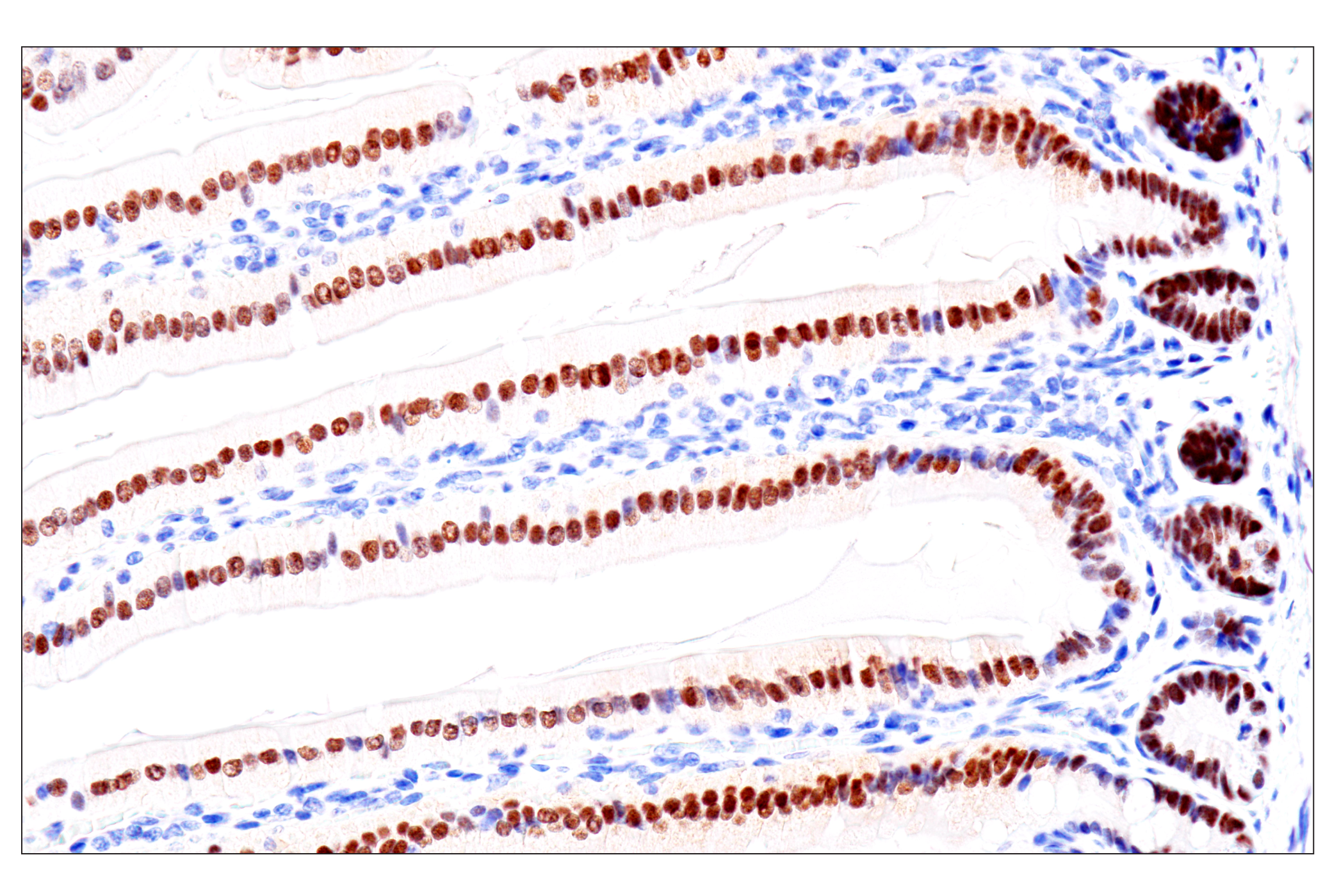
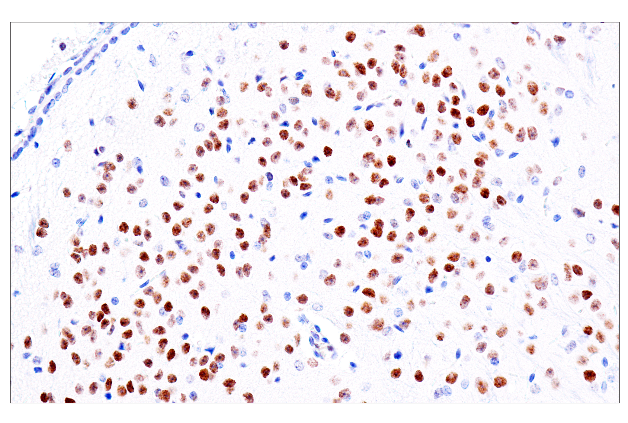
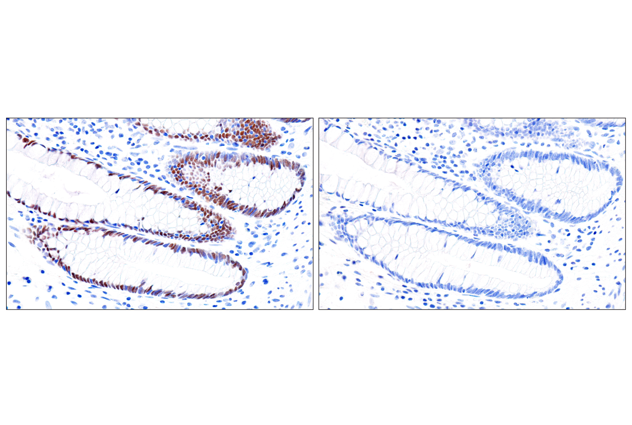
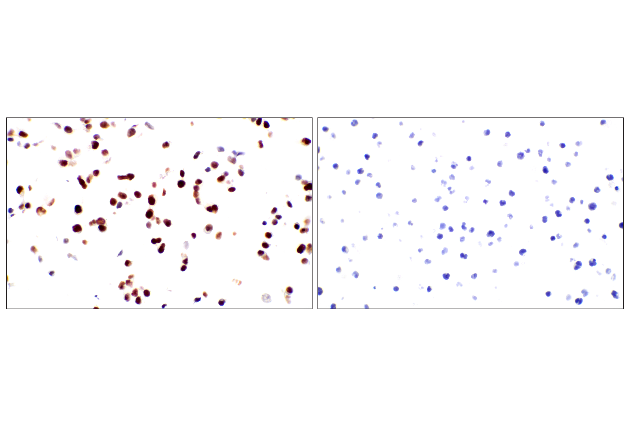
Monoclonal antibodies are produced by immunizing animals with a synthetic peptide corresponding to residues surrounding Pro67 of human NKX2.5 protein, residues near the amino terminus of human GATA-6 protein, a region surrounding Met182 of human MEF2C protein, residues surrounding Phe316 of human α-actinin-1 protein, or residues near the amino terminus of human troponin I protein. Polyclonal antibodies are produced by immunizing animals with a synthetic peptide corresponding to a region surrounding Pro69 of human cardiac troponin T protein or residues of human connexin 43. Polyclonal antibodies are purified by protein A and peptide affinity chromatography.
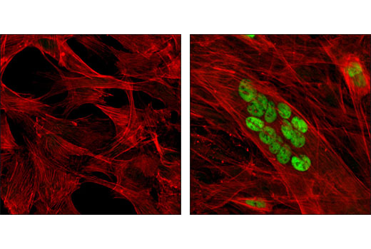
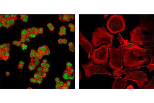
Cardiogenesis is a complex developmental event involving numerous transcription factors. NKX2.5 is a member of the NKX homeobox transcription factor family, which plays an essential role in heart development and is among the earliest factors expressed in the cardiac lineage in developing embryos. Mutations in NKX2.5 are associated with several congenital heart conditions, such as atrial defect with atrioventricular conduction defects (ASD-AVCD) and Tetralogy of Fallot (TOF) (1,2). GATA proteins comprise a group of transcription factors that are related by the presence of conserved zinc finger DNA binding domains, which bind directly to the nucleotide sequence core element GATA (3-5). GATA-6 plays a critical role in endoderm development and knock out of GATA-6 is embryonic lethal due to defects in formation of the heart tube and a failure to develop extraembryonic endoderm (6). MEF2C is a member of the MEF2 (myocyte enhancer factor 2) family of transcription factors. The MEF2 family members were originally described as muscle-specific DNA binding proteins that recognize MEF2 motifs found within the promoters of many muscle-specific genes (7,8). α-Actinin was first recognized as an actin cross-linking protein. The α-actinin protein interacts with a large number of proteins involved in signaling to the cytoskeleton, including those involved in cellular adhesion, migration, and immune cell targeting (9). The muscle isoforms 2 and 3 (ACTN2, ACTN3) localize to the Z-discs of striated muscle and to dense bodies and plaques in smooth muscle (9). Troponin, working in conjunction with tropomyosin, functions as a molecular switch that regulates muscle contraction in response to changes in the intracellular Ca2+ concentration. Troponin consists of three subunits: the Ca2+-binding subunit troponin C (TnC), the tropomyosin-binding subunit troponin T (TnT), and the inhibitory subunit troponin I (TnI) (10). Assays for measuring serum concentrations of cardiac muscle TnT (cTNT), as well as cTnI, have been reported for analyzing cardiac injury. Connexin 43 (Cx43) is a member of the large family of gap junction proteins, which assemble as a hexamer and are transported to the plasma membrane to create a hemichannel that can associate with hemichannels on nearby cells to create cell-to-cell channels. Gap junction communication is important in development and regulation of cell growth. Phosphorylation of Cx43 is important in regulating assembly and function of gap junctions (11,12).
Explore pathways related to this product.
STRING - Known and Predicted Protein-Protein Interactions.
Except as otherwise expressly agreed in a writing signed by a legally authorized representative of CST, the following terms apply to Products provided by CST, its affiliates or its distributors. Any Customer's terms and conditions that are in addition to, or different from, those contained herein, unless separately accepted in writing by a legally authorized representative of CST, are rejected and are of no force or effect.
Products are labeled with For Research Use Only or a similar labeling statement and have not been approved, cleared, or licensed by the FDA or other regulatory foreign or domestic entity, for any purpose. Customer shall not use any Product for any diagnostic or therapeutic purpose, or otherwise in any manner that conflicts with its labeling statement. Products sold or licensed by CST are provided for Customer as the end-user and solely for research and development uses. Any use of Product for diagnostic, prophylactic or therapeutic purposes, or any purchase of Product for resale (alone or as a component) or other commercial purpose, requires a separate license from CST. Customer shall (a) not sell, license, loan, donate or otherwise transfer or make available any Product to any third party, whether alone or in combination with other materials, or use the Products to manufacture any commercial products, (b) not copy, modify, reverse engineer, decompile, disassemble or otherwise attempt to discover the underlying structure or technology of the Products, or use the Products for the purpose of developing any products or services that would compete with CST products or services, (c) not alter or remove from the Products any trademarks, trade names, logos, patent or copyright notices or markings, (d) use the Products solely in accordance with CST Product Terms of Sale and any applicable documentation, and (e) comply with any license, terms of service or similar agreement with respect to any third party products or services used by Customer in connection with the Products.