Revision 1
#97763
Store at -20C
DNA Cytosine Modification Antibody Sampler Kit
1 Kit
(4 x 20 microliters)
877-616-CELL (2355)
877-678-TECH (8324)
3 Trask Lane | Danvers | Massachusetts | 01923 | USA
For Research Use Only. Not for Use in Diagnostic Procedures.
| Product Includes | Product # | Quantity | Mol. Wt | Isotype/Source |
|---|---|---|---|---|
| 5-Methylcytosine (5-mC) (D3S2Z) Rabbit mAb | 28692 | 20 µl | Rabbit IgG | |
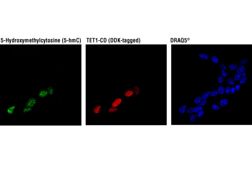
| 5-Hydroxymethylcytosine (5-hmC) (HMC31) Mouse mAb | 51660 | 20 µl | Mouse IgG1 | |
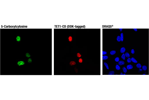
| 5-Carboxylcytosine (5-caC) (D7S8U) Rabbit mAb | 36836 | 20 µl | Rabbit IgG | |
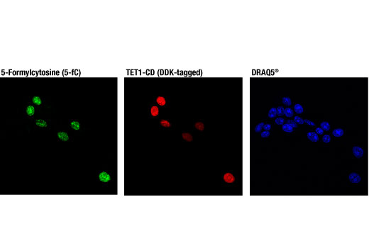
| 5-Formylcytosine (5-fC) (D5D4K) Rabbit mAb | 74178 | 20 µl | Rabbit IgG | |
| Anti-rabbit IgG, HRP-linked Antibody | 7074 | 100 µl | Goat | |
| Anti-mouse IgG, HRP-linked Antibody | 7076 | 100 µl | Horse |
Please visit cellsignal.com for individual component applications, species cross-reactivity, dilutions, protocols, and additional product information.
Description
Storage
Background
TET protein-mediated cytosine hydroxymethylation was initially demonstrated in mouse brain and embryonic stem cells (5, 8). Since then this modification has been discovered in many tissues, with the highest levels found in the brain (9). While 5-fC and 5-caC appear to be short-lived intermediate species, there is mounting evidence showing that 5-hmC is a distinct epigenetic mark with various unique functions (10,11). The modified base itself is stable in vivo and interacts with various readers, including MeCP2 (11,12). The global level of 5-hmC increases during brain development and 5-hmC is enriched at promoter regions and poised enhancers. Furthermore, there is an inverse correlation between levels of 5-hmC and histone H3K9 and H3K27 trimethylation, suggesting a role for 5-hmC in gene activation (12). Lower amounts of 5-hmC have been reported in various cancers, including myeloid leukemia and melanoma (13,14).
Background References
- Hermann, A. et al. (2004) Cell Mol Life Sci 61, 2571-87.
- Turek-Plewa, J. and Jagodziński, P.P. (2005) Cell Mol Biol Lett 10, 631-47.
- Okano, M. et al. (1999) Cell 99, 247-57.
- Li, E. et al. (1992) Cell 69, 915-26.
- Tahiliani, M. et al. (2009) Science 324, 930-5.
- He, Y.F. et al. (2011) Science 333, 1303-7.
- Ito, S. et al. (2011) Science 333, 1300-3.
- Kriaucionis, S. and Heintz, N. (2009) Science 324, 929-30.
- Globisch, D. et al. (2010) PLoS One 5, e15367.
- Gao, Y. et al. (2013) Cell Stem Cell 12, 453-69.
- Mellén, M. et al. (2012) Cell 151, 1417-30.
- Wen, L. et al. (2014) Genome Biol 15, R49.
- Delhommeau, F. et al. (2009) N Engl J Med 360, 2289-301.
- Lian, C.G. et al. (2012) Cell 150, 1135-46.
Trademarks and Patents
Cell Signaling Technology is a trademark of Cell Signaling Technology, Inc.
All other trademarks are the property of their respective owners. Visit cellsignal.com/trademarks for more information.
Limited Uses
Except as otherwise expressly agreed in a writing signed by a legally authorized representative of CST, the following terms apply to Products provided by CST, its affiliates or its distributors. Any Customer's terms and conditions that are in addition to, or different from, those contained herein, unless separately accepted in writing by a legally authorized representative of CST, are rejected and are of no force or effect.
Products are labeled with For Research Use Only or a similar labeling statement and have not been approved, cleared, or licensed by the FDA or other regulatory foreign or domestic entity, for any purpose. Customer shall not use any Product for any diagnostic or therapeutic purpose, or otherwise in any manner that conflicts with its labeling statement. Products sold or licensed by CST are provided for Customer as the end-user and solely for research and development uses. Any use of Product for diagnostic, prophylactic or therapeutic purposes, or any purchase of Product for resale (alone or as a component) or other commercial purpose, requires a separate license from CST. Customer shall (a) not sell, license, loan, donate or otherwise transfer or make available any Product to any third party, whether alone or in combination with other materials, or use the Products to manufacture any commercial products, (b) not copy, modify, reverse engineer, decompile, disassemble or otherwise attempt to discover the underlying structure or technology of the Products, or use the Products for the purpose of developing any products or services that would compete with CST products or services, (c) not alter or remove from the Products any trademarks, trade names, logos, patent or copyright notices or markings, (d) use the Products solely in accordance with CST Product Terms of Sale and any applicable documentation, and (e) comply with any license, terms of service or similar agreement with respect to any third party products or services used by Customer in connection with the Products.
Revision 1

Revision 1

Revision 1
Revision 1