| Cat. # | Size | Qty. | Price |
|---|---|---|---|
| 88548T | 1 Kit (3 x 20 microliters) |
|
| Product Includes | Quantity | Applications | Reactivity | MW(kDa) | Isotype |
|---|---|---|---|---|---|
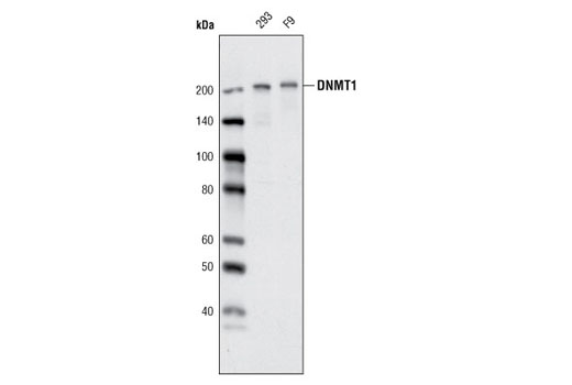
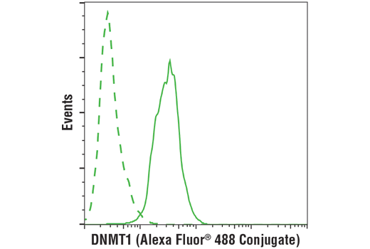
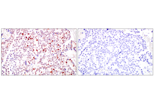
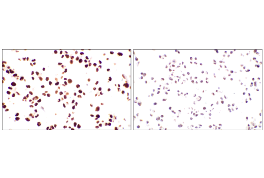
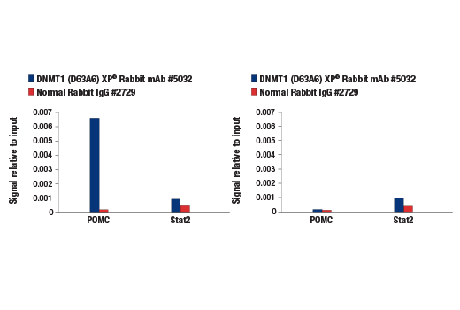
| DNMT1 (D63A6) XP® Rabbit mAb 5032 | 20 µl |
|
H M R Mk | 200 | Rabbit IgG |
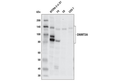
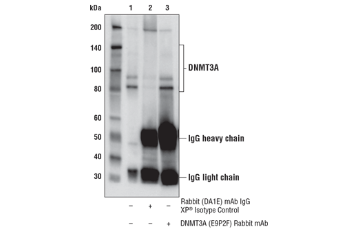
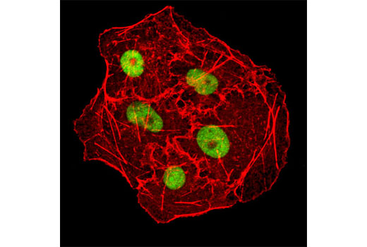
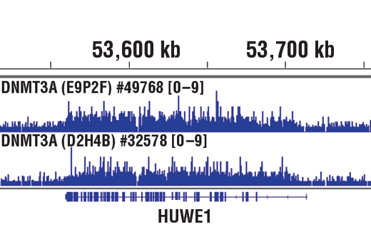
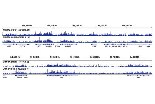
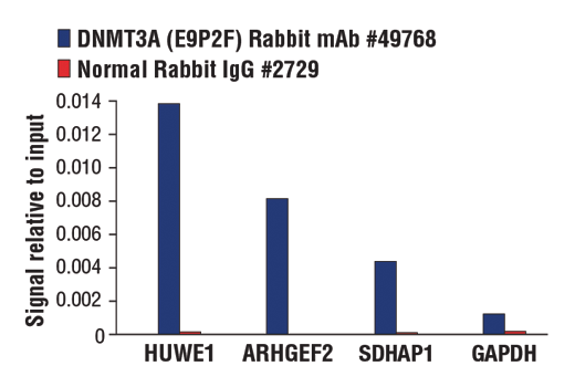
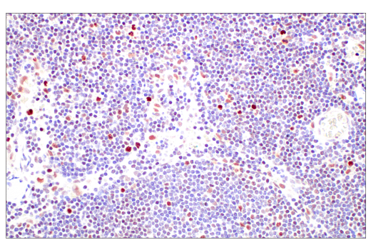
| DNMT3A (E9P2F) Rabbit mAb 49768 | 20 µl |
|
H M R Mk | 85, 95, 130 | Rabbit IgG |
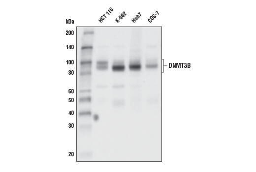
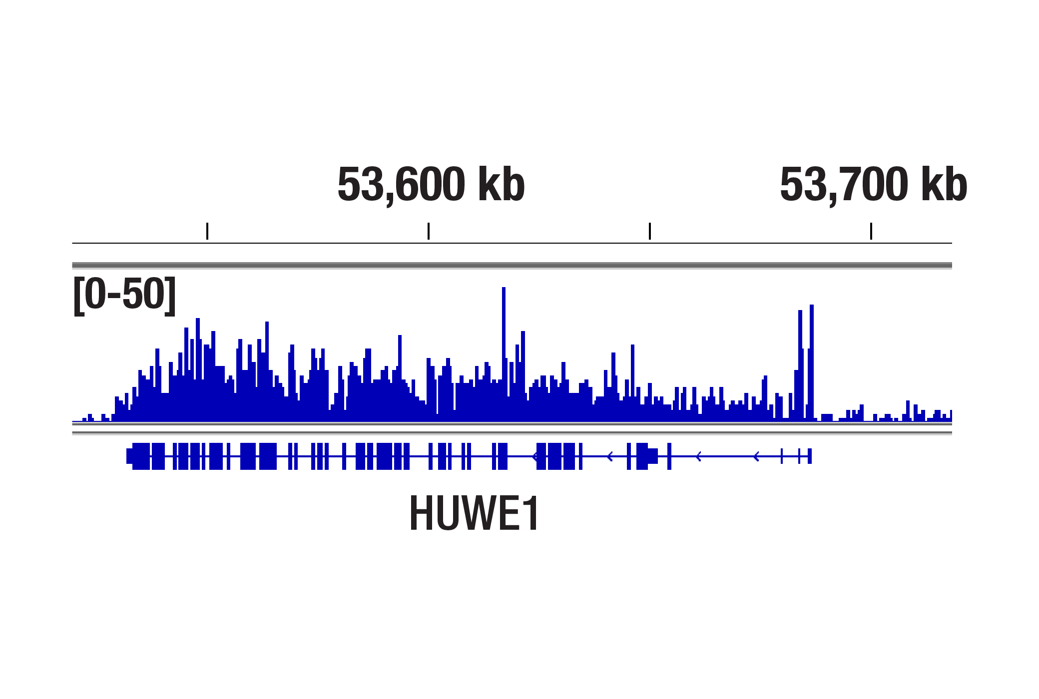
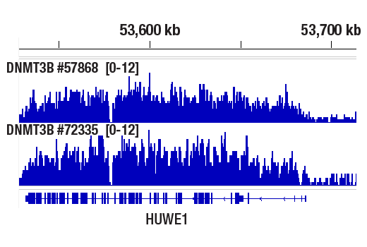
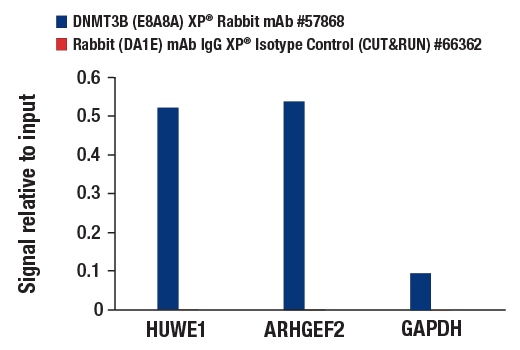
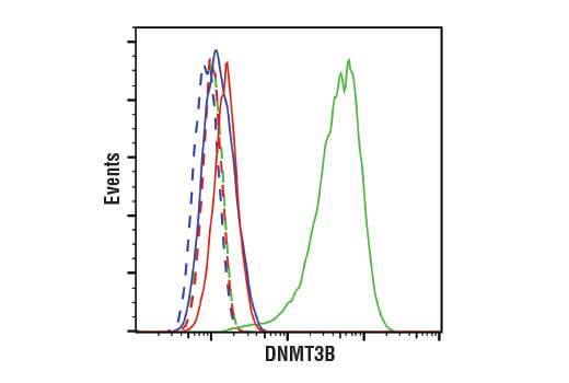
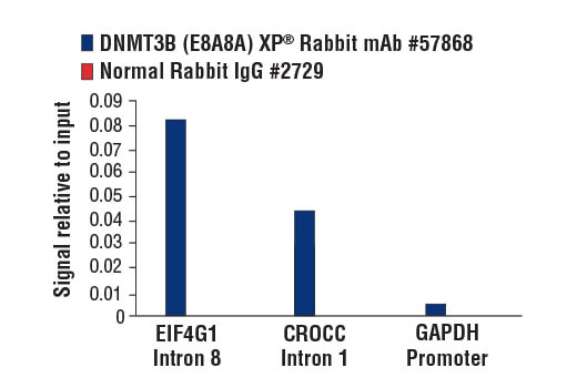
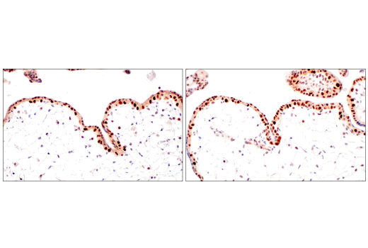
| DNMT3B (E8A8A) XP® Rabbit mAb 57868 | 20 µl |
|
H Mk | 96 | Rabbit IgG |
| Anti-rabbit IgG, HRP-linked Antibody 7074 | 100 µl |
|
Goat |
Product Information
Monoclonal antibodies are produced by immunizing animals with synthetic peptides corresponding to residues surrounding Leu985 of human DNMT1, the amino terminus of human DNMT3B, and recombinant protein specific to the carboxy terminus of human DNMT3A.
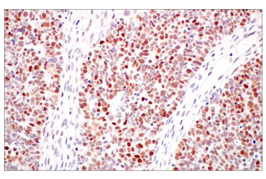
Methylation of DNA at cytosine residues in mammalian cells is a heritable, epigenetic modification that is critical for proper regulation of gene expression, genomic imprinting and development (1,2). Three families of mammalian DNA methyltransferases have been identified: DNMT1, DNMT2, and DNMT3 (1,2). DNMT1 is constitutively expressed in proliferating cells and functions as a maintenance methyltransferase, transferring proper methylation patterns to newly synthesized DNA during replication. DNMT3A and DNMT3B are strongly expressed in embryonic stem cells with reduced expression in adult somatic tissues. DNMT3A and DNMT3B function as de novo methyltransferases that methylate previously unmethylated regions of DNA. DNMT2 is expressed at low levels in adult somatic tissues and its inactivation affects neither de novo nor maintenance DNA methylation. DNMT1, DNMT3A, and DNMT3B together form a protein complex that interacts with histone deacetylases (HDAC1, HDAC2, Sin3A), transcriptional repressor proteins (RB, TAZ-1), and heterochromatin proteins (HP1, SUV39H1) to maintain proper levels of DNA methylation and facilitate gene silencing (3-8). Improper DNA methylation contributes to diseased states such as cancer (1,2). Hypermethylation of promoter CpG islands within tumor suppressor genes correlates with gene silencing and the development of cancer. In addition, hypomethylation of bulk genomic DNA correlates with and may contribute to the onset of cancer. DNMT1, DNMT3A, and DNMT3B are overexpressed in many cancers, including acute and chronic myelogenous leukemias, in addition to colon, breast, and stomach carcinomas (9-12).
Except as otherwise expressly agreed in a writing signed by a legally authorized representative of CST, the following terms apply to Products provided by CST, its affiliates or its distributors. Any Customer's terms and conditions that are in addition to, or different from, those contained herein, unless separately accepted in writing by a legally authorized representative of CST, are rejected and are of no force or effect.
Products are labeled with For Research Use Only or a similar labeling statement and have not been approved, cleared, or licensed by the FDA or other regulatory foreign or domestic entity, for any purpose. Customer shall not use any Product for any diagnostic or therapeutic purpose, or otherwise in any manner that conflicts with its labeling statement. Products sold or licensed by CST are provided for Customer as the end-user and solely for research and development uses. Any use of Product for diagnostic, prophylactic or therapeutic purposes, or any purchase of Product for resale (alone or as a component) or other commercial purpose, requires a separate license from CST. Customer shall (a) not sell, license, loan, donate or otherwise transfer or make available any Product to any third party, whether alone or in combination with other materials, or use the Products to manufacture any commercial products, (b) not copy, modify, reverse engineer, decompile, disassemble or otherwise attempt to discover the underlying structure or technology of the Products, or use the Products for the purpose of developing any products or services that would compete with CST products or services, (c) not alter or remove from the Products any trademarks, trade names, logos, patent or copyright notices or markings, (d) use the Products solely in accordance with CST Product Terms of Sale and any applicable documentation, and (e) comply with any license, terms of service or similar agreement with respect to any third party products or services used by Customer in connection with the Products.