| Cat. # | Size | Qty. | Price |
|---|---|---|---|
| 67935T | 1 Kit |
|
| Product Includes | Quantity | Applications | Reactivity | MW(kDa) | Isotype |
|---|---|---|---|---|---|
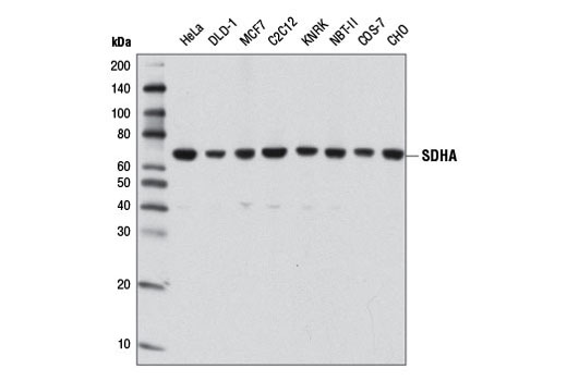
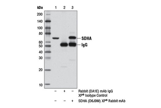
| SDHA (D6J9M) XP® Rabbit mAb 11998 | 20 µl |
|
H M R Hm Mk | 70 | Rabbit IgG |
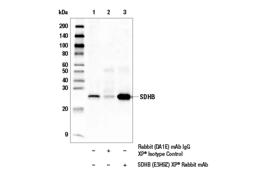
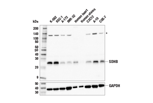
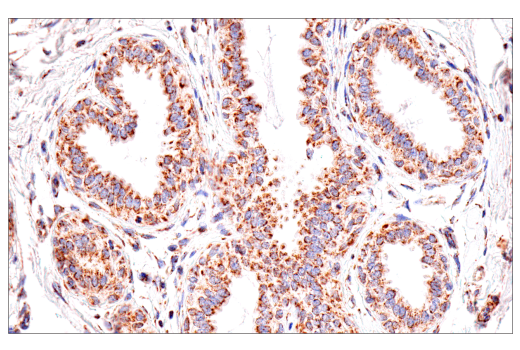
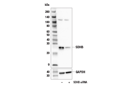
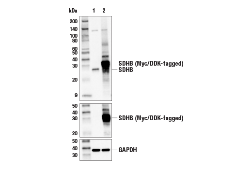
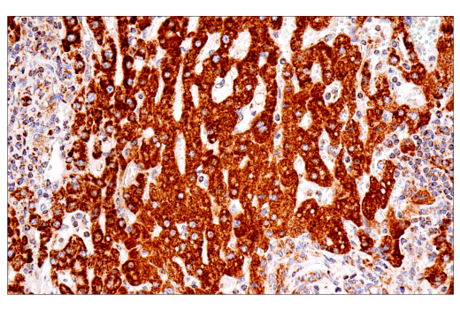
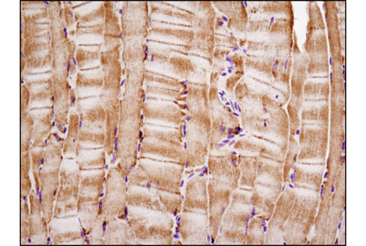
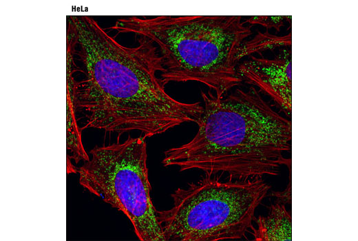
| SDHB (E3H9Z) XP® Rabbit mAb 92649 | 20 µl |
|
H M R Mk | 26 | Rabbit IgG |
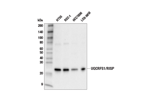
| UQCRFS1/RISP Antibody 95231 | 20 µl |
|
H M R | 23 | Rabbit |
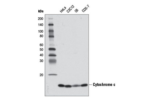
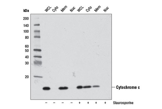
| Cytochrome c (D18C7) Rabbit mAb 11940 | 20 µl |
|
H M R Mk | 14 | Rabbit IgG |
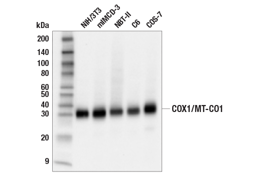
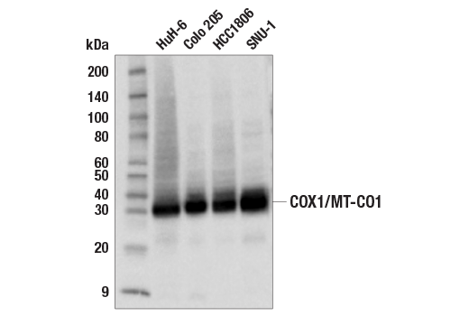
| COX1/MT-CO1 (E2I2R) Rabbit mAb 55159 | 20 µl |
|
H M R Mk | 32 | Rabbit IgG |
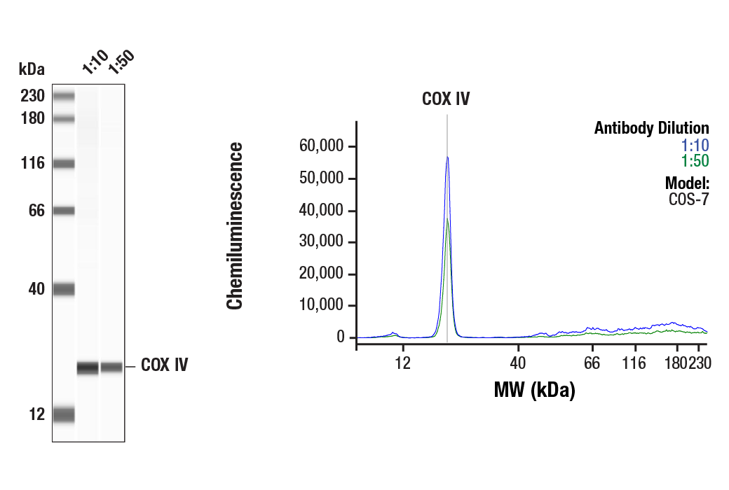
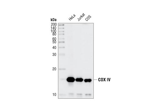
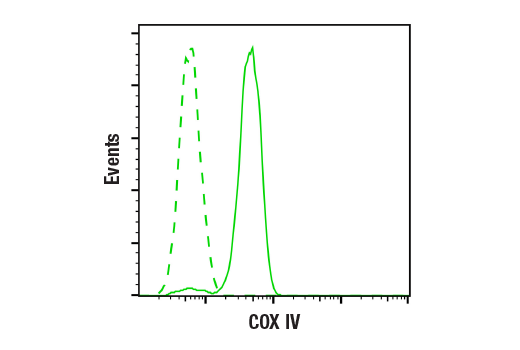
| COX IV (3E11) Rabbit mAb 4850 | 20 µl |
|
H R Mk Z B Pg | 17 | Rabbit IgG |
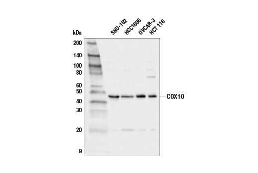
| COX10 (E6K4D) Rabbit mAb 24744 | 20 µl |
|
H Mk | 49 | Rabbit IgG |
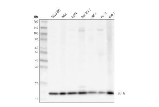
| SDH5 (D1S8D) Rabbit mAb 45849 | 20 µl |
|
H M R Mk | 15 | Rabbit IgG |
| Anti-rabbit IgG, HRP-linked Antibody 7074 | 100 µl |
|
Rab | Goat |
Product Information
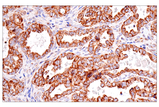
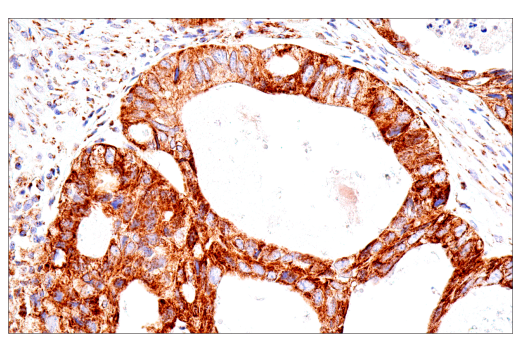
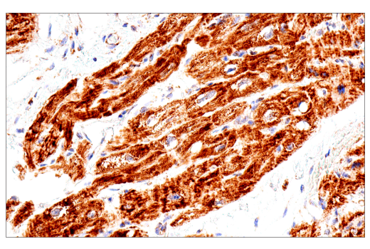
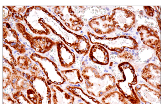
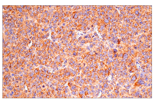
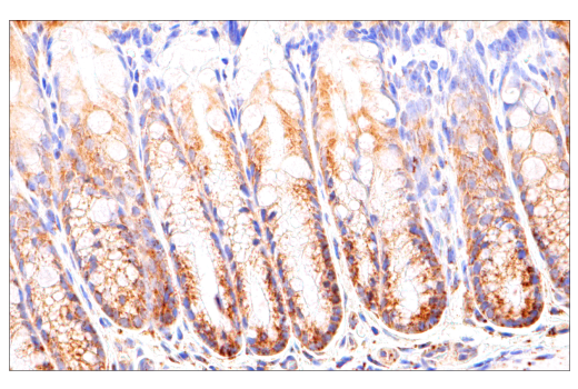
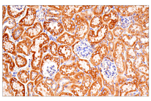
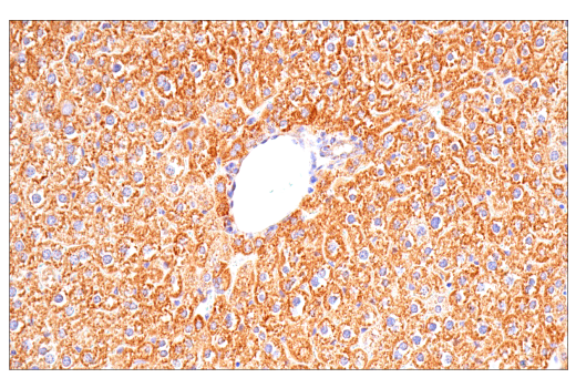
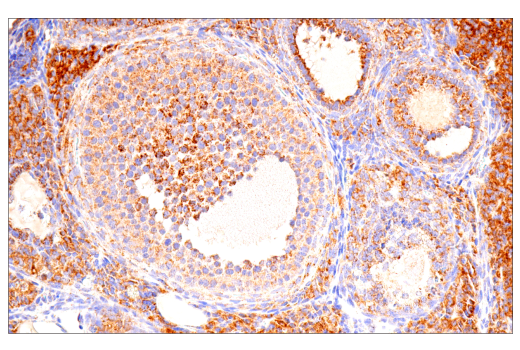
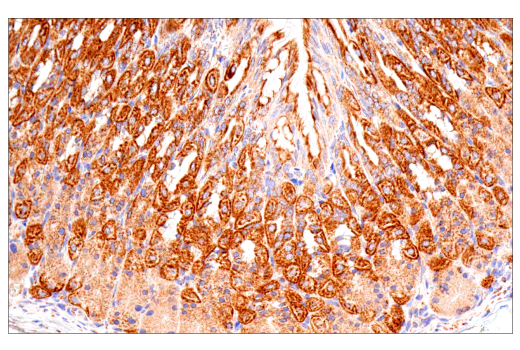
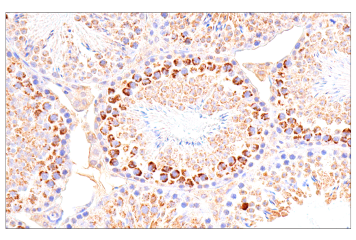
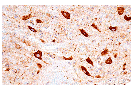
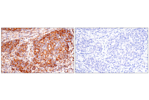
Monoclonal antibodies are produced by immunizing animals with synthetic peptides corresponding to residues surrounding Gly166 of human SDHA protein, Gly208 of human SDHB protein, Pro72 of human cytochrome c protein, Lys29 of human COX IV protein, Asp31 of human COX10 protein, Ala155 of human SDH5 protein, and near the carboxy terminus of human COX1/MT-CO1 protein.
Polyclonal antibodies are produced by immunizing animals with a synthetic peptide corresponding to residues surrounding Pro86 of human UQCRFS1/RISP protein. Antibodies are purified by peptide affinity chromatography.
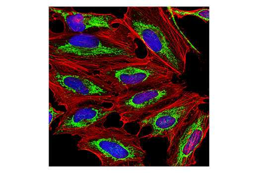
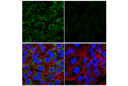
Succinate dehydrogenase (SDH), also known as Complex II or succinate:quinone oxidoreductase, is a key component of the citric acid cycle and the electron transport chain (ETC) (1). Specifically, it is involved in the oxidation of succinate (2). SDH consists of four subunits: SDHA, SDHB, SDHC, and SDHD (3). Ubiquinol-cytochrome c reductase iron-sulfur subunit (UQCRFS1), also known as Rieske iron-sulfur protein (RISP), is a component of Complex III in the mitochondrial ETC. UQCRFS1/RISP and two other subunits, cytochrome b (MT-CYB) and cytochrome c1 (CYC1), are essential for the catalytic activity of Complex III (4). Cytochrome c is a well conserved electron transport protein and is part of the respiratory chain localized to mitochondrial intermembrane space (5). Upon apoptotic stimulation, cytochrome c released from mitochondria associates with procaspase-9 (47 kDa)/Apaf-1. This complex processes caspase-9 from inactive proenzyme to its active form (6). This event further triggers caspase-3 activation and eventually leads to apoptosis (7). The mitochondrial ETC comprises multiple protein complexes, including cytochrome c oxidase. Cytochrome c oxidase catalyzes the reduction of oxygen to water. This process is coupled with pumping protons from the mitochondrial matrix into mitochondrial intermembrane space, contributing to the proton gradient used for ATP synthesis (8). Cytochrome c oxidase consists of 3 mitochondrial DNA-encoded subunits (COX1/MT-CO1, COX2/MT-CO2, and COX3/MT-CO3) and multiple nuclear DNA-encoded subunits (9). Research studies show that the mRNAs of the mitochondrially encoded oxidative phosphorylation subunits, including COX1/MT-CO1, decline significantly during aging (10). Cytochrome c oxidase (COX) is a hetero-oligomeric enzyme consisting of 13 subunits localized to the inner mitochondrial membrane (11-13). It is the terminal enzyme complex in the respiratory chain, catalyzing the reduction of molecular oxygen to water coupled to the translocation of protons across the mitochondrial inner membrane to drive ATP synthesis. The 3 largest subunits forming the catalytic core are encoded by mitochondrial DNA, while the other smaller subunits, including COX IV, are nuclear-encoded. Research studies have shown that deficiency in COX activity correlates with a number of human diseases (14). COX10 is an assembly factor for cytochrome c oxidase (COX, also known as Complex IV) in the mitochondrial ETC (15,16). Studies show that, when the gene encoding the β2-adrenergic receptor (Adrb2) is deleted, increased oxidative phosphorylation in endothelial cells inhibits angiogenesis. Deletion of Cox10 prevents the metabolic switch to oxidative phosphorylation in endothelial cells deleted of Adrb2, causing angiogenesis and cancer progression (16). In addition, COX10 contributes to T cell quiescence exit and is critical for T cell activation (17). Succinate dehydrogenase subunit 5 (SDH5, SDHAF2) is a subunit of the succinate dehydrogenase (SDH) protein complex responsible for the oxidation of succinate during the citric acid cycle. Mitochondrial SDH5 associates with the catalytic subunit of succinate dehydrogenase and is required for adding FAD cofactor to the SDH catalytic subunit (18). Mutations in the corresponding SDHAF2 gene are associated with hereditary head and neck paragangliomas, an autosomal disorder characterized by the development of tumors with increased penetrance over time (18). Additional research studies show that SDH5 is involved in regulation of lung cancer metastasis mediated by the glycogen synthase kinase 3β and β-catenin signaling pathways (19).
Explore pathways related to this product.
STRING - Known and Predicted Protein-Protein Interactions.
Except as otherwise expressly agreed in a writing signed by a legally authorized representative of CST, the following terms apply to Products provided by CST, its affiliates or its distributors. Any Customer's terms and conditions that are in addition to, or different from, those contained herein, unless separately accepted in writing by a legally authorized representative of CST, are rejected and are of no force or effect.
Products are labeled with For Research Use Only or a similar labeling statement and have not been approved, cleared, or licensed by the FDA or other regulatory foreign or domestic entity, for any purpose. Customer shall not use any Product for any diagnostic or therapeutic purpose, or otherwise in any manner that conflicts with its labeling statement. Products sold or licensed by CST are provided for Customer as the end-user and solely for research and development uses. Any use of Product for diagnostic, prophylactic or therapeutic purposes, or any purchase of Product for resale (alone or as a component) or other commercial purpose, requires a separate license from CST. Customer shall (a) not sell, license, loan, donate or otherwise transfer or make available any Product to any third party, whether alone or in combination with other materials, or use the Products to manufacture any commercial products, (b) not copy, modify, reverse engineer, decompile, disassemble or otherwise attempt to discover the underlying structure or technology of the Products, or use the Products for the purpose of developing any products or services that would compete with CST products or services, (c) not alter or remove from the Products any trademarks, trade names, logos, patent or copyright notices or markings, (d) use the Products solely in accordance with CST Product Terms of Sale and any applicable documentation, and (e) comply with any license, terms of service or similar agreement with respect to any third party products or services used by Customer in connection with the Products.