Revision 1
#53898
Store at -20C
ER Homeostasis Antibody Sampler Kit
1 Kit
(9 x 20 microliters)
877-616-CELL (2355)
877-678-TECH (8324)
3 Trask Lane | Danvers | Massachusetts | 01923 | USA
For Research Use Only. Not for Use in Diagnostic Procedures.
| Product Includes | Product # | Quantity | Mol. Wt | Isotype/Source |
|---|---|---|---|---|
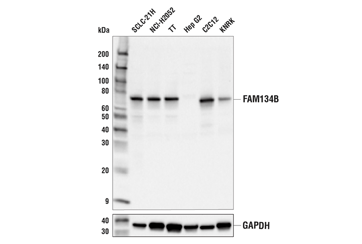
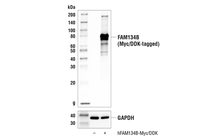
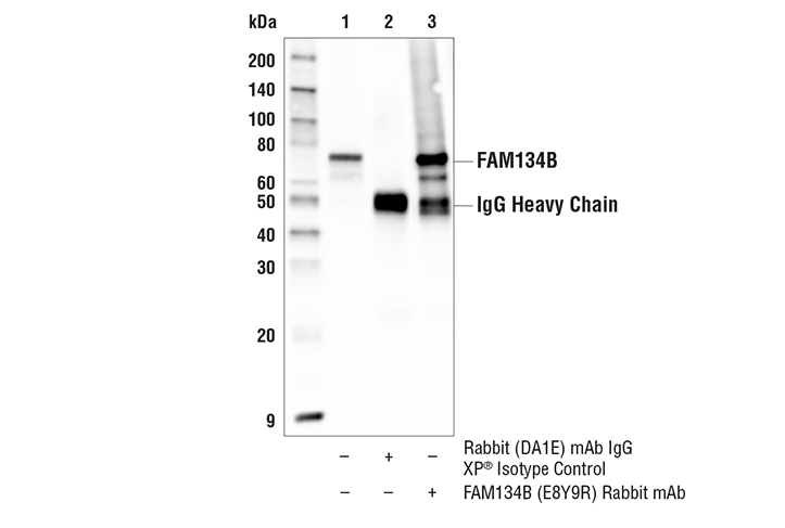
| FAM134B (E8Y9R) Rabbit Monoclonal Antibody | 83414 | 20 µl | 70 kDa | Rabbit IgG |
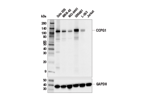
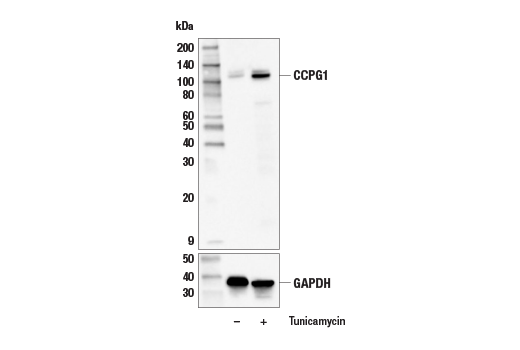
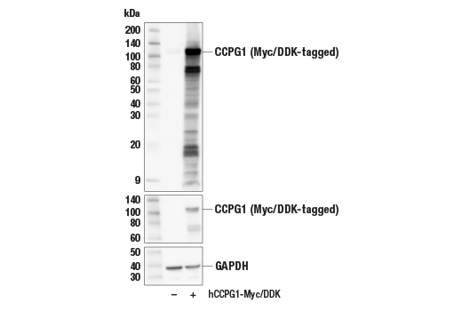
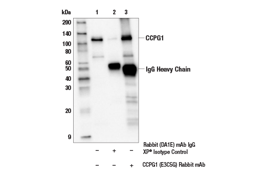
| CCPG1 (E3C5G) Rabbit Monoclonal Antibody | 80158 | 20 µl | 105-120 kDa | Rabbit IgG |
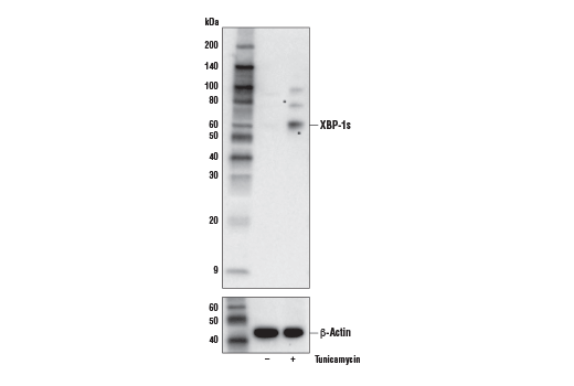
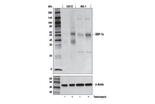
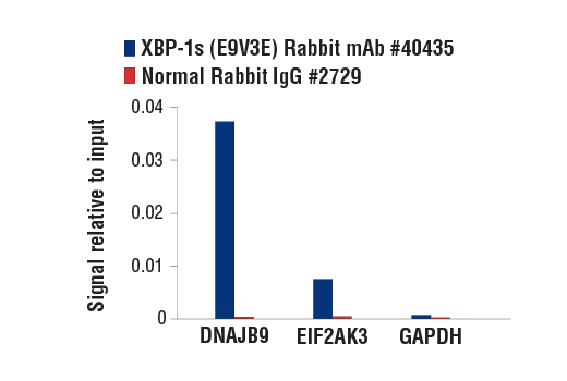
| XBP-1s (E9V3E) Rabbit Monoclonal Antibody | 40435 | 20 µl | 60 (human), 55 (mouse/rat) kDa | Rabbit IgG |
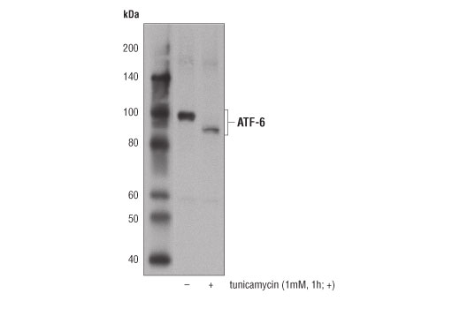
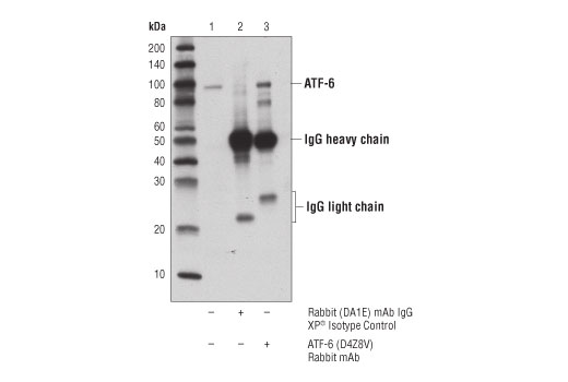
| ATF-6 (D4Z8V) Rabbit Monoclonal Antibody | 65880 | 20 µl | 90-100 kDa | Rabbit IgG |
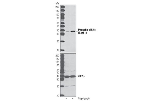
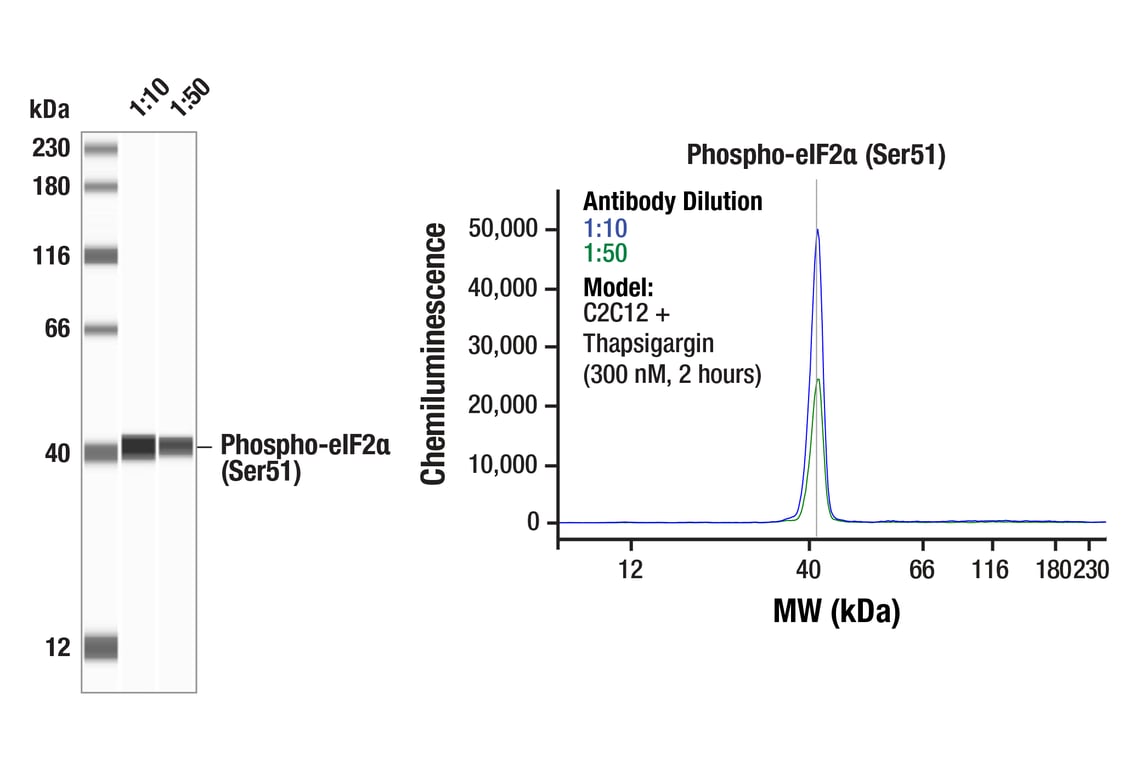
| Phospho-eIF2 alpha (Ser51) (D9G8) Rabbit Monoclonal Antibody | 3398 | 20 µl | 38 kDa | Rabbit IgG |
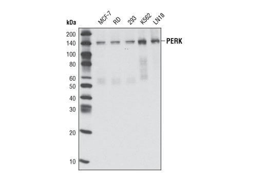
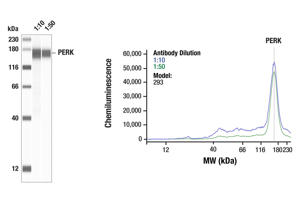
| PERK (C33E10) Rabbit Monoclonal Antibody | 3192 | 20 µl | 140 kDa | Rabbit IgG |
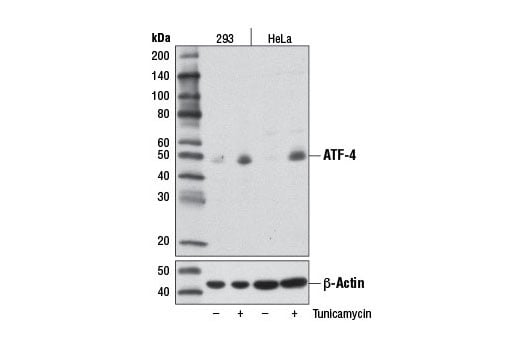
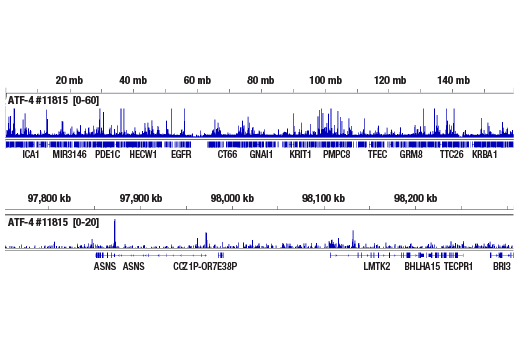
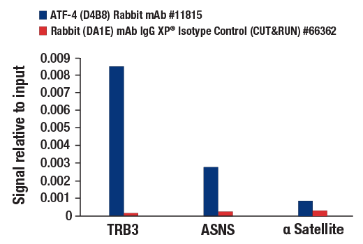
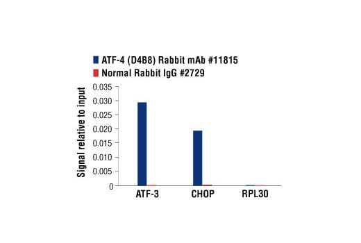
| ATF-4 (D4B8) Rabbit Monoclonal Antibody | 11815 | 20 µl | 49 kDa | Rabbit IgG |
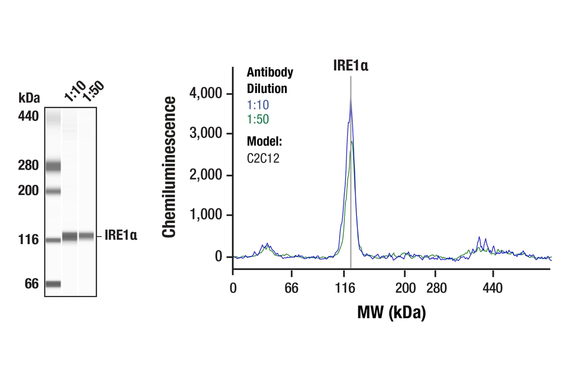
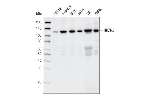
| IRE1 alpha (14C10) Rabbit Monoclonal Antibody | 3294 | 20 µl | 130 kDa | Rabbit IgG |
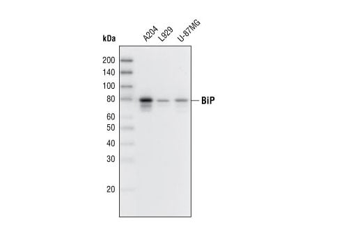
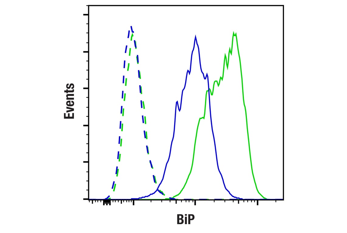
| BiP (C50B12) Rabbit Monoclonal Antibody | 3177 | 20 µl | 78 kDa | Rabbit IgG |
| Anti-rabbit IgG, HRP-linked Antibody | 7074 | 100 µl | Goat |
Please visit cellsignal.com for individual component applications, species cross-reactivity, dilutions, protocols, and additional product information.
Description
Storage
Background
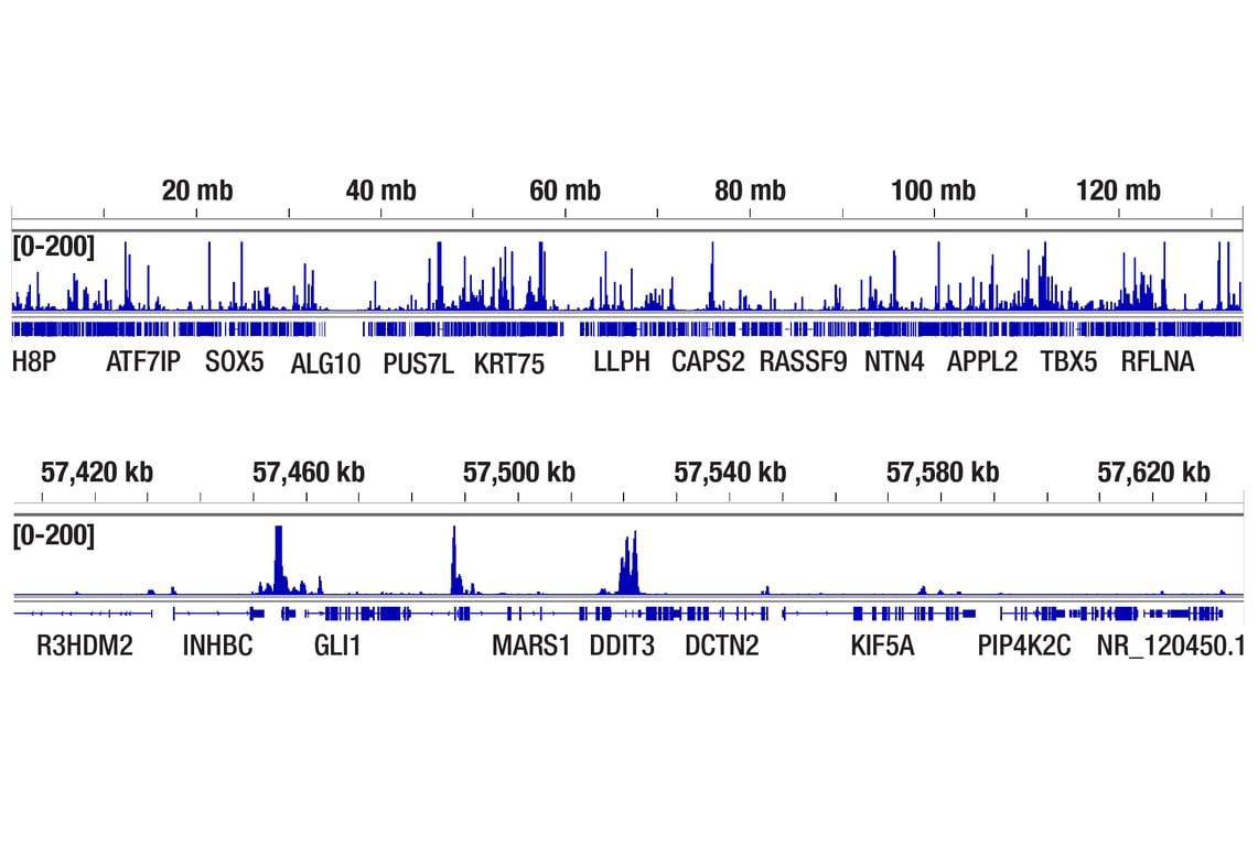
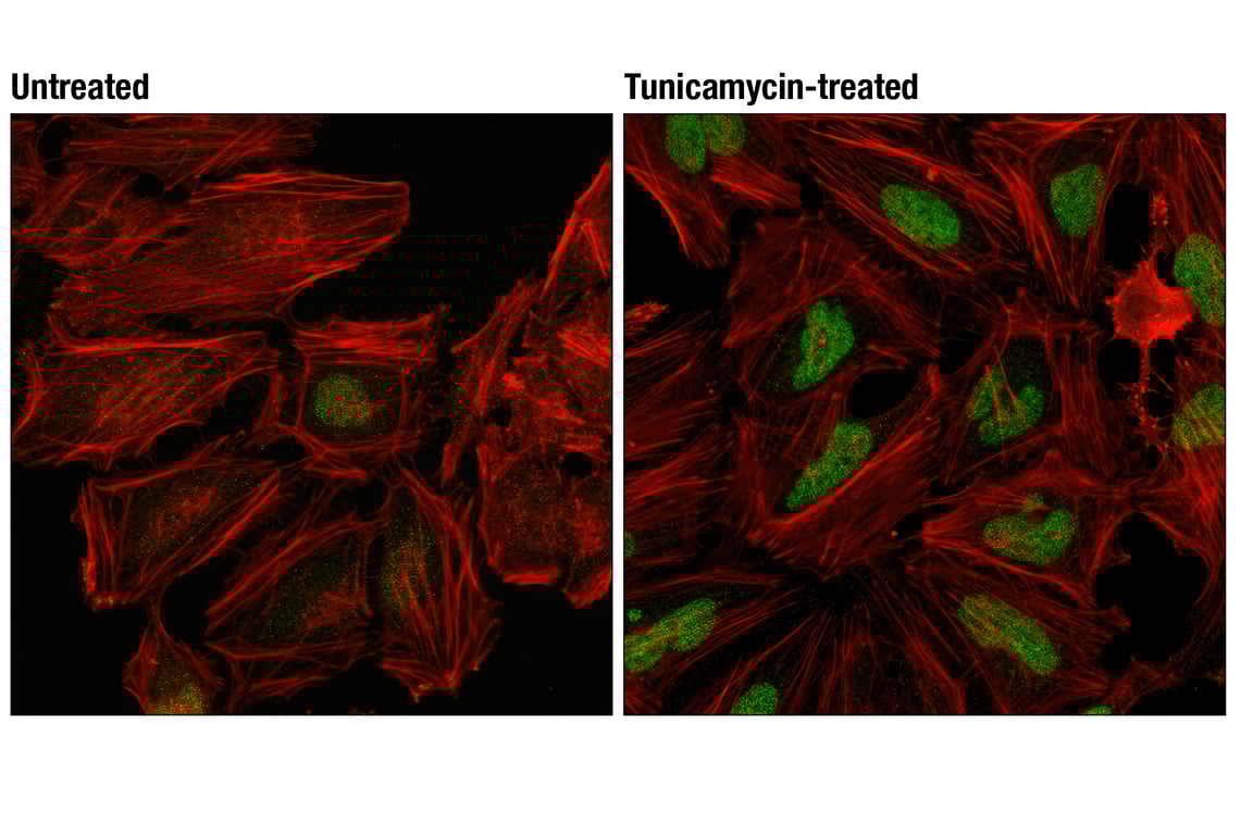
Subsequent to ER expansion triggered by the UPR, cells may trigger a process of ER-phagy, the degradation of ER fragments through autophagy (3). Autophagy is a process for the bulk degradation of cytoplasmic components by a double membrane autophagosome fusing to the lysosome. Selective autophagy, like ER-phagy, permits the degradation of specific targets. This process generally involves specific cargo receptors containing LIR or GIM domains targeting bound cargo to the autophagosome through interactions with LC3 or GABARAP, respectively. FAM134B was the first ER-phagy receptor discovered. Loss of FAM134B can sensitize cells to apoptosis when challenged by nutrient deprivation or ER stress stimuli. CCPG1 is another ER-phagy cargo receptor that associates with FIP200, a component of the ULK1 complex facilitating ER trafficking to autophagosomes. Importantly, CCPG1 is transcriptionally regulated by ER stress. Taken together, signaling from the UPR and ER-phagy help regulate ER homeostasis. Defects in this process may contribute to pathological conditions, including metabolic and neurological disorders, cancer, and defense against infectious diseases (3).
Trademarks and Patents
Cell Signaling Technology is a trademark of Cell Signaling Technology, Inc.
U.S. Patent No. 7,429,487, foreign equivalents, and child patents deriving therefrom.
All other trademarks are the property of their respective owners. Visit cellsignal.com/trademarks for more information.
Limited Uses
Except as otherwise expressly agreed in a writing signed by a legally authorized representative of CST, the following terms apply to Products provided by CST, its affiliates or its distributors. Any Customer's terms and conditions that are in addition to, or different from, those contained herein, unless separately accepted in writing by a legally authorized representative of CST, are rejected and are of no force or effect.
Products are labeled with For Research Use Only or a similar labeling statement and have not been approved, cleared, or licensed by the FDA or other regulatory foreign or domestic entity, for any purpose. Customer shall not use any Product for any diagnostic or therapeutic purpose, or otherwise in any manner that conflicts with its labeling statement. Products sold or licensed by CST are provided for Customer as the end-user and solely for research and development uses. Any use of Product for diagnostic, prophylactic or therapeutic purposes, or any purchase of Product for resale (alone or as a component) or other commercial purpose, requires a separate license from CST. Customer shall (a) not sell, license, loan, donate or otherwise transfer or make available any Product to any third party, whether alone or in combination with other materials, or use the Products to manufacture any commercial products, (b) not copy, modify, reverse engineer, decompile, disassemble or otherwise attempt to discover the underlying structure or technology of the Products, or use the Products for the purpose of developing any products or services that would compete with CST products or services, (c) not alter or remove from the Products any trademarks, trade names, logos, patent or copyright notices or markings, (d) use the Products solely in accordance with CST Product Terms of Sale and any applicable documentation, and (e) comply with any license, terms of service or similar agreement with respect to any third party products or services used by Customer in connection with the Products.
Revision 1
Revision 1
Revision 1
Revision 1

Revision 1
Revision 1

Revision 1
Revision 1
Revision 1

Revision 1
Revision 1
Revision 1
Revision 1
Revision 1
Revision 1