Revision 3
#45850
Store at -20C
877-616-CELL (2355)
877-678-TECH (8324)
3 Trask Lane | Danvers | Massachusetts | 01923 | USA
For Research Use Only. Not for Use in Diagnostic Procedures.
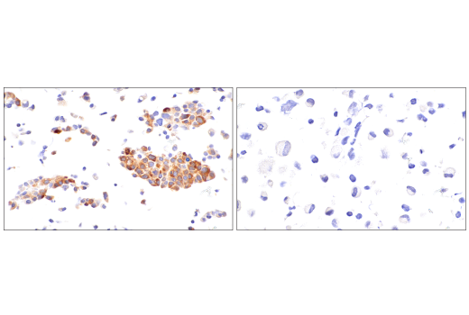
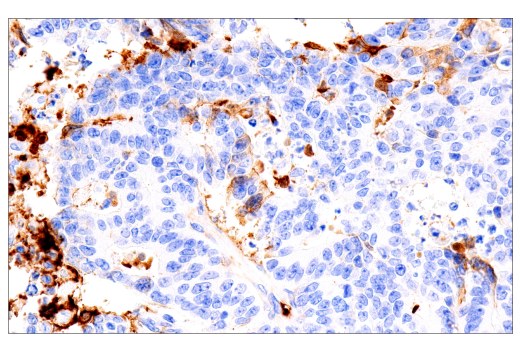
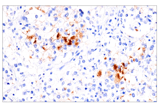
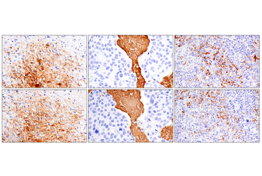
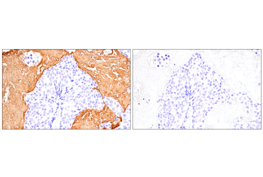
Applications:
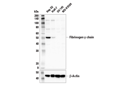
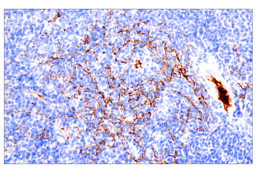
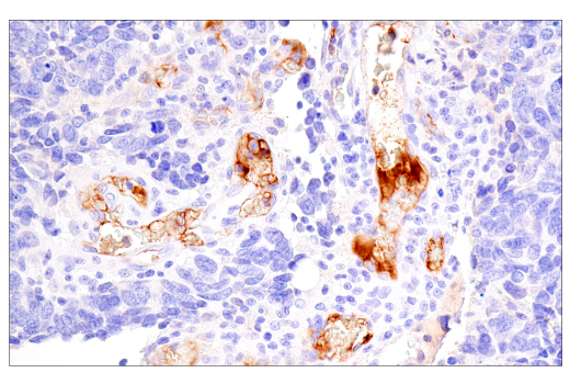
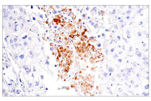
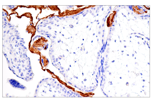
W, IHC-Bond, IHC-P
Reactivity:
H
Sensitivity:
Endogenous
MW (kDa):
50
Source/Isotype:
Rabbit IgG
UniProt ID:
#P02679
Entrez-Gene Id:
2266
Product Usage Information
| Application | Dilution |
|---|---|
| Western Blotting | 1:1000 |
| IHC Leica Bond | 1:200 - 1:800 |
| Immunohistochemistry (Paraffin) | 1:200 - 1:800 |
Storage
For a carrier free (BSA and azide free) version of this product see product #38915.
Specificity/Sensitivity
Source / Purification
Background
Upon engagement of the clotting cascade, thrombin-mediated cleavage of the fibrinogen Aα and Bβ chains creates fibrin monomers and a platform for the polymerization of fibrin monomers into protofibrils and an insoluble web of stable fibrin fibers (4,5). Whereas thrombin is the enzyme that drives fibrin polymerization and clot formation, plasmin is the enzymatic counterpart that facilitates fibrinolysis and clot breakdown (6).
In addition to its position as a central node in the normal coagulation cascade, dysregulated fibrin deposition has been observed in pathological conditions such as cancer and viral infection (7,8).
The fibrinogen gamma chain is expressed as two isoforms that result from alternative mRNA splicing, the γA chain and the γ' chain. Research studies have shown that the fibrinogen gamma chain plays important roles in shaping the architecture of fibrin clots and modulates the innate immune response through interaction with its receptor, CD11b, expressed on neutrophils and monocytes (9,10).
Background References
- Chapin, J.C. and Hajjar, K.A. (2015) Blood Rev 29, 17-24.
- Dalmon, J. et al. (1993) Mol Cell Biol 13, 1183-93.
- Huber, P. et al. (1990) J Biol Chem 265, 5695-701.
- Lord, S.T. (2011) Arterioscler Thromb Vasc Biol 31, 494-9.
- Yang, Z. et al. (2000) Proc Natl Acad Sci U S A 97, 14156-61.
- Cesarman-Maus, G. and Hajjar, K.A. (2005) Br J Haematol 129, 307-21.
- Fernandez, P.M. et al. (2004) Semin Thromb Hemost 30, 31-44.
- Merad, M. and Martin, J.C. (2020) Nat Rev Immunol 20, 355-362.
- Yakovlev, S. et al. (2000) Biochemistry 39, 15721-9.
- Siebenlist, K.R. and Mosesson, M.W. (1994) J Biol Chem 269, 28414-9.
Species Reactivity
Species reactivity is determined by testing in at least one approved application (e.g., western blot).
Western Blot Buffer
IMPORTANT: For western blots, incubate membrane with diluted primary antibody in 5% w/v nonfat dry milk, 1X TBS, 0.1% Tween® 20 at 4°C with gentle shaking, overnight.
Applications Key
W: Western Blotting IHC-Bond: IHC Leica Bond IHC-P: Immunohistochemistry (Paraffin)
Cross-Reactivity Key
H: Human
Trademarks and Patents
Cell Signaling Technology is a trademark of Cell Signaling Technology, Inc.
SignalStain is a registered trademark of Cell Signaling Technology, Inc.
All other trademarks are the property of their respective owners. Visit cellsignal.com/trademarks for more information.
Limited Uses
Except as otherwise expressly agreed in a writing signed by a legally authorized representative of CST, the following terms apply to Products provided by CST, its affiliates or its distributors. Any Customer's terms and conditions that are in addition to, or different from, those contained herein, unless separately accepted in writing by a legally authorized representative of CST, are rejected and are of no force or effect.
Products are labeled with For Research Use Only or a similar labeling statement and have not been approved, cleared, or licensed by the FDA or other regulatory foreign or domestic entity, for any purpose. Customer shall not use any Product for any diagnostic or therapeutic purpose, or otherwise in any manner that conflicts with its labeling statement. Products sold or licensed by CST are provided for Customer as the end-user and solely for research and development uses. Any use of Product for diagnostic, prophylactic or therapeutic purposes, or any purchase of Product for resale (alone or as a component) or other commercial purpose, requires a separate license from CST. Customer shall (a) not sell, license, loan, donate or otherwise transfer or make available any Product to any third party, whether alone or in combination with other materials, or use the Products to manufacture any commercial products, (b) not copy, modify, reverse engineer, decompile, disassemble or otherwise attempt to discover the underlying structure or technology of the Products, or use the Products for the purpose of developing any products or services that would compete with CST products or services, (c) not alter or remove from the Products any trademarks, trade names, logos, patent or copyright notices or markings, (d) use the Products solely in accordance with CST Product Terms of Sale and any applicable documentation, and (e) comply with any license, terms of service or similar agreement with respect to any third party products or services used by Customer in connection with the Products.
Revision 3
Revision 3
Revision 3
Revision 3