Revision 6
#89572
Store at -20C
877-616-CELL (2355)
877-678-TECH (8324)
3 Trask Lane | Danvers | Massachusetts | 01923 | USA
For Research Use Only. Not for Use in Diagnostic Procedures.
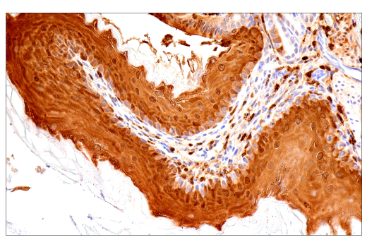
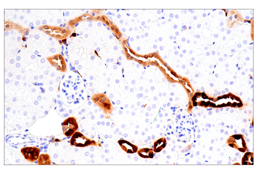
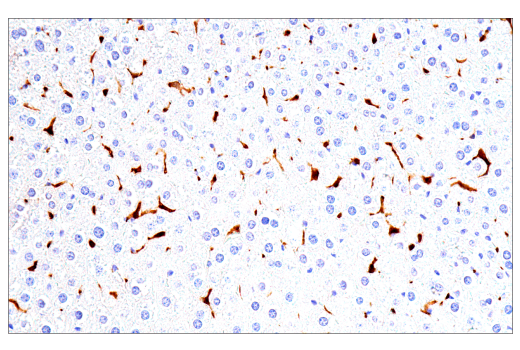
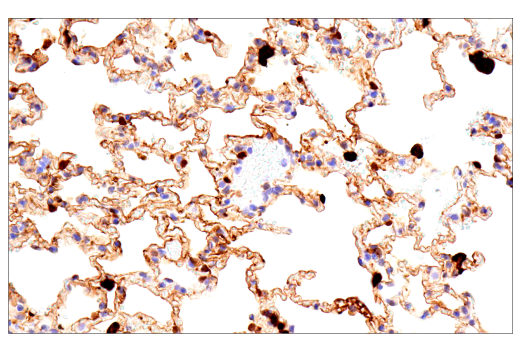
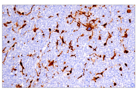
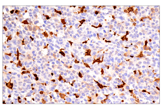
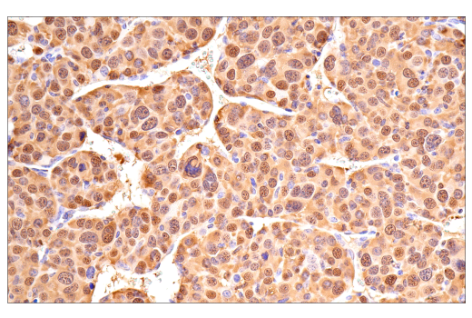
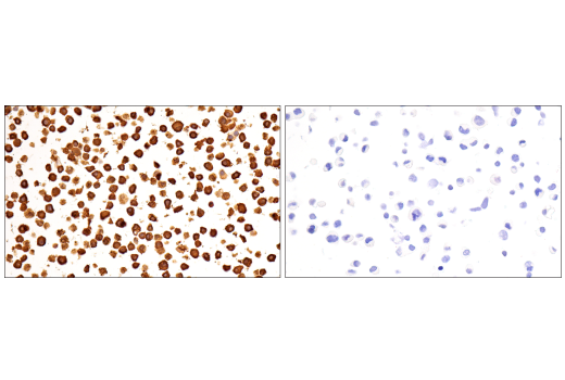
Applications:
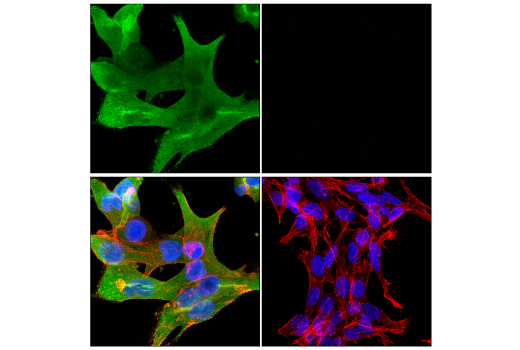
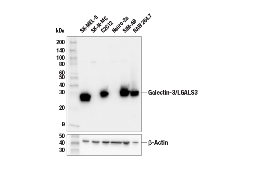
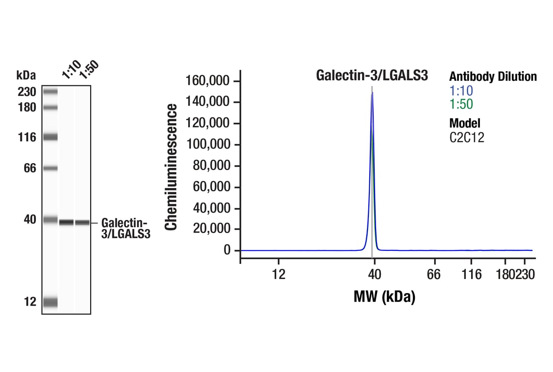
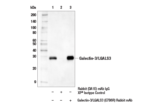
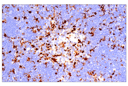
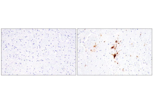
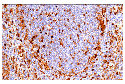
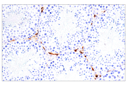
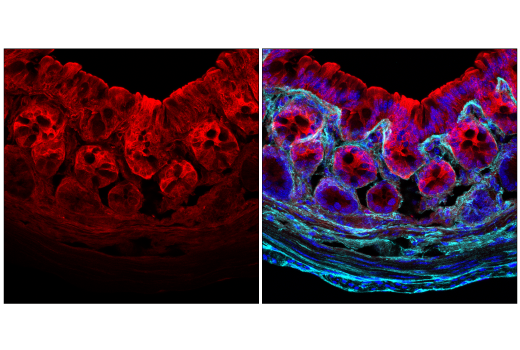
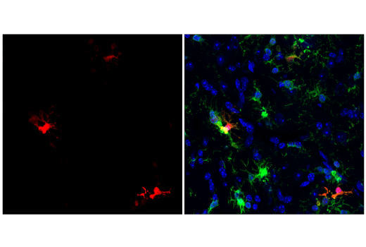
W, W-S, IP, IHC-P, IF-F, IF-IC
Reactivity:
H M R
Sensitivity:
Endogenous
MW (kDa):
28
Source/Isotype:
Rabbit IgG
UniProt ID:
#P16110
Entrez-Gene Id:
16854
Product Usage Information
| Application | Dilution |
|---|---|
| Western Blotting | 1:1000 |
| Simple Western™ | 1:10 - 1:50 |
| Immunoprecipitation | 1:50 |
| Immunohistochemistry (Paraffin) | 1:50 - 1:200 |
| Immunofluorescence (Frozen) | 1:100 - 1:400 |
| Immunofluorescence (Immunocytochemistry) | 1:800 - 1:3200 |
Storage
For a carrier free (BSA and azide free) version of this product see product #76015.
Specificity/Sensitivity
Source / Purification
Background
Galectin-3/LGALS3 is involved in several diverse biological functions. Galectin-3/LGALS3 binds IgE (8). Galectin-3/LGALS3 is an unusual protein in that it can be found both extracellularly and intracellularly. Intracellularly, Galectin-3/LGALS3 can localize to the cytoplasm, nucleus, or both, depending on cell type and experimental conditions. Nuclear Galectin-3/LGALS3 has been identified as a pre-mRNA splicing factor (9). Galectin-3/LGALS3 production has been shown to increase during inflammation and in obesity, and the protein itself can have an inflammatory effect under certain conditions (10). Galectin-3/LGALS3 forms a complex with α3, β1 integrin to act as a surface receptor on endothelial cells for the NG2 proteoglycan, which triggers cell motility and angiogenesis (11). In addition to these functions, Galectin-3/LGALS3 is also a required factor for the terminal differentiation of epithelial cells (12).
Background References
- Barondes, S.H. et al. (1994) Cell 76, 597-8.
- Barondes, S.H. et al. (1994) J Biol Chem 269, 20807-10.
- Offner, H. et al. (1990) J Neuroimmunol 28, 177-84.
- Wells, V. and Mallucci, L. (1991) Cell 64, 91-7.
- Filer, A. et al. (2009) Arthritis Rheum 60, 1604-14.
- Perillo, N.L. et al. (1995) Nature 378, 736-9.
- Cooper, D.N. et al. (1991) J Cell Biol 115, 1437-48.
- Platzer, B. et al. (2011) Immunol Lett 141, 36-44.
- Haudek, K.C. et al. (2010) Biochim Biophys Acta 1800, 181-9.
- Pang, J. et al. (2013) PLoS One 8, e57915.
- Fukushi, J. et al. (2004) Mol Biol Cell 15, 3580-90.
- Hikita, C. et al. (2000) J Cell Biol 151, 1235-46.
Species Reactivity
Species reactivity is determined by testing in at least one approved application (e.g., western blot).
Western Blot Buffer
IMPORTANT: For western blots, incubate membrane with diluted primary antibody in 5% w/v nonfat dry milk, 1X TBS, 0.1% Tween® 20 at 4°C with gentle shaking, overnight.
Applications Key
W: Western Blotting W-S: Simple Western™ IP: Immunoprecipitation IHC-P: Immunohistochemistry (Paraffin) IF-F: Immunofluorescence (Frozen) IF-IC: Immunofluorescence (Immunocytochemistry)
Cross-Reactivity Key
H: Human M: Mouse R: Rat
Trademarks and Patents
Cell Signaling Technology is a trademark of Cell Signaling Technology, Inc.
Alexa Fluor is a registered trademark of Life Technologies Corporation.
All other trademarks are the property of their respective owners. Visit cellsignal.com/trademarks for more information.
Limited Uses
Except as otherwise expressly agreed in a writing signed by a legally authorized representative of CST, the following terms apply to Products provided by CST, its affiliates or its distributors. Any Customer's terms and conditions that are in addition to, or different from, those contained herein, unless separately accepted in writing by a legally authorized representative of CST, are rejected and are of no force or effect.
Products are labeled with For Research Use Only or a similar labeling statement and have not been approved, cleared, or licensed by the FDA or other regulatory foreign or domestic entity, for any purpose. Customer shall not use any Product for any diagnostic or therapeutic purpose, or otherwise in any manner that conflicts with its labeling statement. Products sold or licensed by CST are provided for Customer as the end-user and solely for research and development uses. Any use of Product for diagnostic, prophylactic or therapeutic purposes, or any purchase of Product for resale (alone or as a component) or other commercial purpose, requires a separate license from CST. Customer shall (a) not sell, license, loan, donate or otherwise transfer or make available any Product to any third party, whether alone or in combination with other materials, or use the Products to manufacture any commercial products, (b) not copy, modify, reverse engineer, decompile, disassemble or otherwise attempt to discover the underlying structure or technology of the Products, or use the Products for the purpose of developing any products or services that would compete with CST products or services, (c) not alter or remove from the Products any trademarks, trade names, logos, patent or copyright notices or markings, (d) use the Products solely in accordance with CST Product Terms of Sale and any applicable documentation, and (e) comply with any license, terms of service or similar agreement with respect to any third party products or services used by Customer in connection with the Products.
Revision 6
Revision 6
Revision 6
Revision 6

Revision 6
Revision 6
Revision 6