Revision 3
#54330
Store at -20C
877-616-CELL (2355)
877-678-TECH (8324)
3 Trask Lane | Danvers | Massachusetts | 01923 | USA
For Research Use Only. Not for Use in Diagnostic Procedures.
Applications:
W, IHC-Bond, IHC-P, FC-FP
Reactivity:
H
Sensitivity:
Endogenous
MW (kDa):
9-40
Source/Isotype:
Rabbit IgG
UniProt ID:
#O00182
Entrez-Gene Id:
3965
Product Usage Information
| Application | Dilution |
|---|---|
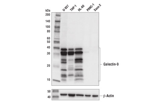
| Western Blotting | 1:1000 |
| IHC Leica Bond | 1:800 |
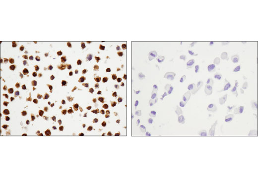
| Immunohistochemistry (Paraffin) | 1:800 |
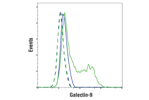
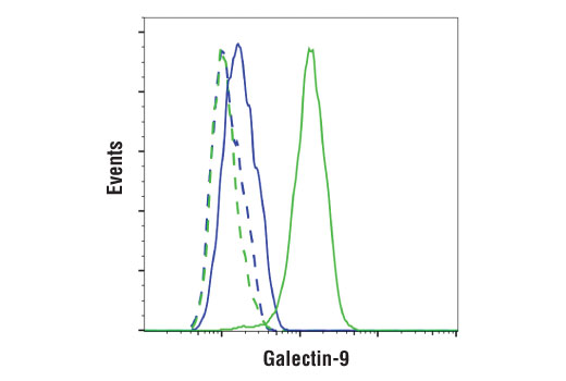
| Flow Cytometry (Fixed/Permeabilized) | 1:1600 |
Storage
For a carrier free (BSA and azide free) version of this product see product #71325.
Specificity/Sensitivity
Source / Purification
Background
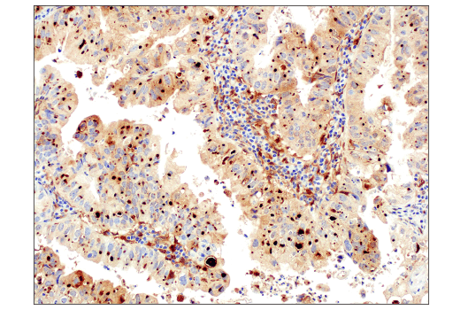
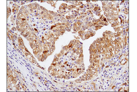
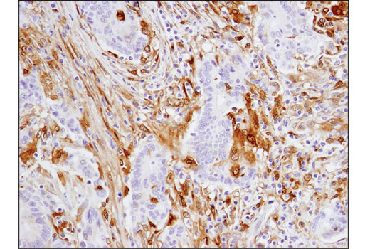
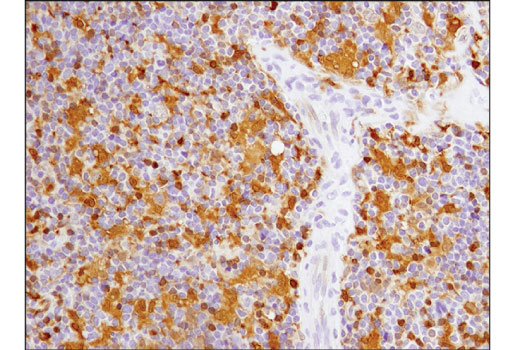
Galectin-9 is induced by proinflammatory stimuli, including IFN-γ, TNF-α, and TLR ligands, and regulates various immune responses through interaction with its ligand TIM-3 (8, 9). Binding of galectin-9 to TIM-3 expressed by Th1 CD4 T cells resulted in T cell death (9). On the other hand, galectin-9 treatment of tumor-bearing mice increased the number of IFN-γ-producing TIM-3+ CD8 T cells and TIM-3+ dendritic cells (10). Transgenic overexpression of either TIM-3 or galectin-9 in mice led to an increase in cells with a myeloid-derived suppressor cell phenotype and inhibition of immune responses (11). CD44 is also proposed to be a receptor for galectin-9, and interaction of galectin-9 with CD44 expressed by induced regulatory T (iTreg) cells enhanced the stability of function of iTreg cells. In addition, galectin-9 was recently demonstrated to bind Dectin-1 expressed by pancreatic ductal adenocarcinoma-infiltrating macrophages, resulting in tolerogenic macrophage reprogramming and suppression of anti-tumor immunity. Increased galectin-9 expression has been observed in several cancer types, including lung, liver, breast, and kidney (12). Alternative splicing of the galectin-9 transcript leads to several isoforms (13).
Background References
- Barondes, S.H. et al. (1994) Cell 76, 597-8.
- Barondes, S.H. et al. (1994) J Biol Chem 269, 20807-10.
- Offner, H. et al. (1990) J Neuroimmunol 28, 177-84.
- Wells, V. and Mallucci, L. (1991) Cell 64, 91-7.
- Filer, A. et al. (2009) Arthritis Rheum 60, 1604-14.
- Perillo, N.L. et al. (1995) Nature 378, 736-9.
- Cooper, D.N. et al. (1991) J Cell Biol 115, 1437-48.
- Gieseke, F. et al. (2013) Eur J Immunol 43, 2741-9.
- Zhu, C. et al. (2005) Nat Immunol 6, 1245-52.
- Nagahara, K. et al. (2008) J Immunol 181, 7660-9.
- Dardalhon, V. et al. (2010) J Immunol 185, 1383-92.
- Heusschen, R. et al. (2014) Biochim Biophys Acta 1842, 284-92.
- Heusschen, R. et al. (2013) Biol Reprod 88, 22.
Species Reactivity
Species reactivity is determined by testing in at least one approved application (e.g., western blot).
Western Blot Buffer
IMPORTANT: For western blots, incubate membrane with diluted primary antibody in 5% w/v BSA, 1X TBS, 0.1% Tween® 20 at 4°C with gentle shaking, overnight.
Applications Key
W: Western Blotting IHC-Bond: IHC Leica Bond IHC-P: Immunohistochemistry (Paraffin) FC-FP: Flow Cytometry (Fixed/Permeabilized)
Cross-Reactivity Key
H: Human
Trademarks and Patents
Cell Signaling Technology is a trademark of Cell Signaling Technology, Inc.
Alexa Fluor is a registered trademark of Life Technologies Corporation.
All other trademarks are the property of their respective owners. Visit cellsignal.com/trademarks for more information.
Limited Uses
Except as otherwise expressly agreed in a writing signed by a legally authorized representative of CST, the following terms apply to Products provided by CST, its affiliates or its distributors. Any Customer's terms and conditions that are in addition to, or different from, those contained herein, unless separately accepted in writing by a legally authorized representative of CST, are rejected and are of no force or effect.
Products are labeled with For Research Use Only or a similar labeling statement and have not been approved, cleared, or licensed by the FDA or other regulatory foreign or domestic entity, for any purpose. Customer shall not use any Product for any diagnostic or therapeutic purpose, or otherwise in any manner that conflicts with its labeling statement. Products sold or licensed by CST are provided for Customer as the end-user and solely for research and development uses. Any use of Product for diagnostic, prophylactic or therapeutic purposes, or any purchase of Product for resale (alone or as a component) or other commercial purpose, requires a separate license from CST. Customer shall (a) not sell, license, loan, donate or otherwise transfer or make available any Product to any third party, whether alone or in combination with other materials, or use the Products to manufacture any commercial products, (b) not copy, modify, reverse engineer, decompile, disassemble or otherwise attempt to discover the underlying structure or technology of the Products, or use the Products for the purpose of developing any products or services that would compete with CST products or services, (c) not alter or remove from the Products any trademarks, trade names, logos, patent or copyright notices or markings, (d) use the Products solely in accordance with CST Product Terms of Sale and any applicable documentation, and (e) comply with any license, terms of service or similar agreement with respect to any third party products or services used by Customer in connection with the Products.
Revision 3
Revision 3
Revision 3