Revision 12
#5852
Store at -20C
877-616-CELL (2355)
877-678-TECH (8324)
3 Trask Lane | Danvers | Massachusetts | 01923 | USA
For Research Use Only. Not for Use in Diagnostic Procedures.
Applications:
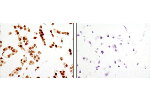
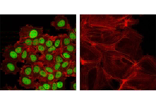
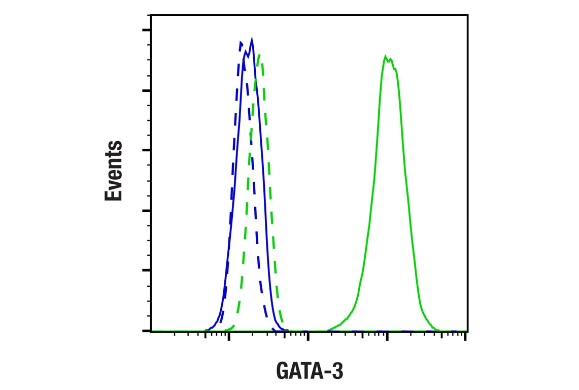
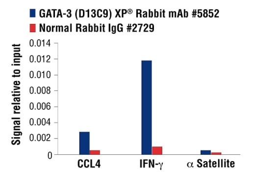
W, W-S, IHC-P, IF-IC, FC-FP, ChIP, C&R
Reactivity:
H M
Sensitivity:
Endogenous
MW (kDa):
48
Source/Isotype:
Rabbit IgG
UniProt ID:
#P23771
Entrez-Gene Id:
2625
Product Usage Information
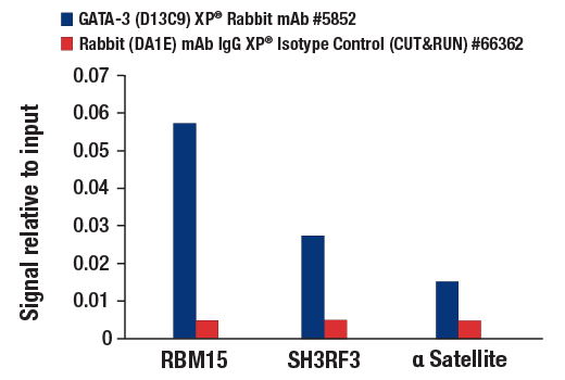
The CUT&RUN dilution was determined using CUT&RUN Assay Kit #86652.
| Application | Dilution |
|---|---|
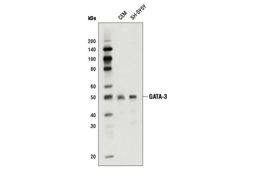
| Western Blotting | 1:1000 |
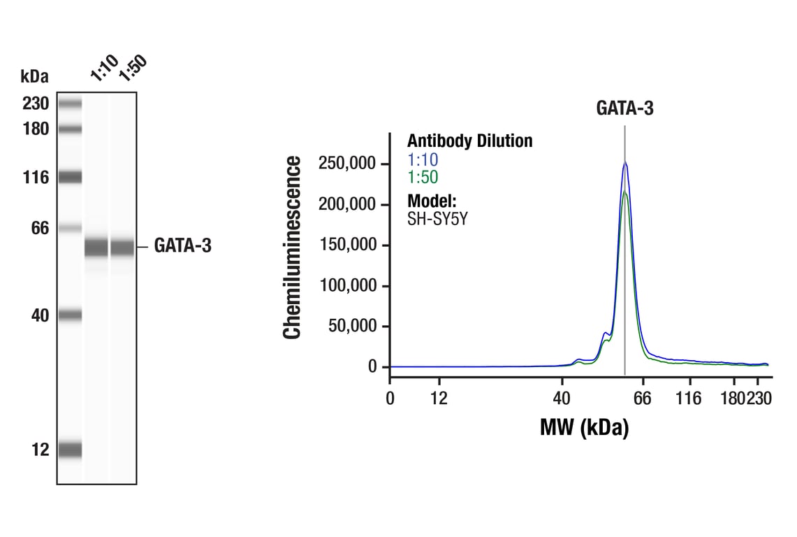
| Simple Western™ | 1:10 - 1:50 |
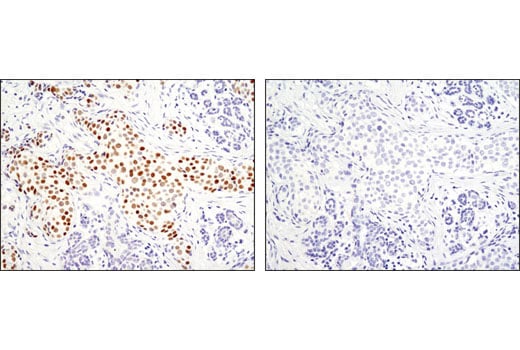
| Immunohistochemistry (Paraffin) | 1:1600 - 1:6400 |
| Immunofluorescence (Immunocytochemistry) | 1:800 - 1:3200 |
| Flow Cytometry (Fixed/Permeabilized) | 1:200 - 1:800 |
| Chromatin IP | 1:100 |
| CUT&RUN | 1:50 |
Storage
For a carrier free (BSA and azide free) version of this product see product #10630.
Specificity/Sensitivity
Species predicted to react based on 100% sequence homology
Source / Purification
Background
GATA-3 is a critical regulator of development of various systems in both mouse and human (4). GATA-3 mouse embryos die between E11 and E12 due to growth retardation and deformities in the brain and spinal cord (5). The function of GATA-3 has been extensively studied in T cell development and has recently been shown to be a downstream target of Notch in Notch-mediated differentiation of TH2 cells (6,7). It is expressed in both hematopoietic and non-hematopoietic tissues, including the kidney, skin, mammary gland, and central nervous system (8-10). Decreased expression of GATA-3 in luminal breast cancer is associated with poor clinical outcome. GATA-3 expression level may therefore be a promising prognostic biomarker (11). Haploinsufficiency of GATA-3 results in Barakat syndome in humans, a condition characterized by sensorineural deafness and renal dysplasia (12).
Background References
- Ko, L.J. and Engel, J.D. (1993) Mol Cell Biol 13, 4011-22.
- Merika, M. and Orkin, S.H. (1993) Mol Cell Biol 13, 3999-4010.
- Lowry, J.A. and Atchley, W.R. (2000) J Mol Evol 50, 103-15.
- Debacker, C. et al. (1999) Mech Dev 85, 183-7.
- Pandolfi, P.P. et al. (1995) Nat Genet 11, 40-4.
- Ho, I.C. et al. (2009) Nat Rev Immunol 9, 125-35.
- Amsen, D. et al. (2007) Immunity 27, 89-99.
- Grote, D. et al. (2008) PLoS Genet 4, e1000316.
- Kaufman, C.K. et al. (2003) Genes Dev 17, 2108-22.
- Kouros-Mehr, H. et al. (2006) Cell 127, 1041-55.
- Chou, J. et al. (2010) J Cell Physiol 222, 42-9.
- Van Esch, H. et al. (2000) Nature 406, 419-22.
Species Reactivity
Species reactivity is determined by testing in at least one approved application (e.g., western blot).
Western Blot Buffer
IMPORTANT: For western blots, incubate membrane with diluted primary antibody in 5% w/v BSA, 1X TBS, 0.1% Tween® 20 at 4°C with gentle shaking, overnight.
Applications Key
W: Western Blotting W-S: Simple Western™ IHC-P: Immunohistochemistry (Paraffin) IF-IC: Immunofluorescence (Immunocytochemistry) FC-FP: Flow Cytometry (Fixed/Permeabilized) ChIP: Chromatin IP C&R: CUT&RUN
Cross-Reactivity Key
H: Human M: Mouse
Trademarks and Patents
Cell Signaling Technology is a trademark of Cell Signaling Technology, Inc.
All other trademarks are the property of their respective owners. Visit cellsignal.com/trademarks for more information.
Limited Uses
Except as otherwise expressly agreed in a writing signed by a legally authorized representative of CST, the following terms apply to Products provided by CST, its affiliates or its distributors. Any Customer's terms and conditions that are in addition to, or different from, those contained herein, unless separately accepted in writing by a legally authorized representative of CST, are rejected and are of no force or effect.
Products are labeled with For Research Use Only or a similar labeling statement and have not been approved, cleared, or licensed by the FDA or other regulatory foreign or domestic entity, for any purpose. Customer shall not use any Product for any diagnostic or therapeutic purpose, or otherwise in any manner that conflicts with its labeling statement. Products sold or licensed by CST are provided for Customer as the end-user and solely for research and development uses. Any use of Product for diagnostic, prophylactic or therapeutic purposes, or any purchase of Product for resale (alone or as a component) or other commercial purpose, requires a separate license from CST. Customer shall (a) not sell, license, loan, donate or otherwise transfer or make available any Product to any third party, whether alone or in combination with other materials, or use the Products to manufacture any commercial products, (b) not copy, modify, reverse engineer, decompile, disassemble or otherwise attempt to discover the underlying structure or technology of the Products, or use the Products for the purpose of developing any products or services that would compete with CST products or services, (c) not alter or remove from the Products any trademarks, trade names, logos, patent or copyright notices or markings, (d) use the Products solely in accordance with CST Product Terms of Sale and any applicable documentation, and (e) comply with any license, terms of service or similar agreement with respect to any third party products or services used by Customer in connection with the Products.
Revision 12
Revision 12
Revision 12
Revision 12