Revision 1
#24642
Store at -20C
GATA Transcription Factor Antibody Sampler Kit
1 Kit
(5 x 20 microliters)
877-616-CELL (2355)
877-678-TECH (8324)
3 Trask Lane | Danvers | Massachusetts | 01923 | USA
For Research Use Only. Not for Use in Diagnostic Procedures.
| Product Includes | Product # | Quantity | Mol. Wt | Isotype/Source |
|---|---|---|---|---|
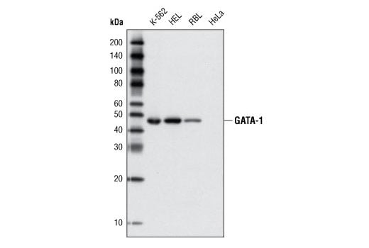
| GATA-1 (D52H6) Rabbit Monoclonal Antibody | 3535 | 20 µl | 43 kDa | Rabbit IgG |
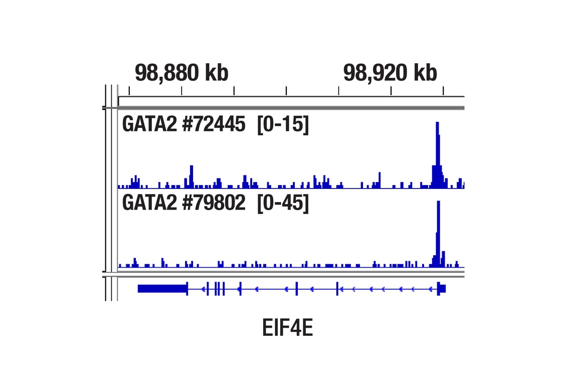
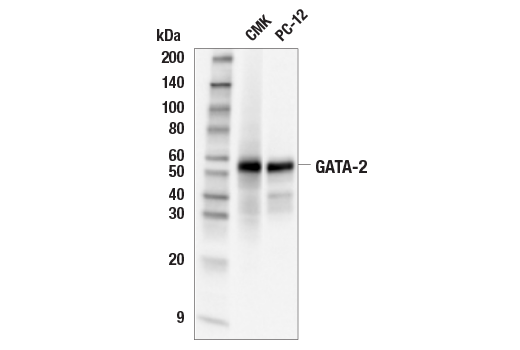
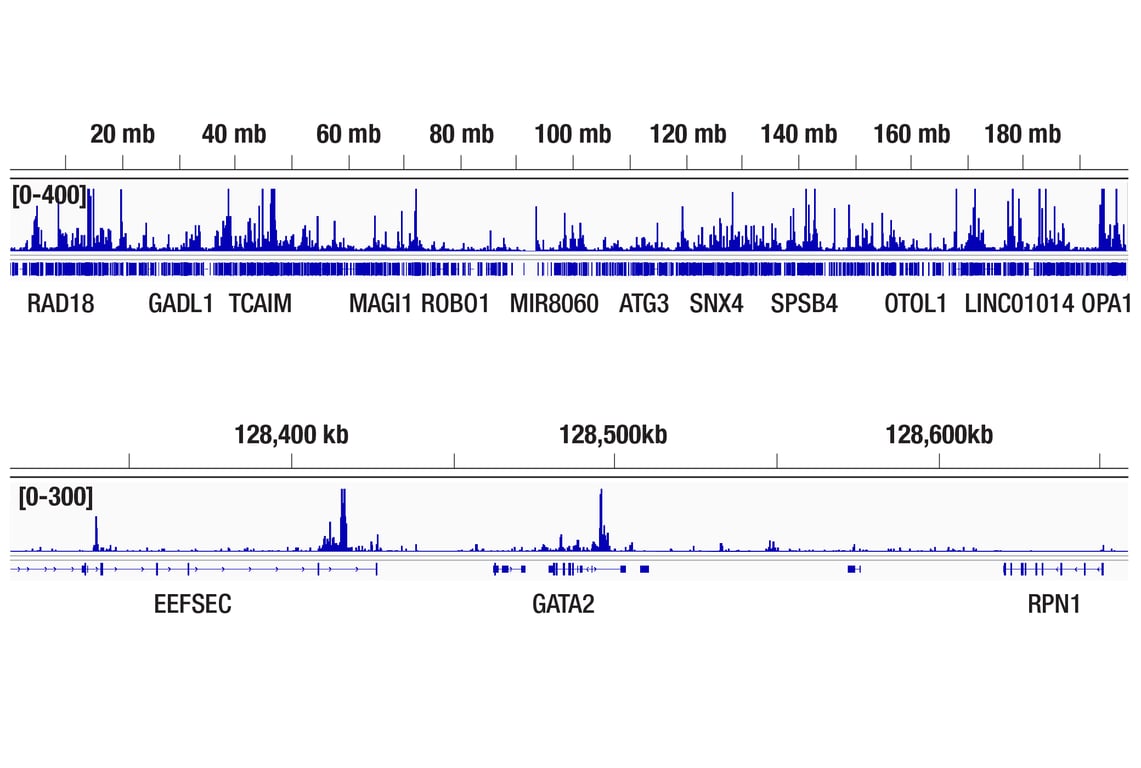
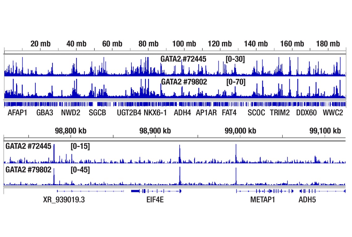
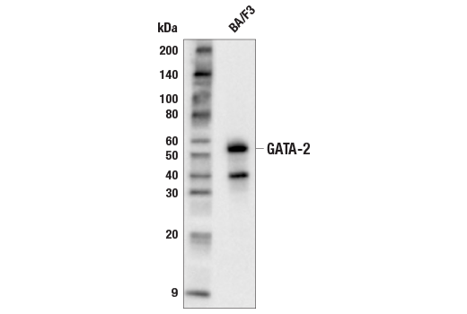
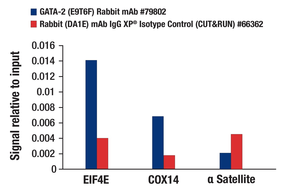
| GATA-2 (E9T6F) Rabbit Monoclonal Antibody | 79802 | 20 µl | 51 kDa | Rabbit IgG |
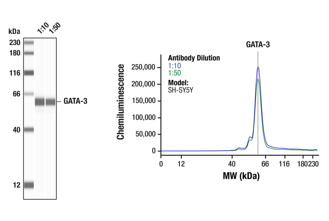
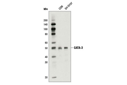
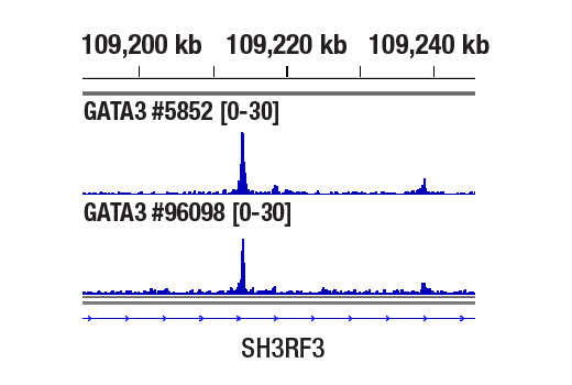
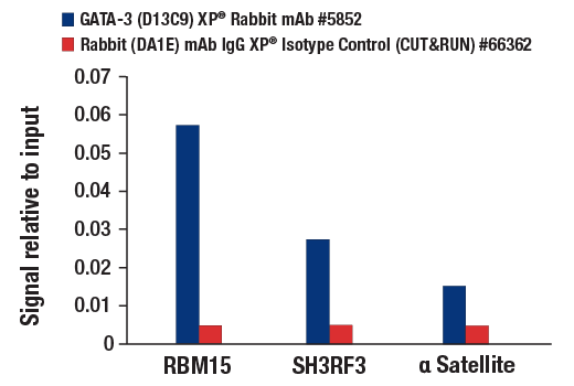
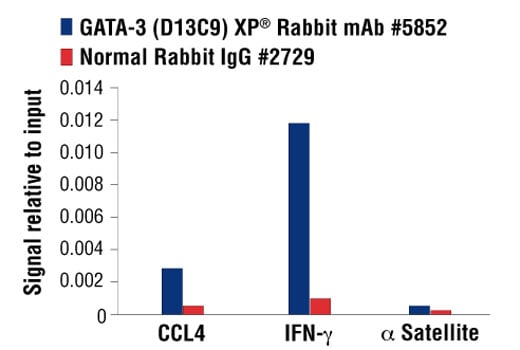
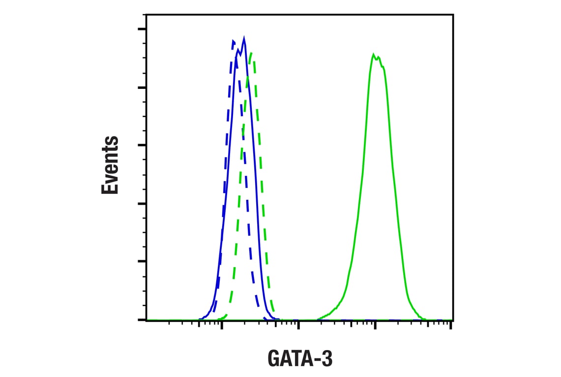
| GATA-3 (D13C9) Rabbit Monoclonal Antibody | 5852 | 20 µl | 48 kDa | Rabbit IgG |
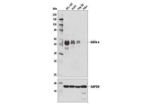
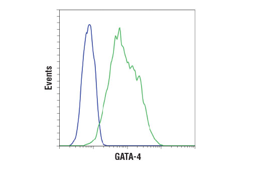
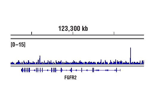
| GATA-4 (D3A3M) Rabbit Monoclonal Antibody | 36966 | 20 µl | 55 kDa | Rabbit IgG |
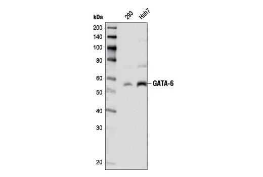
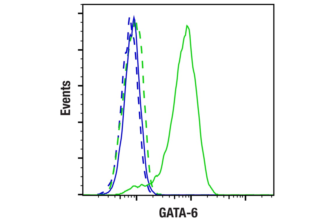
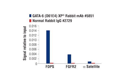
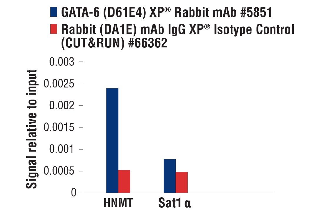
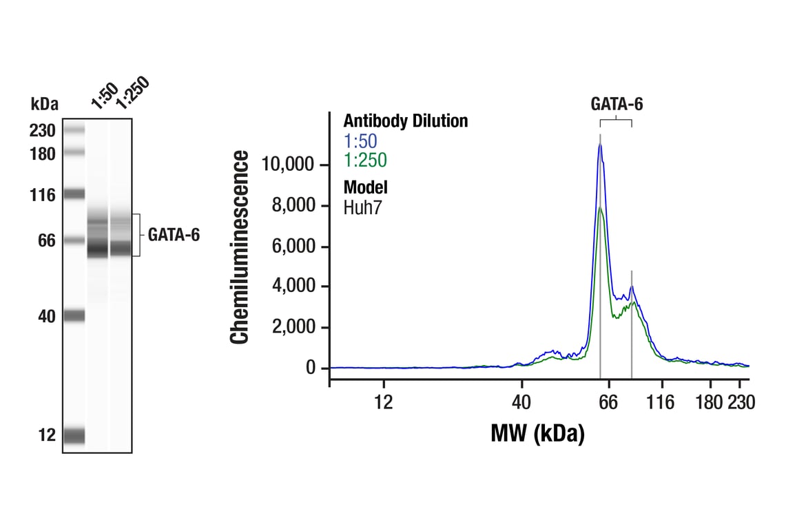
| GATA-6 (D61E4) Rabbit Monoclonal Antibody | 5851 | 20 µl | 55 kDa | Rabbit IgG |
| Anti-rabbit IgG, HRP-linked Antibody | 7074 | 100 µl | Goat |
Please visit cellsignal.com for individual component applications, species cross-reactivity, dilutions, protocols, and additional product information.
Description
Storage
Background
Background References
- Ko, L.J. and Engel, J.D. (1993) Mol Cell Biol 13, 4011-22.
- Merika, M. and Orkin, S.H. (1993) Mol Cell Biol 13, 3999-4010.
- Lowry, J.A. and Atchley, W.R. (2000) J Mol Evol 50, 103-15.
- Tremblay, M. et al. (2018) Development 145, dev164384. doi: 10.1242/dev.164384.
- Pevny, L. et al. (1991) Nature 349, 257-60.
- Fujiwara, Y. et al. (1996) Proc Natl Acad Sci USA 93, 12355-8.
- Wechsler, J. et al. (2002) Nat Genet 32, 148-52.
- Cantor, A.B. (2005) Int J Hematol 81, 378-84.
- Mehaffey, M.G. et al. (2001) Blood 98, 2681-8.
- Tubman, V.N. et al. (2007) Blood 109, 3297-9.
- Tong, Q. et al. (2003) Drug News Perspect 16, 585-8.
- Tsai, F.Y. et al. (1994) Nature 371, 221-6.
- Tong, Q. et al. (2005) Mol Cell Biol 25, 706-15.
- Tsai, J. et al. (2005) EMBO Rep 6, 879-84.
- El Wakil, A. et al. (2006) Development 133, 2155-65.
- Debacker, C. et al. (1999) Mech Dev 85, 183-7.
- Grote, D. et al. (2008) PLoS Genet 4, e1000316.
- Kaufman, C.K. et al. (2003) Genes Dev 17, 2108-22.
- Kouros-Mehr, H. et al. (2006) Cell 127, 1041-55.
- Pandolfi, P.P. et al. (1995) Nat Genet 11, 40-4.
- Ho, I.C. et al. (2009) Nat Rev Immunol 9, 125-35.
- Amsen, D. et al. (2007) Immunity 27, 89-99.
- Gan, L. et al. (2014) Gene Expr Patterns 16, 8-22.
- Yang, Y.Q. et al. (2013) Hum Mutat 34, 1662-71.
- Li, R.G. et al. (2013) Biochem Biophys Res Commun 439, 591-6.
- Li, J. et al. (2014) Gene 548, 174-81.
- Mohan, R.A. et al. (2014) Am J Med Genet A 164A, 2732-8.
- Walker, E.M. et al. (2014) Dev Biol 392, 283-94.
- LaVoie, H.A. (2014) Biol Reprod 91, 38.
- Cai, K.Q. et al. (2008) Dev Dyn 237, 2820-9.
- Charron, F. and Nemer, M. (1999) Semin Cell Dev Biol 10, 85-91.
- Haveri, H. et al. (2008) BMC Gastroenterol 8, 9.
Trademarks and Patents
Cell Signaling Technology is a trademark of Cell Signaling Technology, Inc.
All other trademarks are the property of their respective owners. Visit cellsignal.com/trademarks for more information.
Limited Uses
Except as otherwise expressly agreed in a writing signed by a legally authorized representative of CST, the following terms apply to Products provided by CST, its affiliates or its distributors. Any Customer's terms and conditions that are in addition to, or different from, those contained herein, unless separately accepted in writing by a legally authorized representative of CST, are rejected and are of no force or effect.
Products are labeled with For Research Use Only or a similar labeling statement and have not been approved, cleared, or licensed by the FDA or other regulatory foreign or domestic entity, for any purpose. Customer shall not use any Product for any diagnostic or therapeutic purpose, or otherwise in any manner that conflicts with its labeling statement. Products sold or licensed by CST are provided for Customer as the end-user and solely for research and development uses. Any use of Product for diagnostic, prophylactic or therapeutic purposes, or any purchase of Product for resale (alone or as a component) or other commercial purpose, requires a separate license from CST. Customer shall (a) not sell, license, loan, donate or otherwise transfer or make available any Product to any third party, whether alone or in combination with other materials, or use the Products to manufacture any commercial products, (b) not copy, modify, reverse engineer, decompile, disassemble or otherwise attempt to discover the underlying structure or technology of the Products, or use the Products for the purpose of developing any products or services that would compete with CST products or services, (c) not alter or remove from the Products any trademarks, trade names, logos, patent or copyright notices or markings, (d) use the Products solely in accordance with CST Product Terms of Sale and any applicable documentation, and (e) comply with any license, terms of service or similar agreement with respect to any third party products or services used by Customer in connection with the Products.
Revision 1
Revision 1
Revision 1

Revision 1
Revision 1
Revision 1

Revision 1
Revision 1
Revision 1
Revision 1
Revision 1
Revision 1
Revision 1
Revision 1
Revision 1
Revision 1
Revision 1
Revision 1
Revision 1
Revision 1
Revision 1
Revision 1
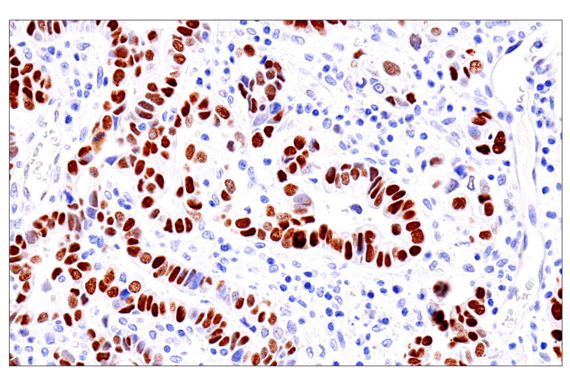
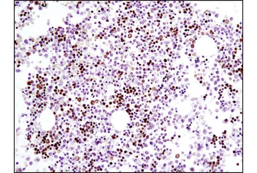
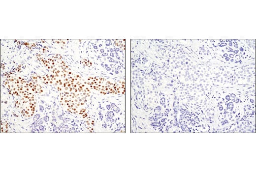
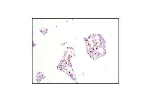
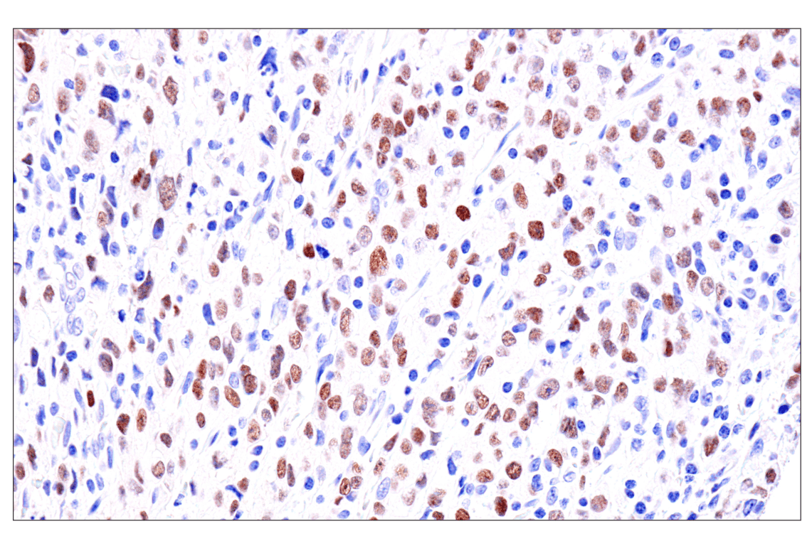
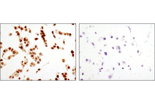
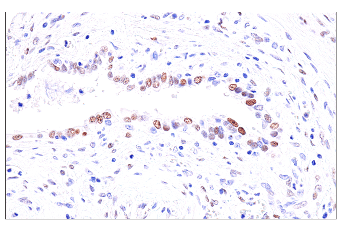
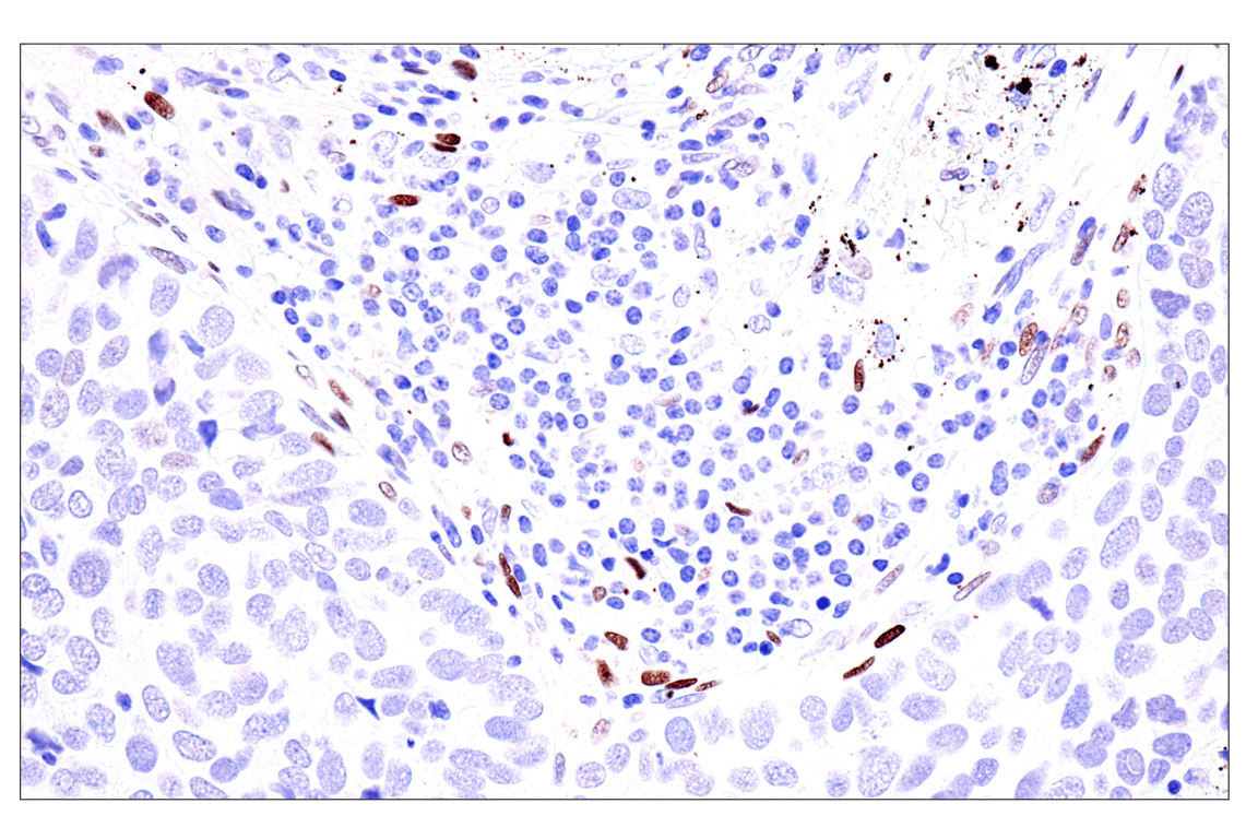
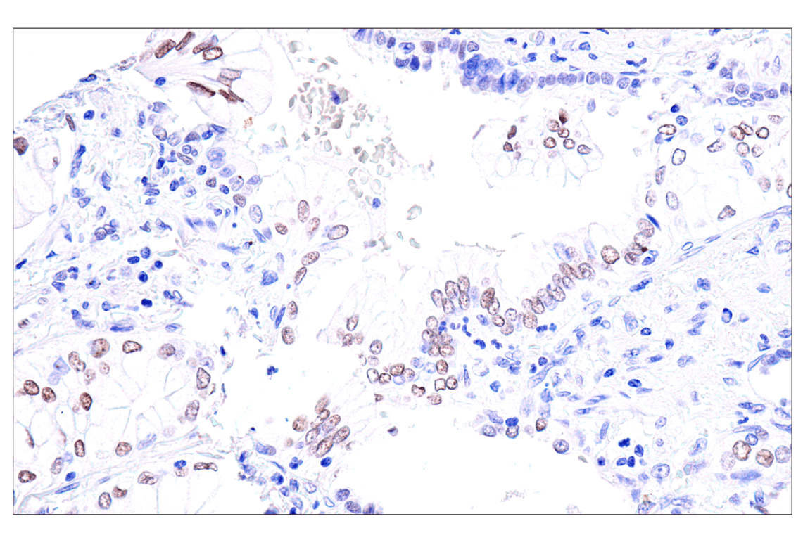
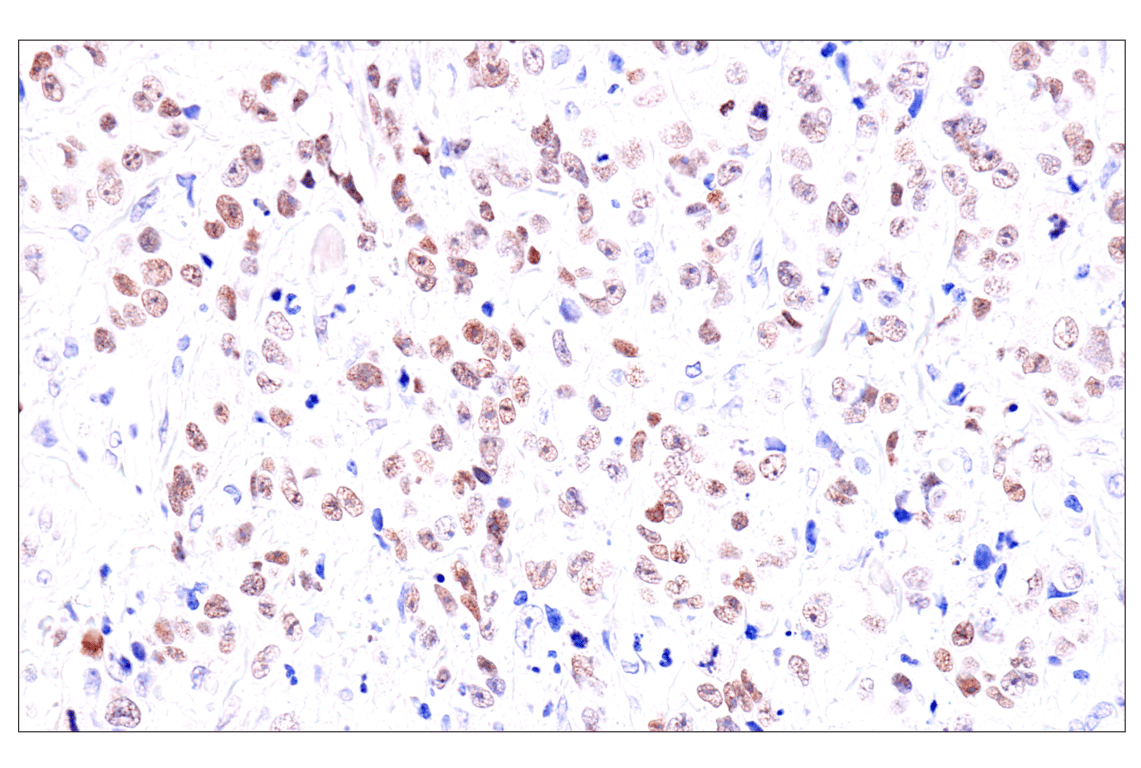
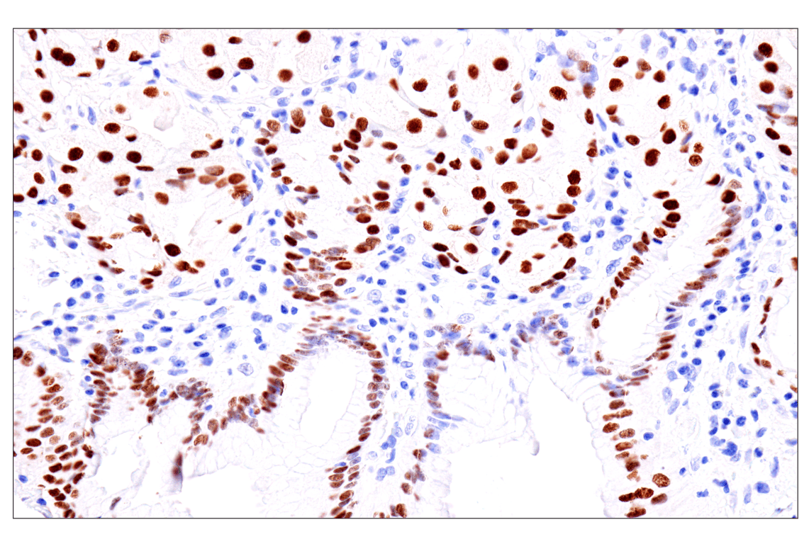
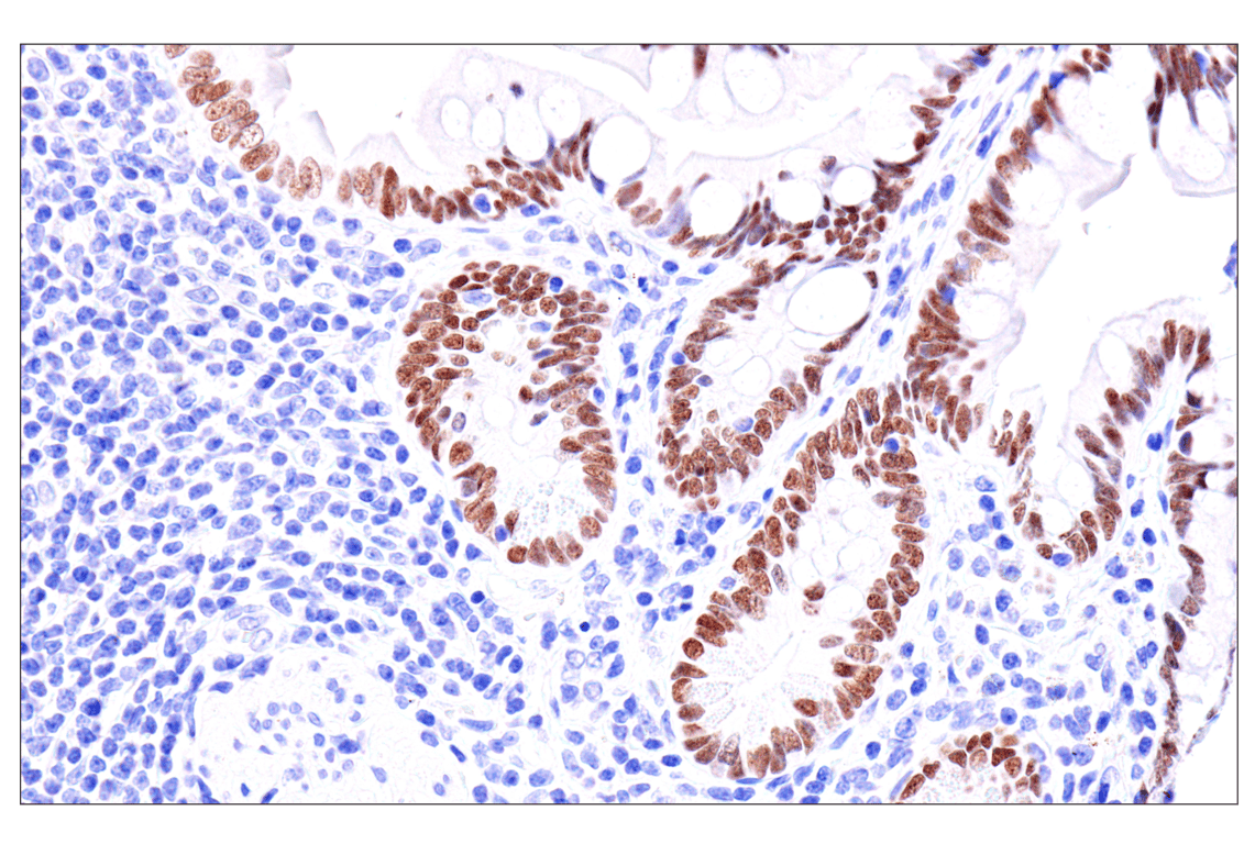
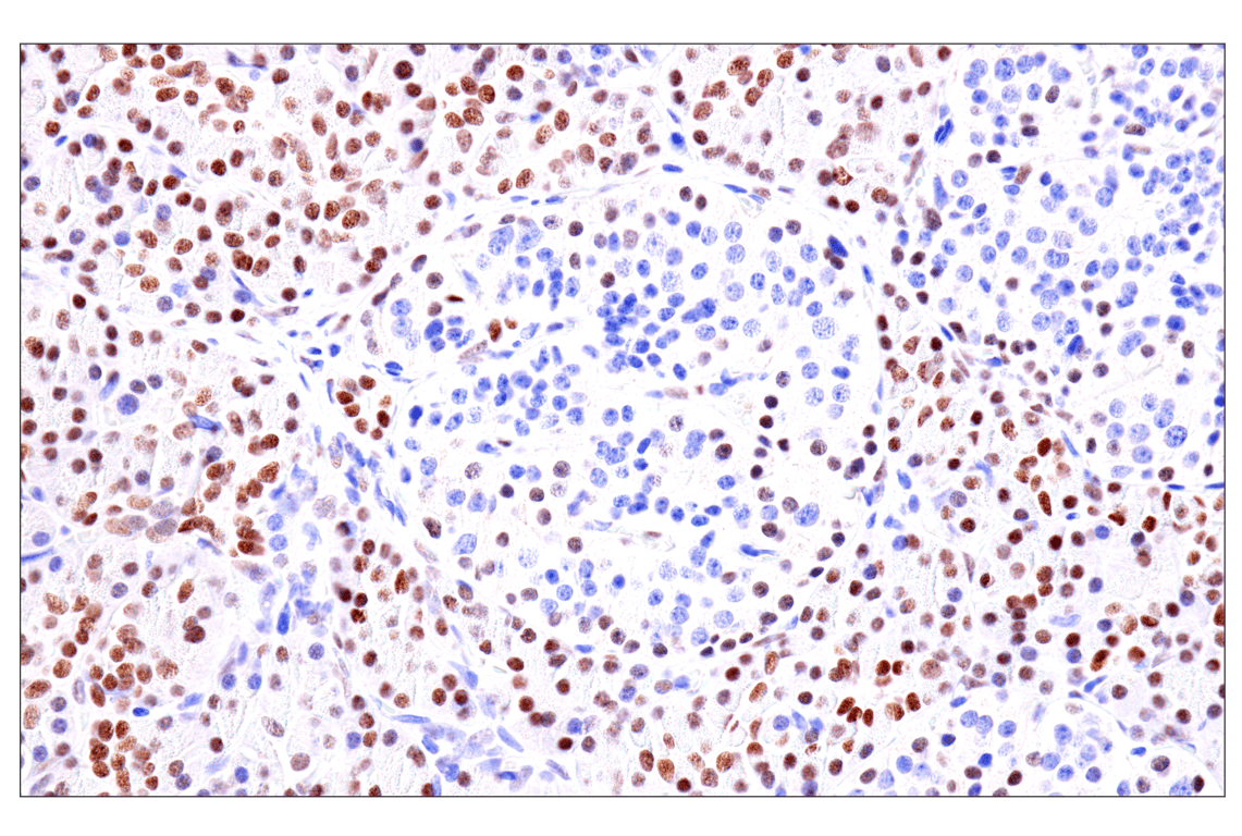
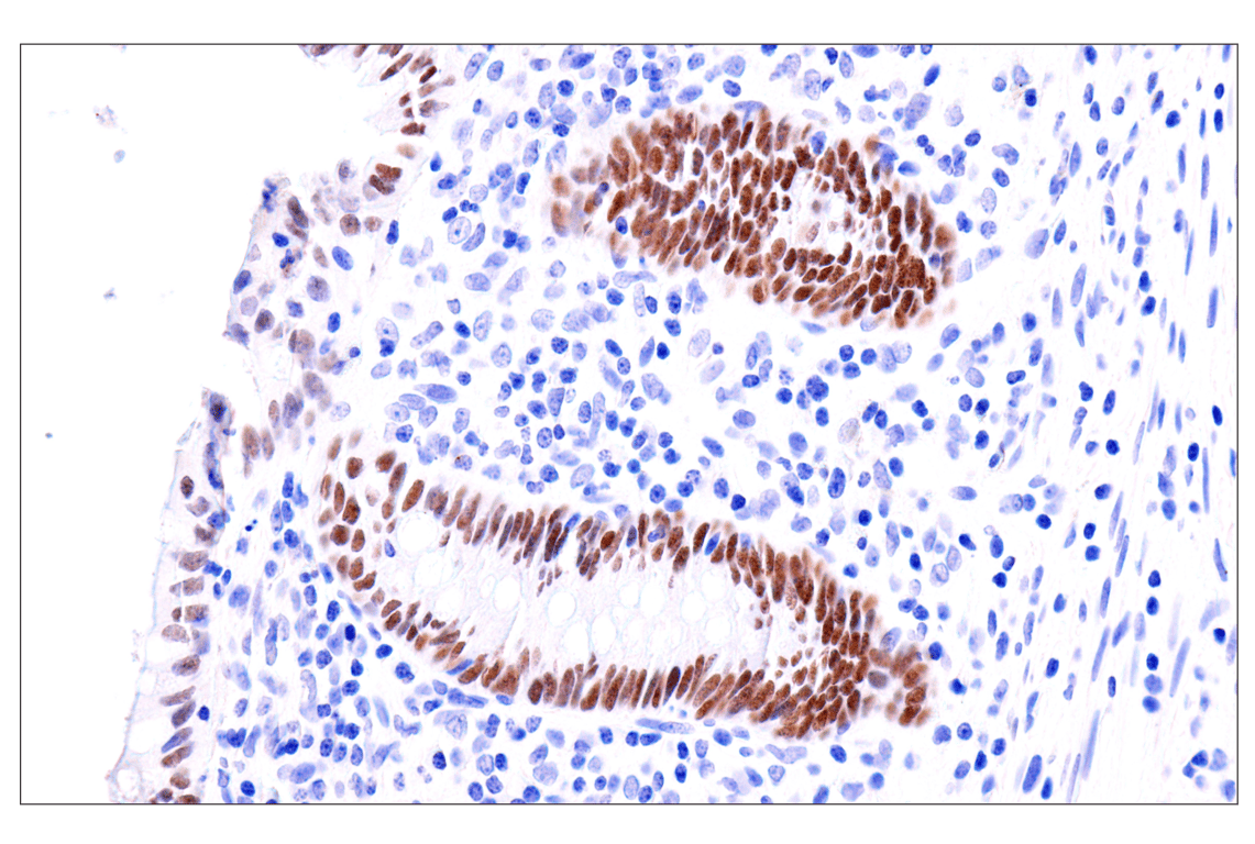
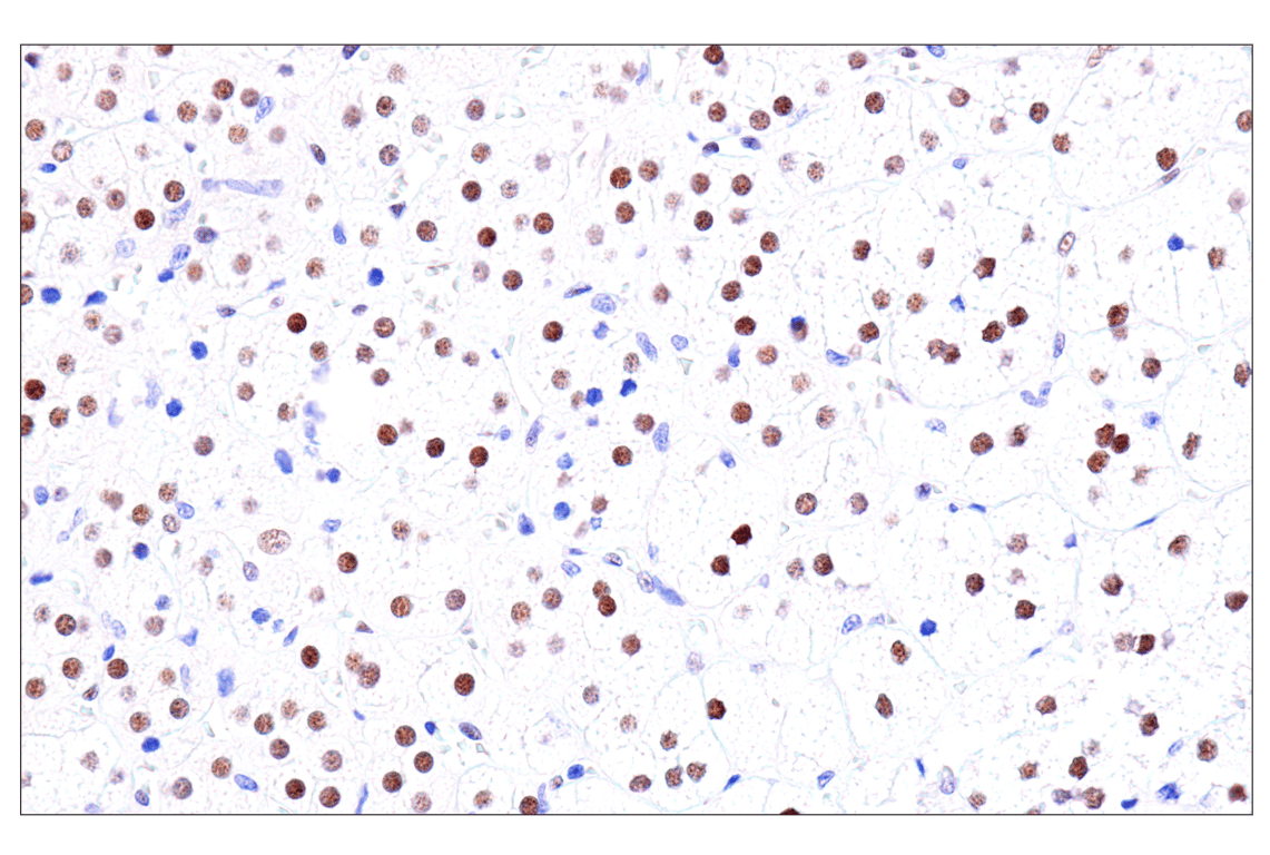
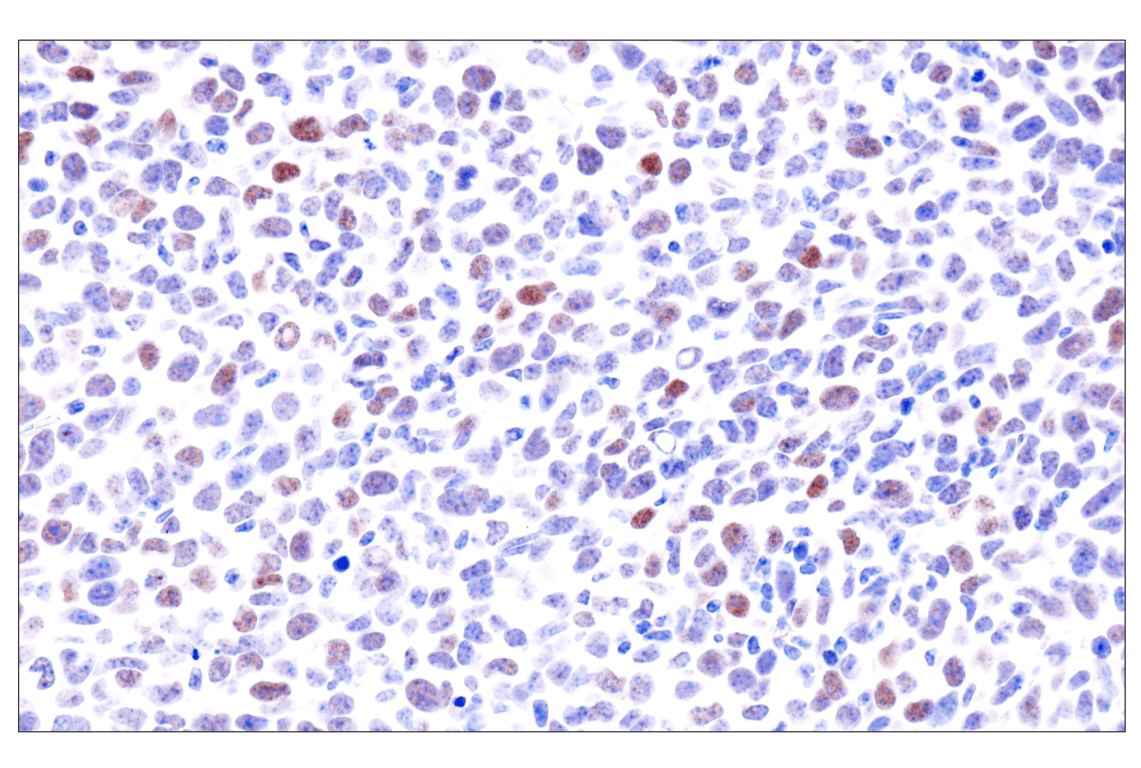
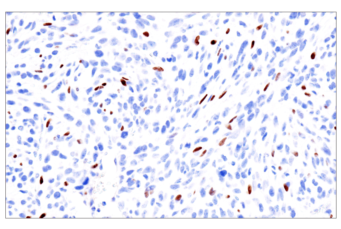
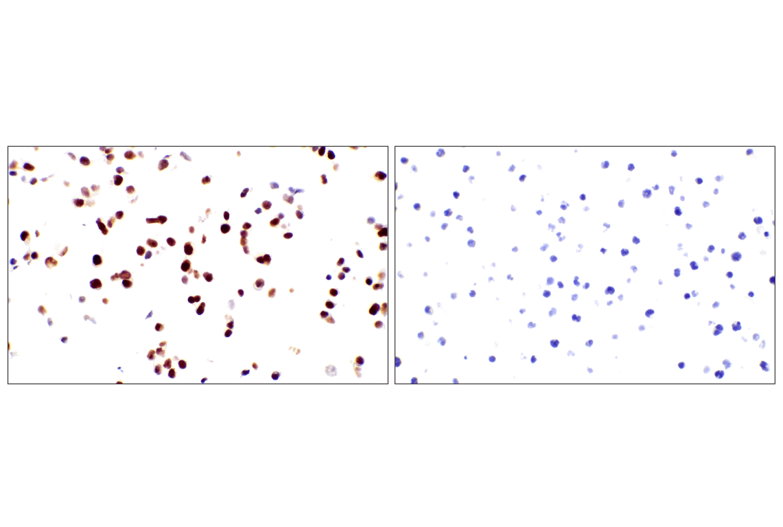
Immunohistochemical analysis of paraffin-embedded HuH-7 cell pellet (left, positive) and SUP-B15 cell pellet (right, negative) using GATA-6 (D61E4) XP® Rabbit mAb.
Revision 1