| Cat. # | Size | Qty. | Price |
|---|---|---|---|
| 24642T | 1 Kit (5 x 20 microliters) |
|
| Product Includes | Quantity | Applications | Reactivity | MW(kDa) | Isotype |
|---|---|---|---|---|---|
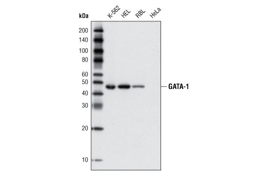
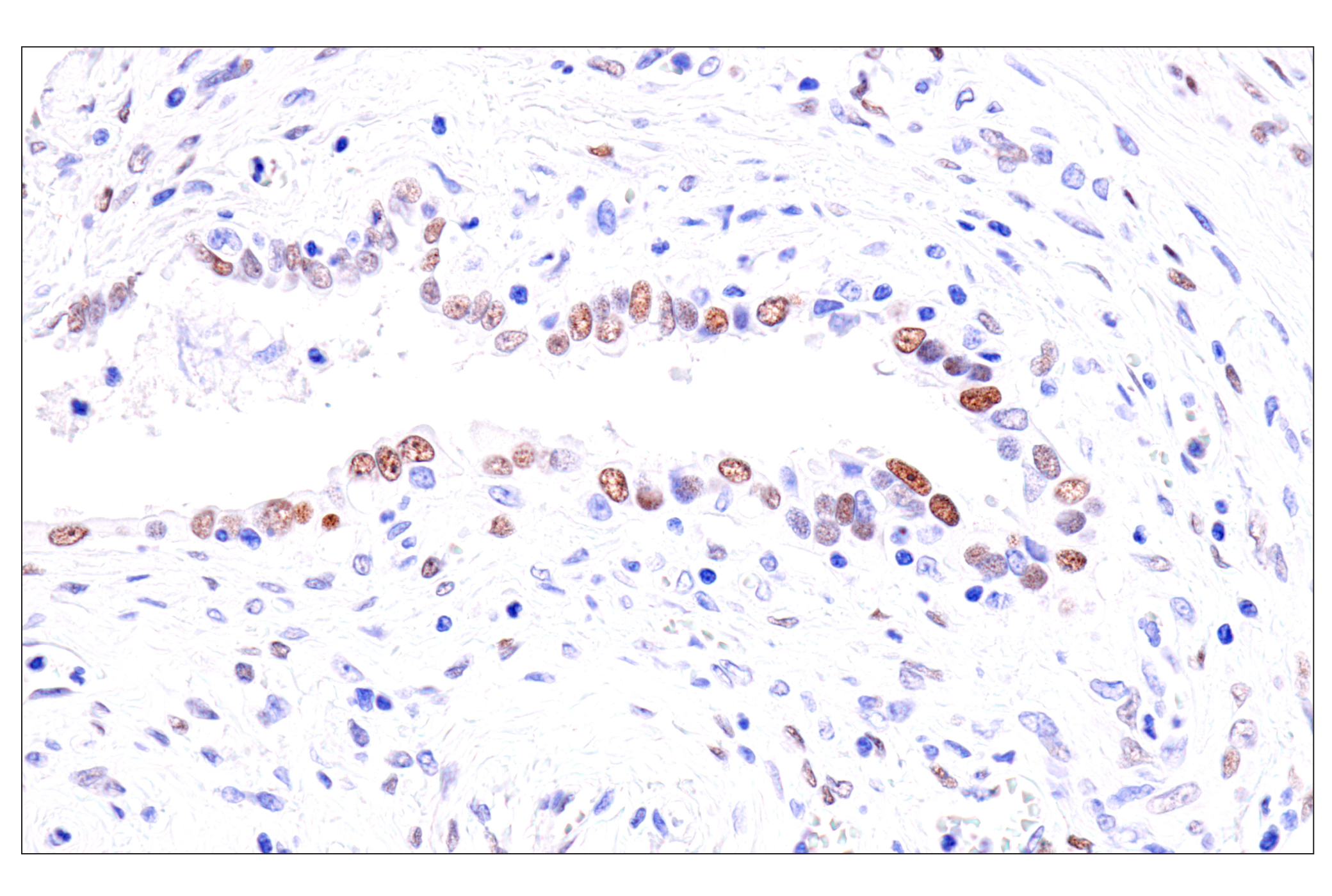
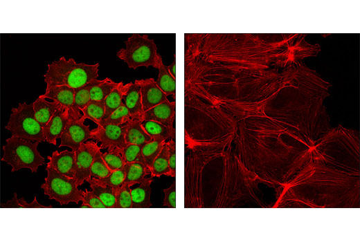
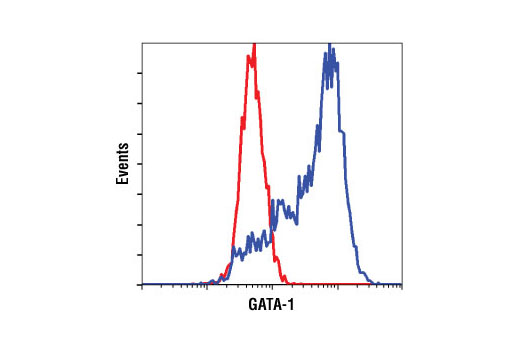
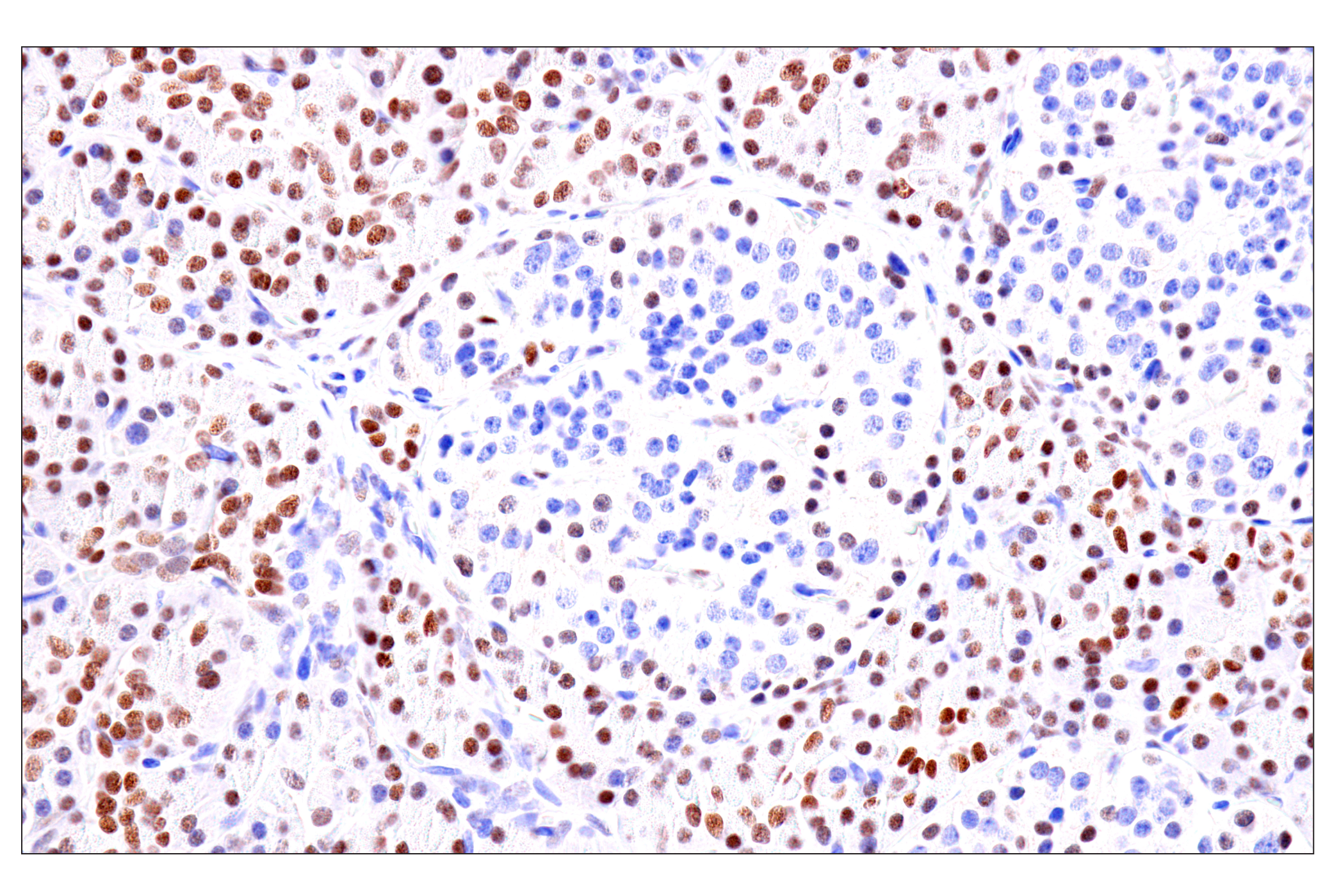
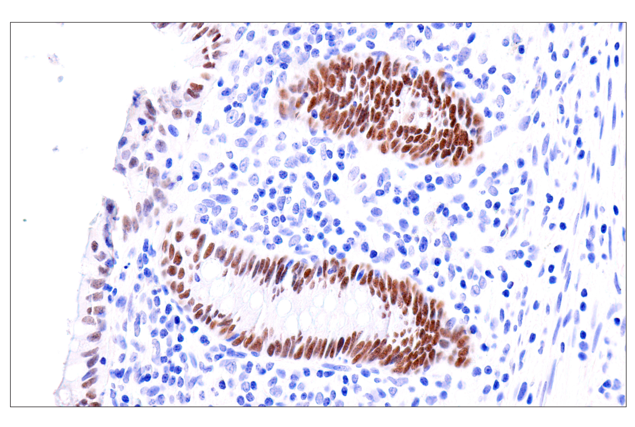
| GATA-1 (D52H6) XP® Rabbit mAb 3535 | 20 µl |
|
H M R | 43 | Rabbit IgG |
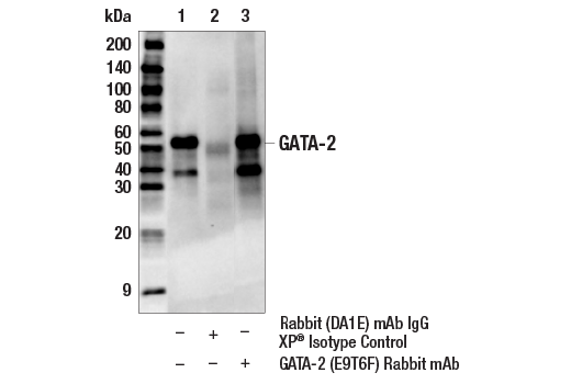
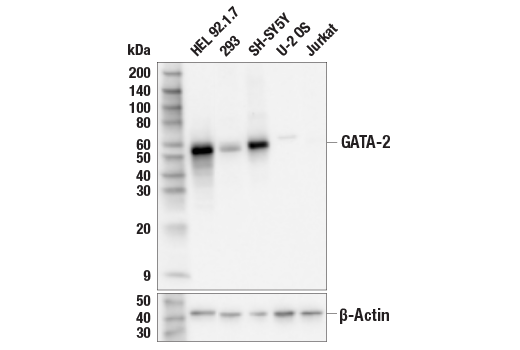
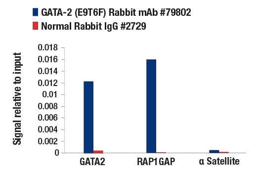
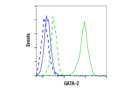
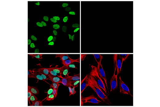
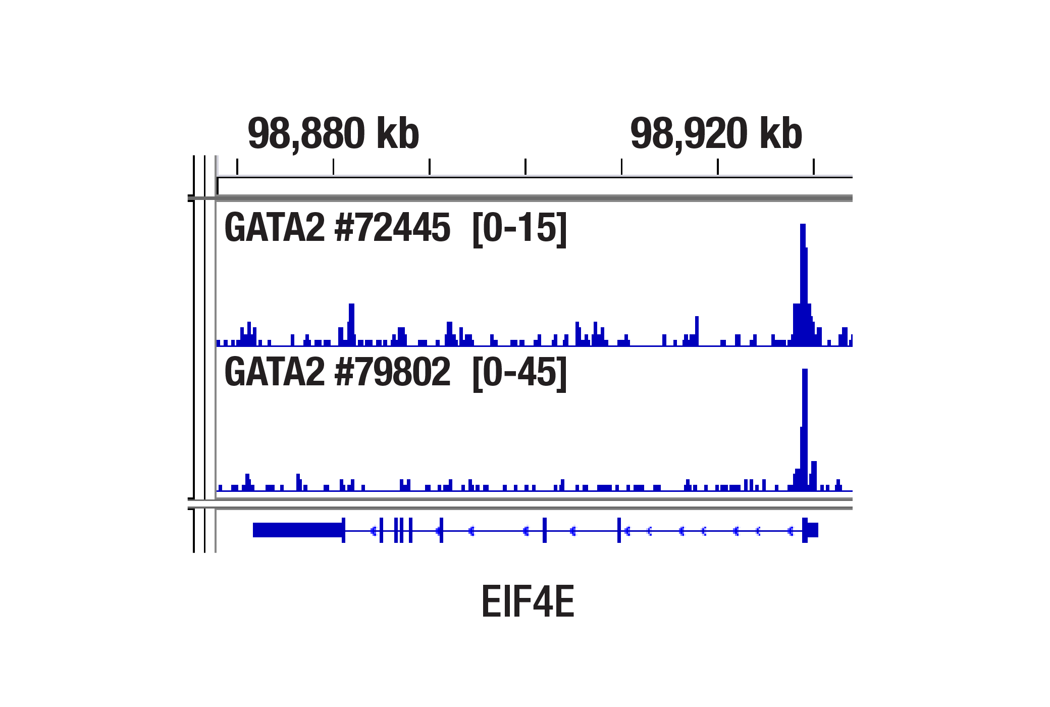
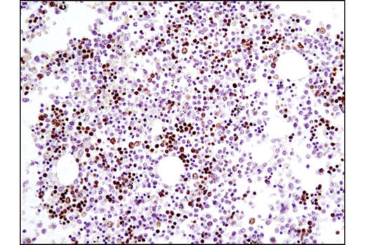
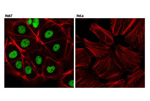
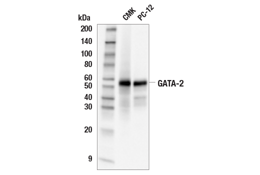
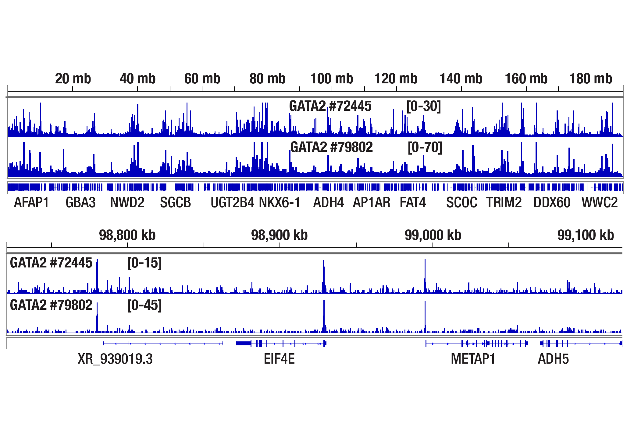
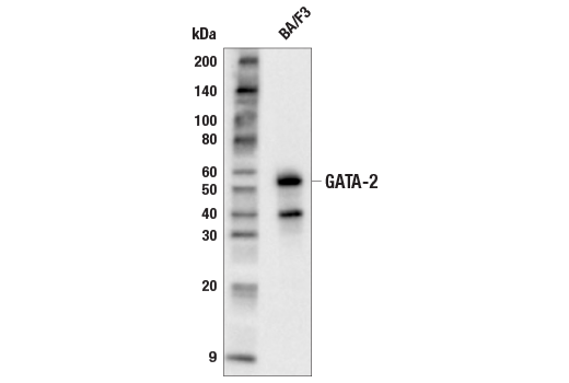
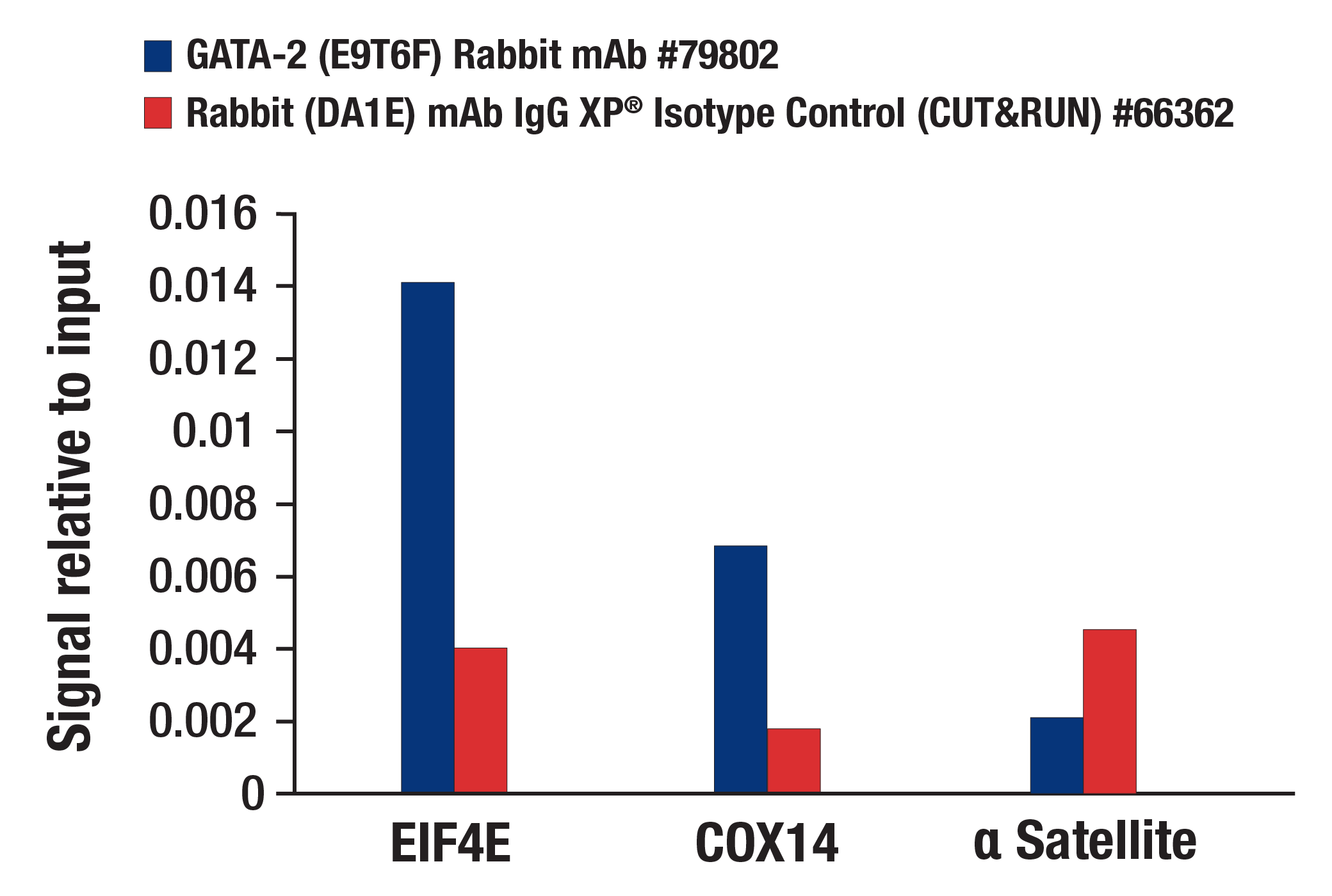
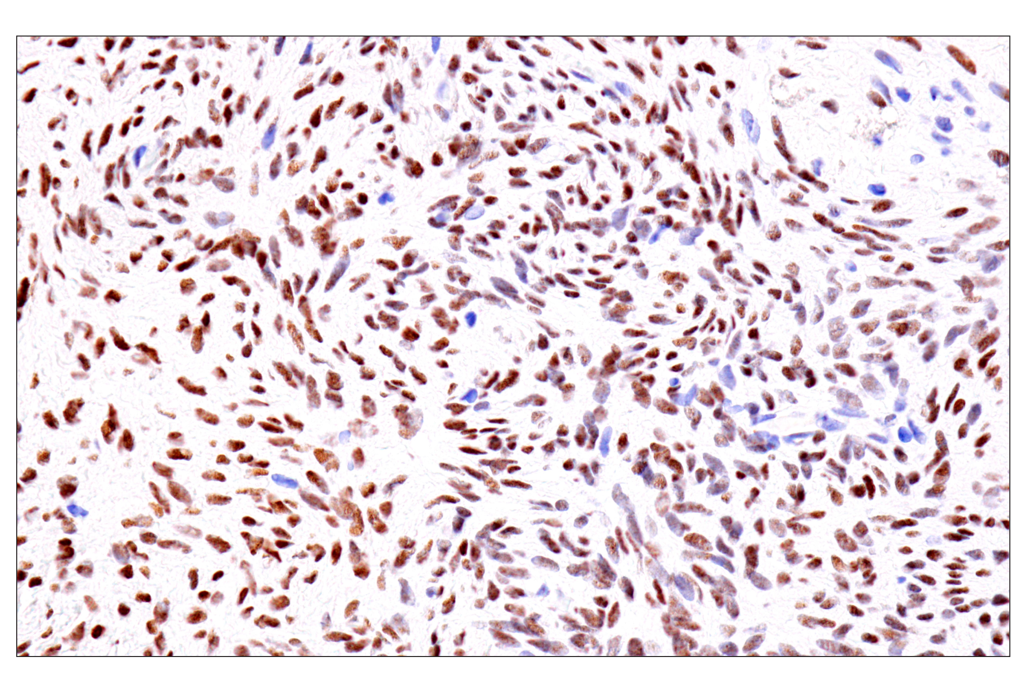
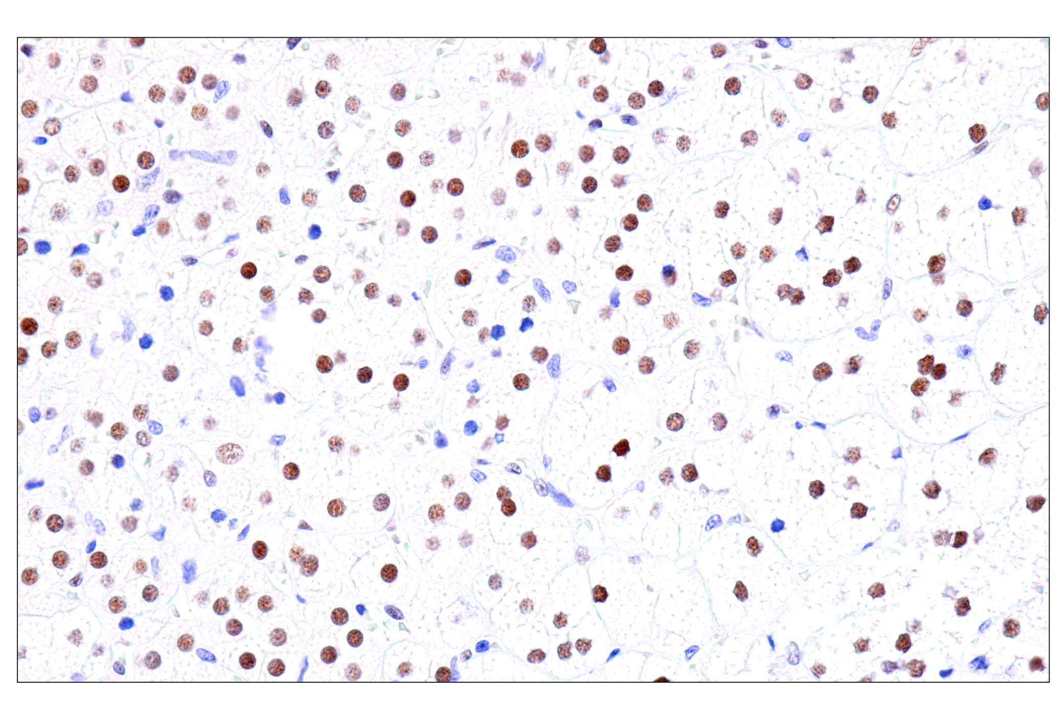
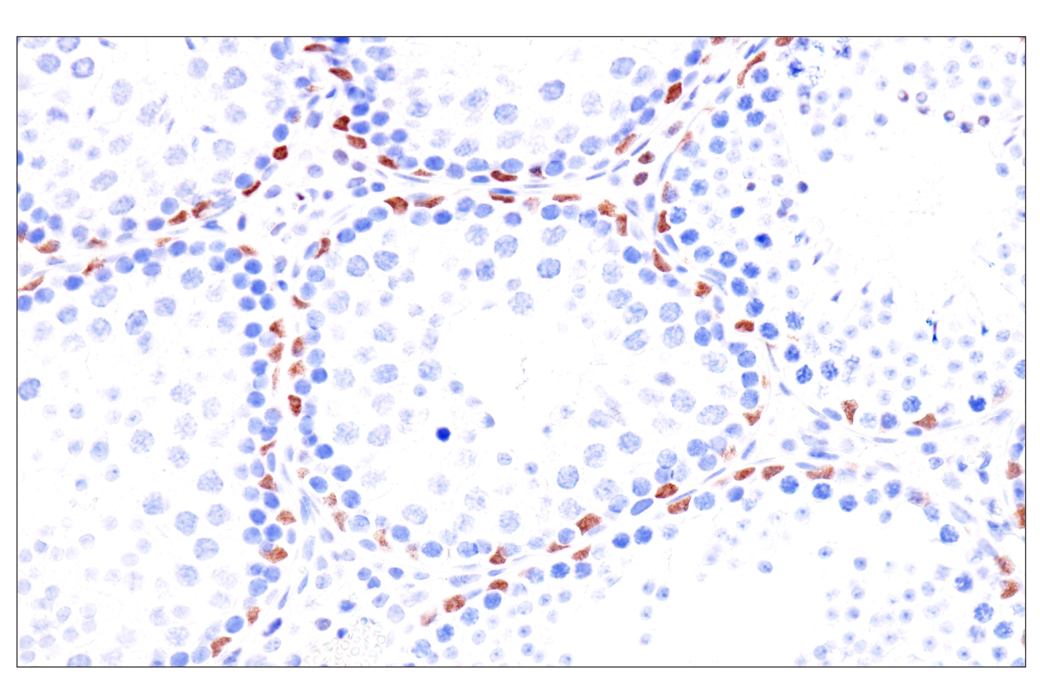
| GATA-2 (E9T6F) Rabbit mAb 79802 | 20 µl |
|
H M R | 51 | Rabbit IgG |
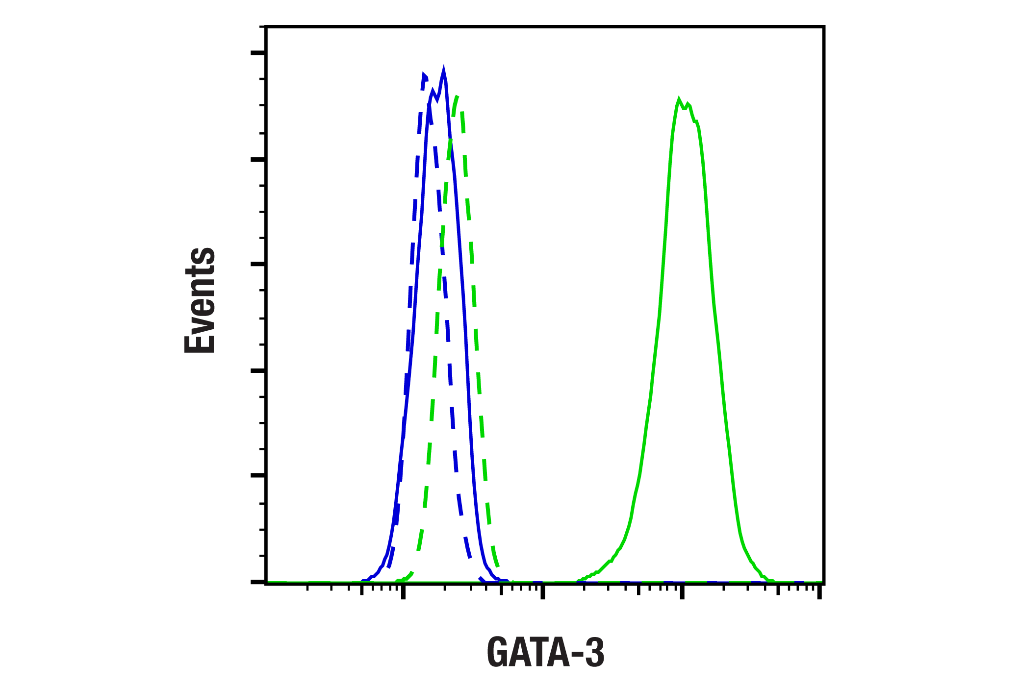
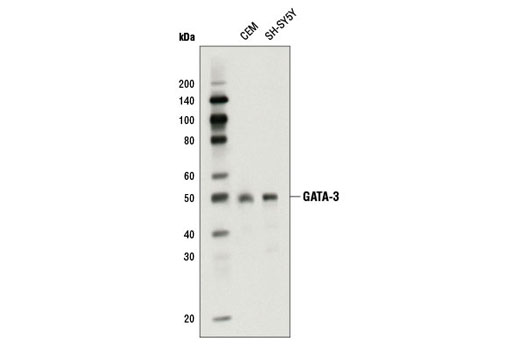
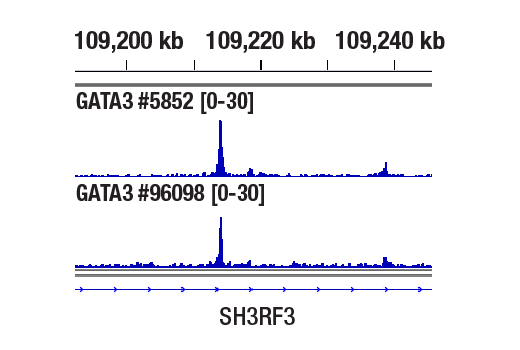
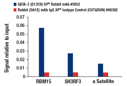
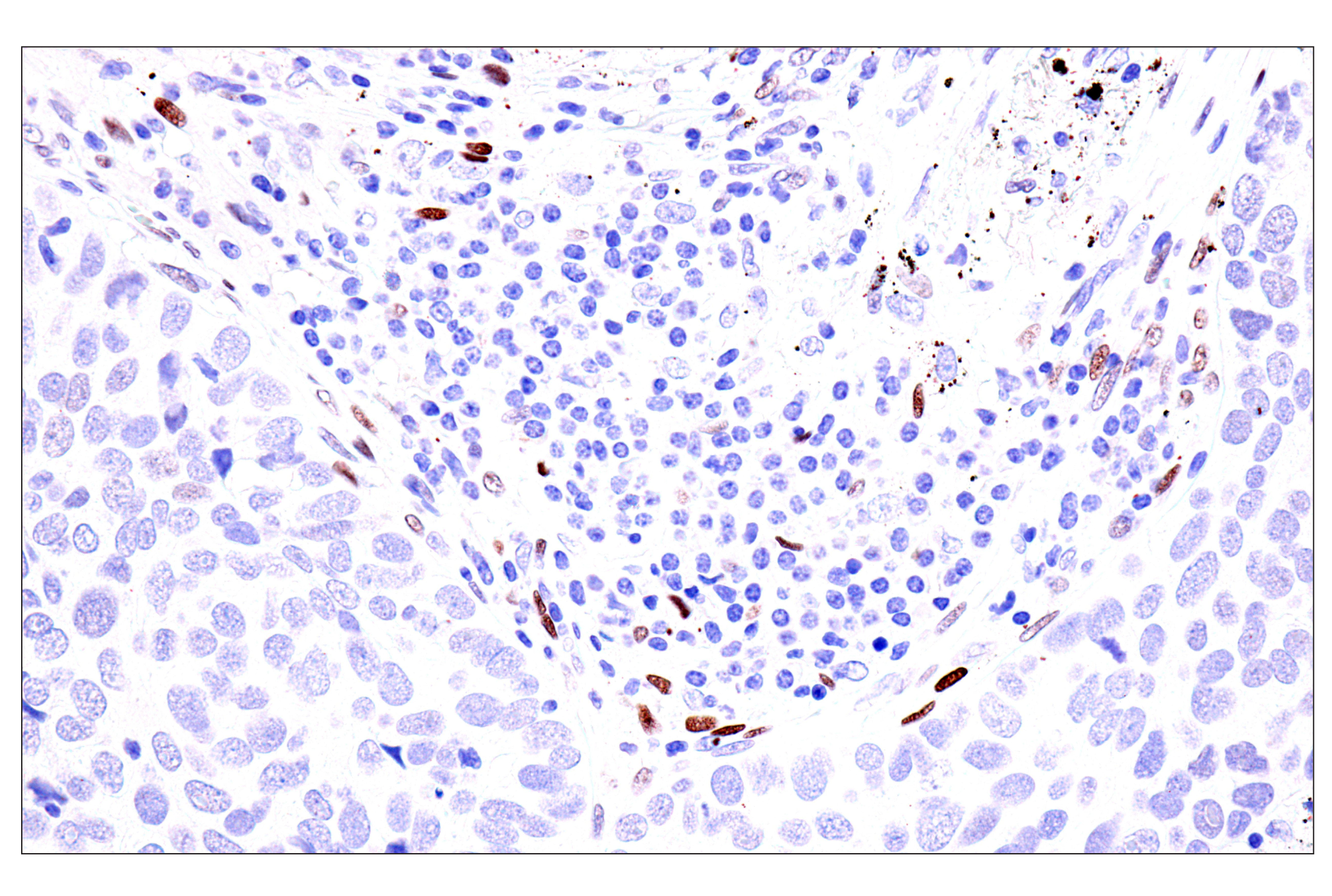
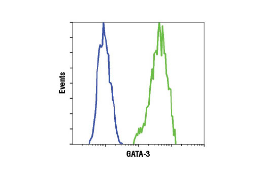
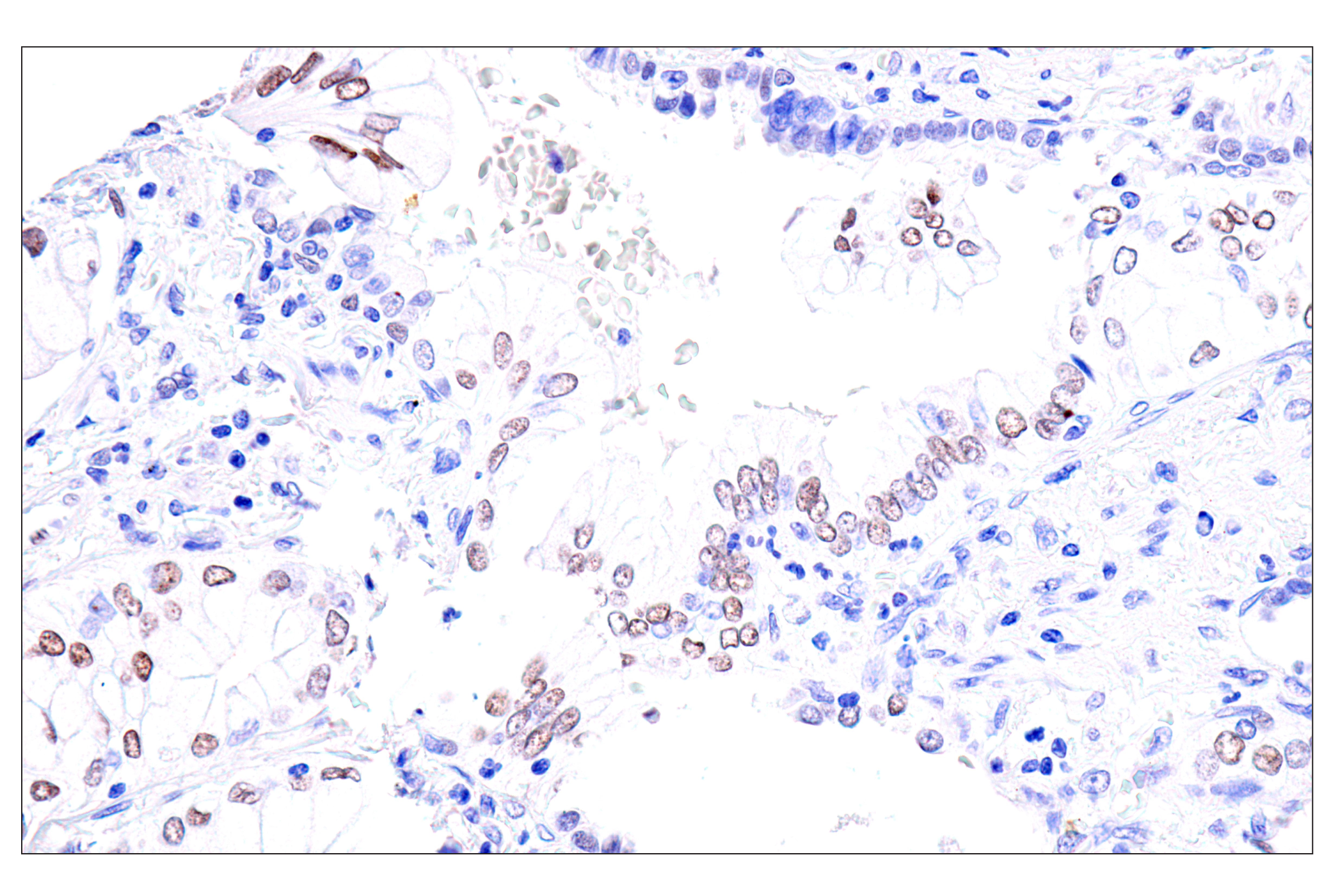
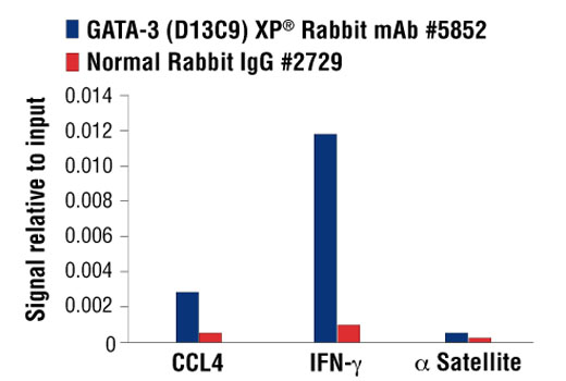
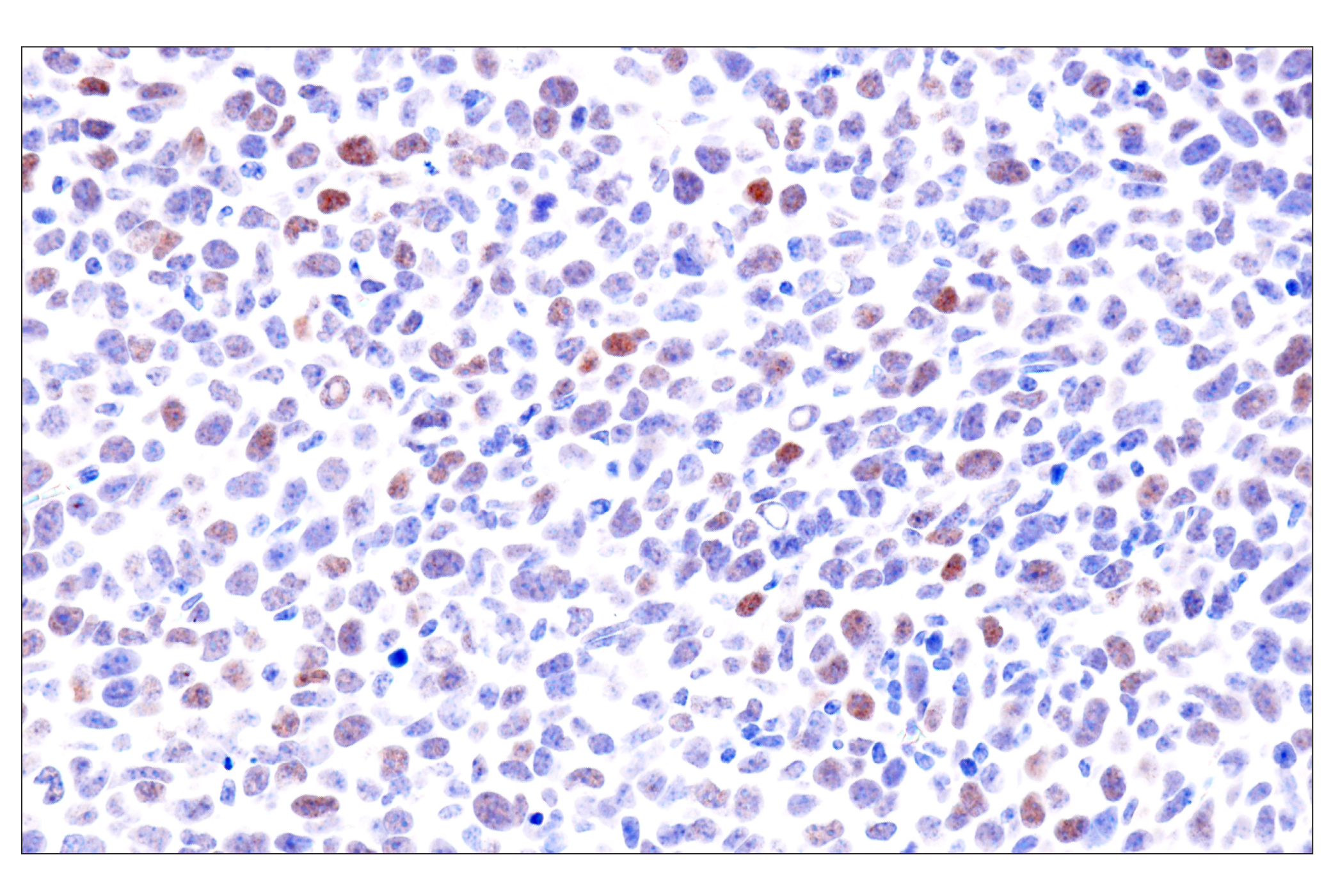
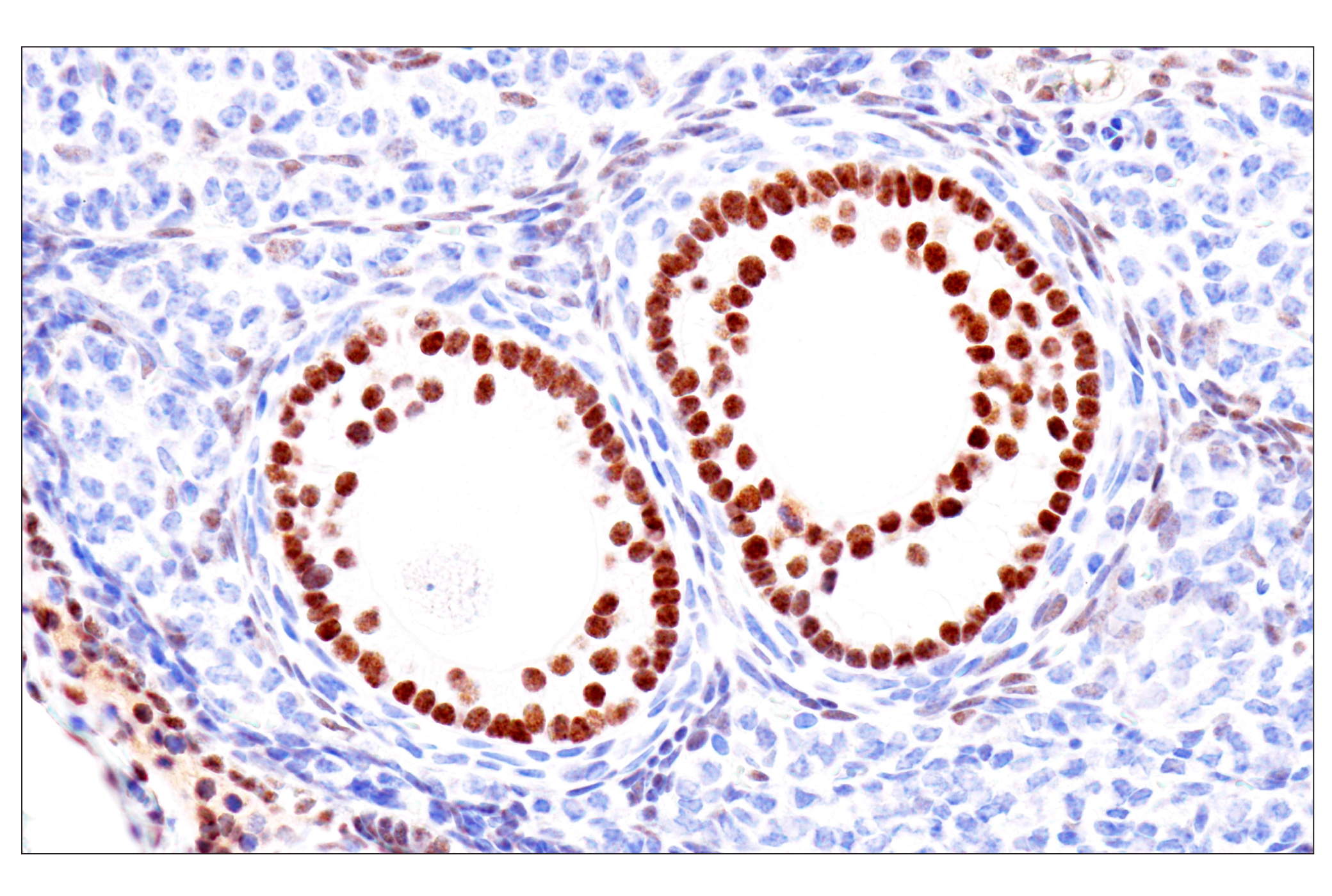
| GATA-3 (D13C9) XP® Rabbit mAb 5852 | 20 µl |
|
H M | 48 | Rabbit IgG |
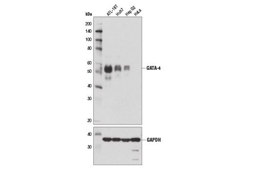
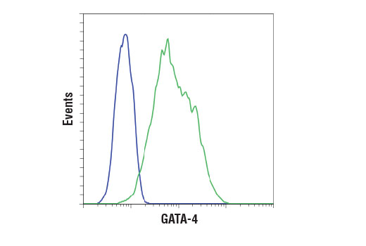
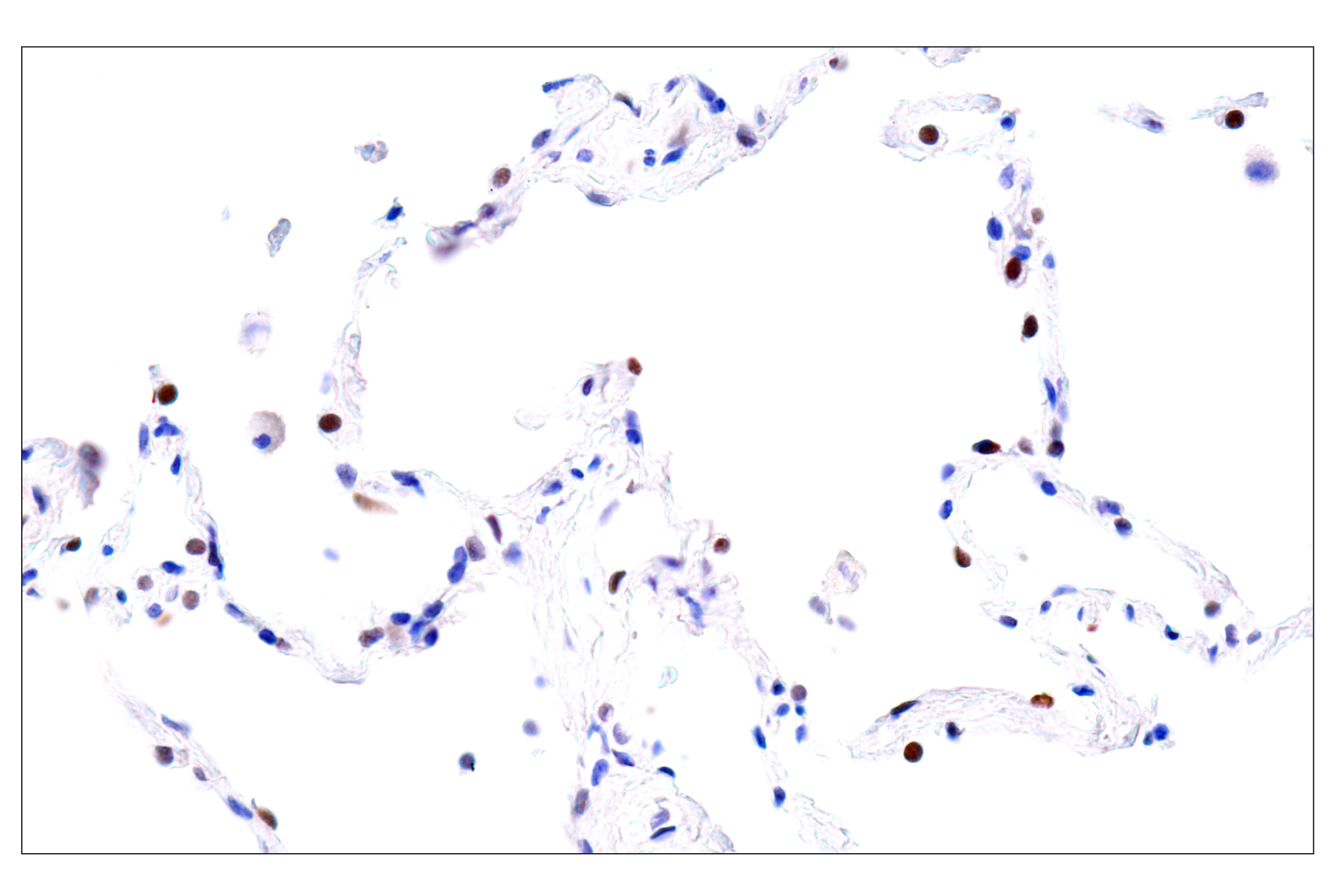
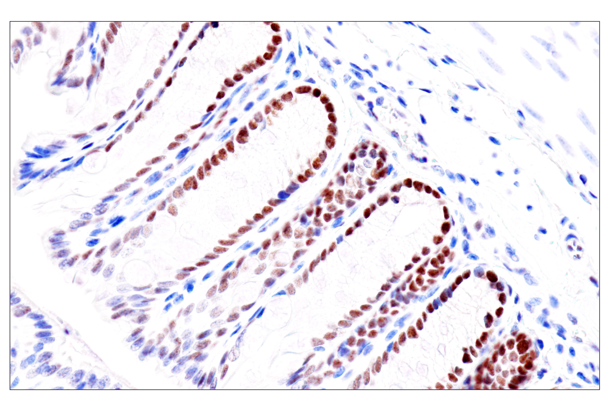
| GATA-4 (D3A3M) Rabbit mAb 36966 | 20 µl |
|
H | 55 | Rabbit IgG |
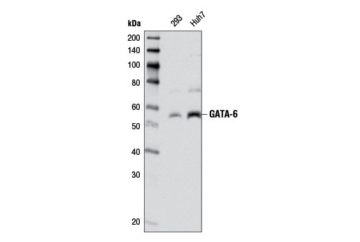
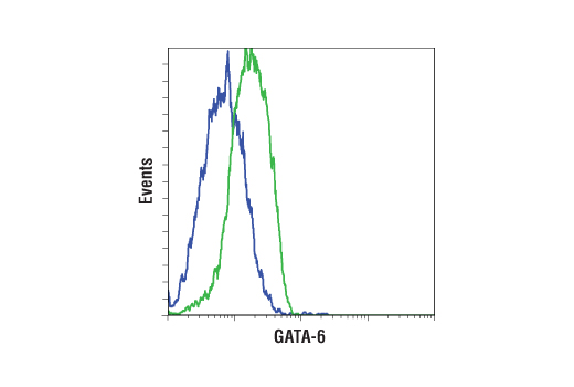
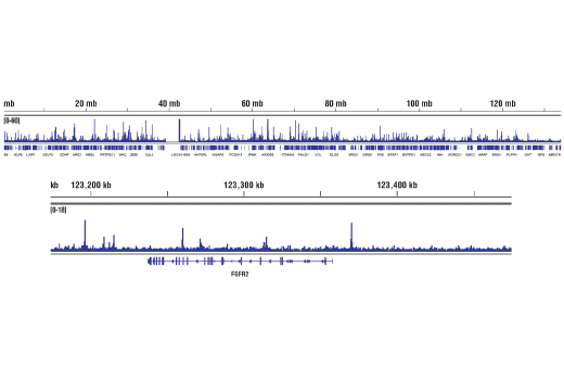
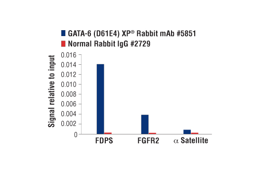
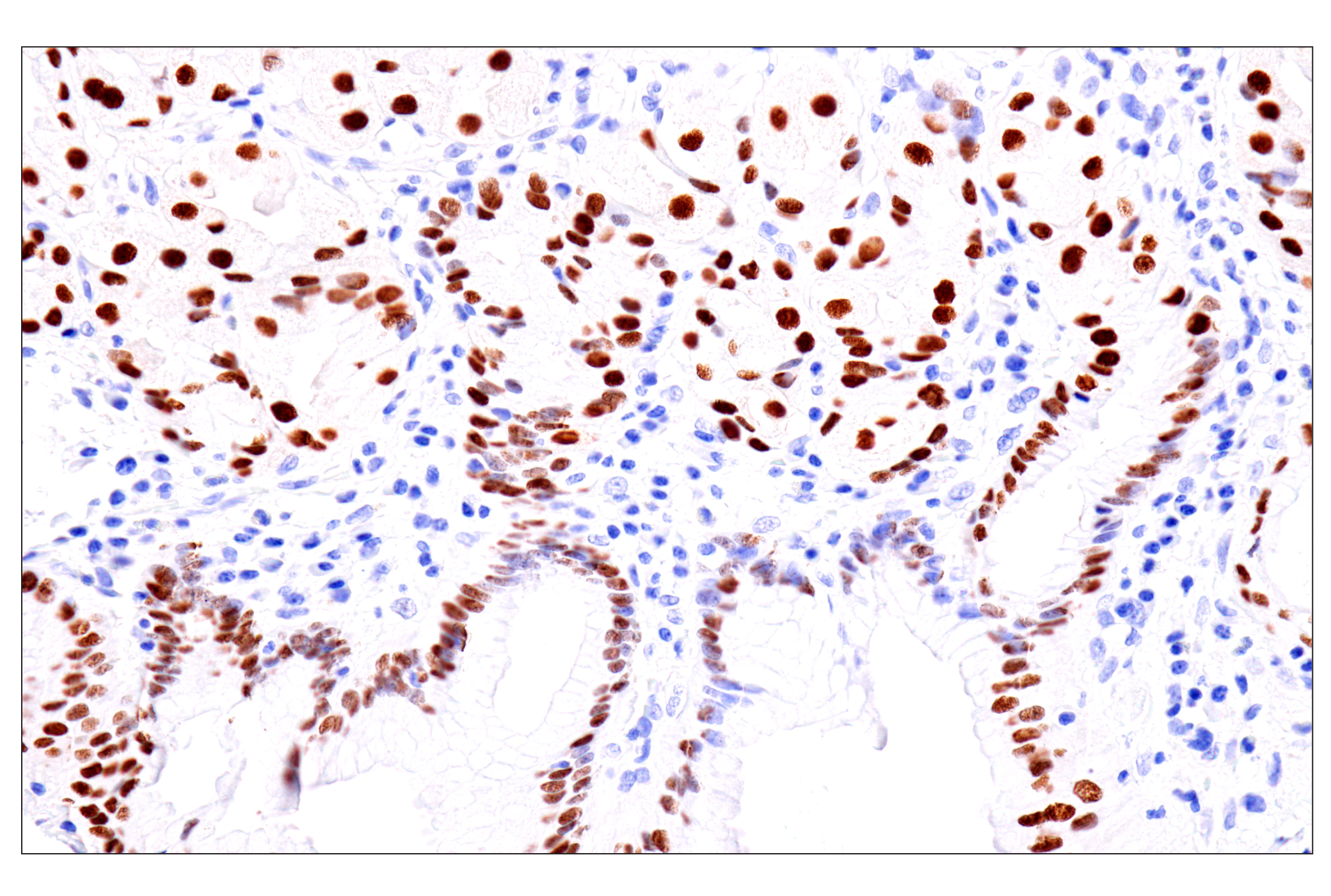
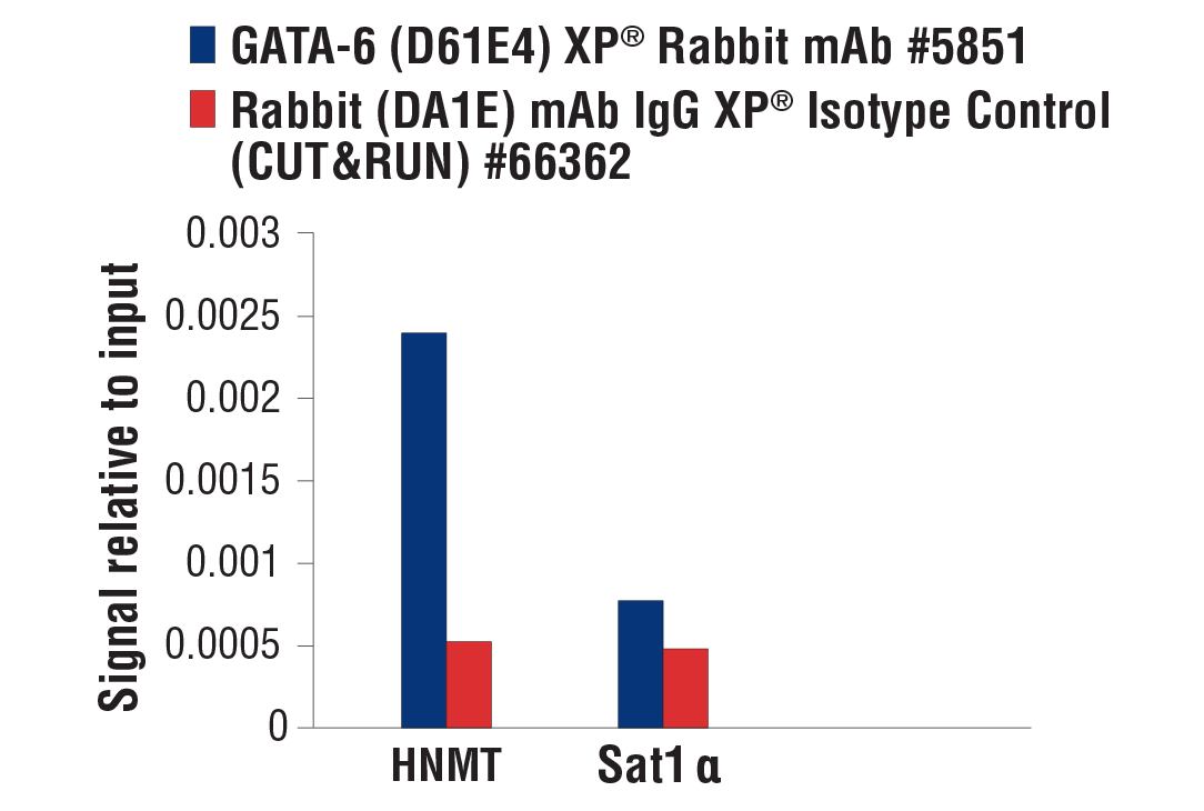
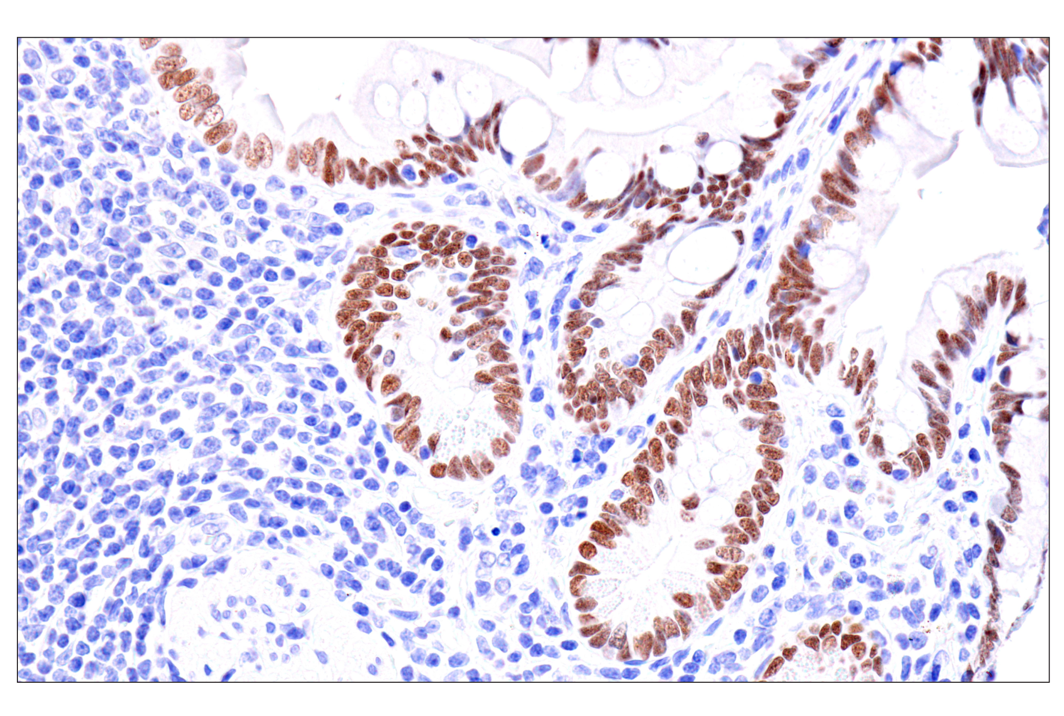
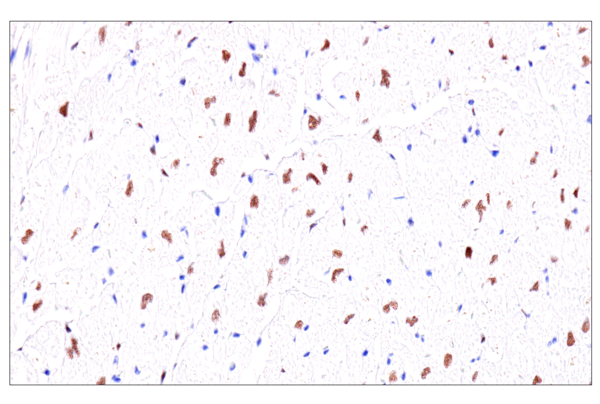
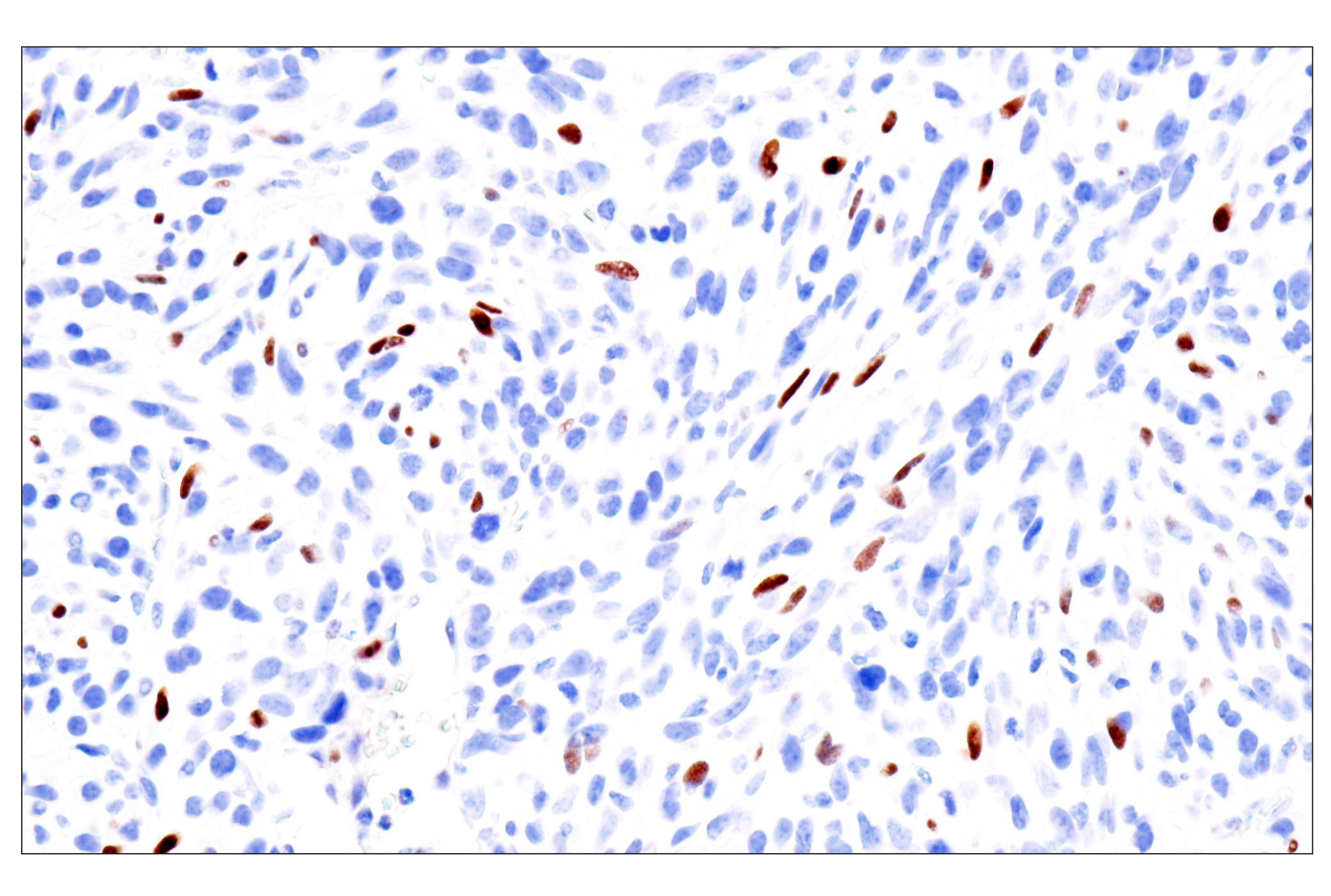
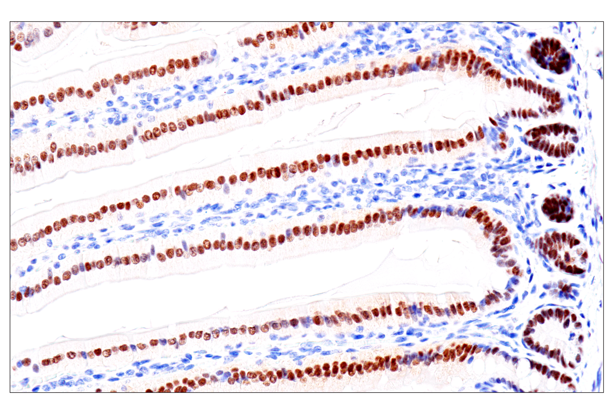
| GATA-6 (D61E4) XP® Rabbit mAb 5851 | 20 µl |
|
H M | 55 | Rabbit IgG |
| Anti-rabbit IgG, HRP-linked Antibody 7074 | 100 µl |
|
Goat |
Product Information
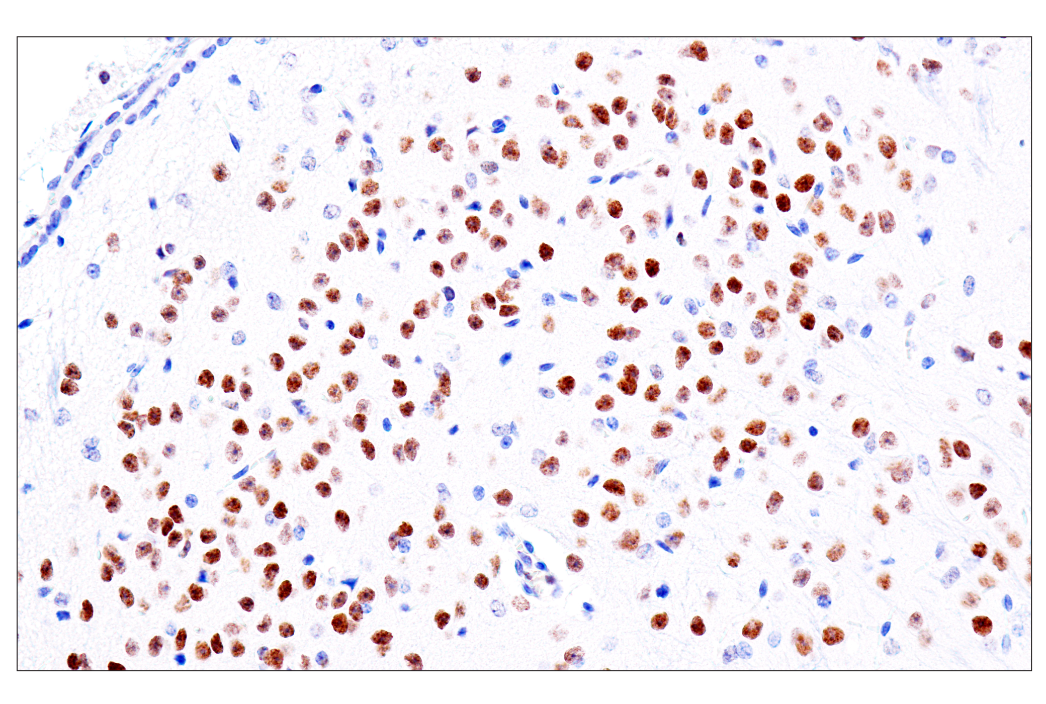
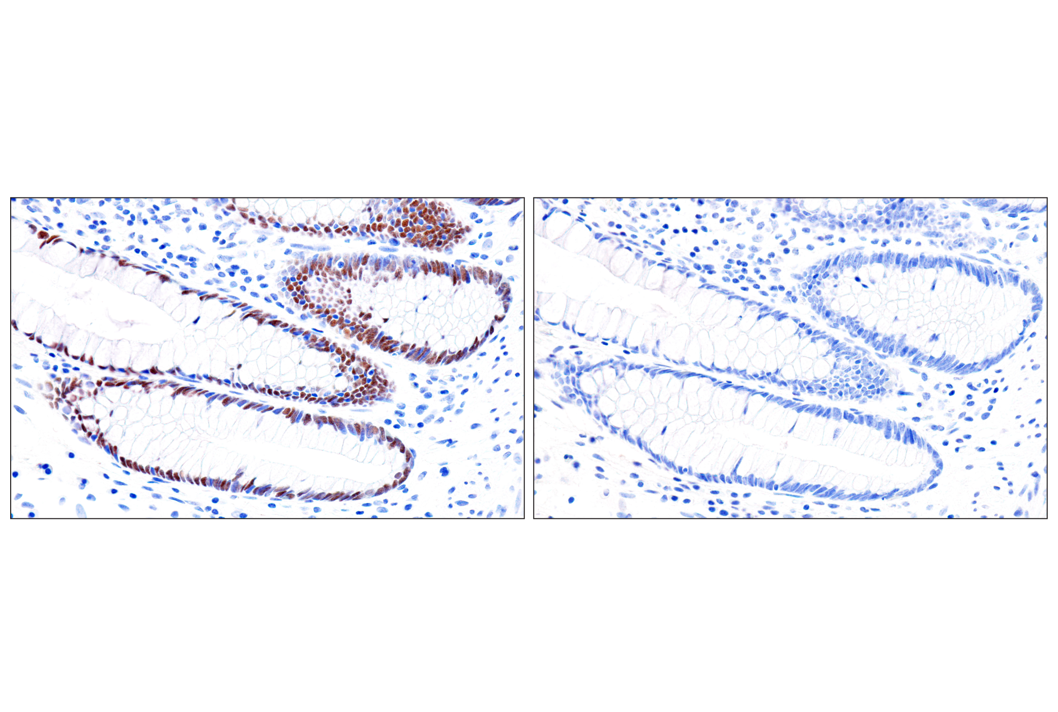
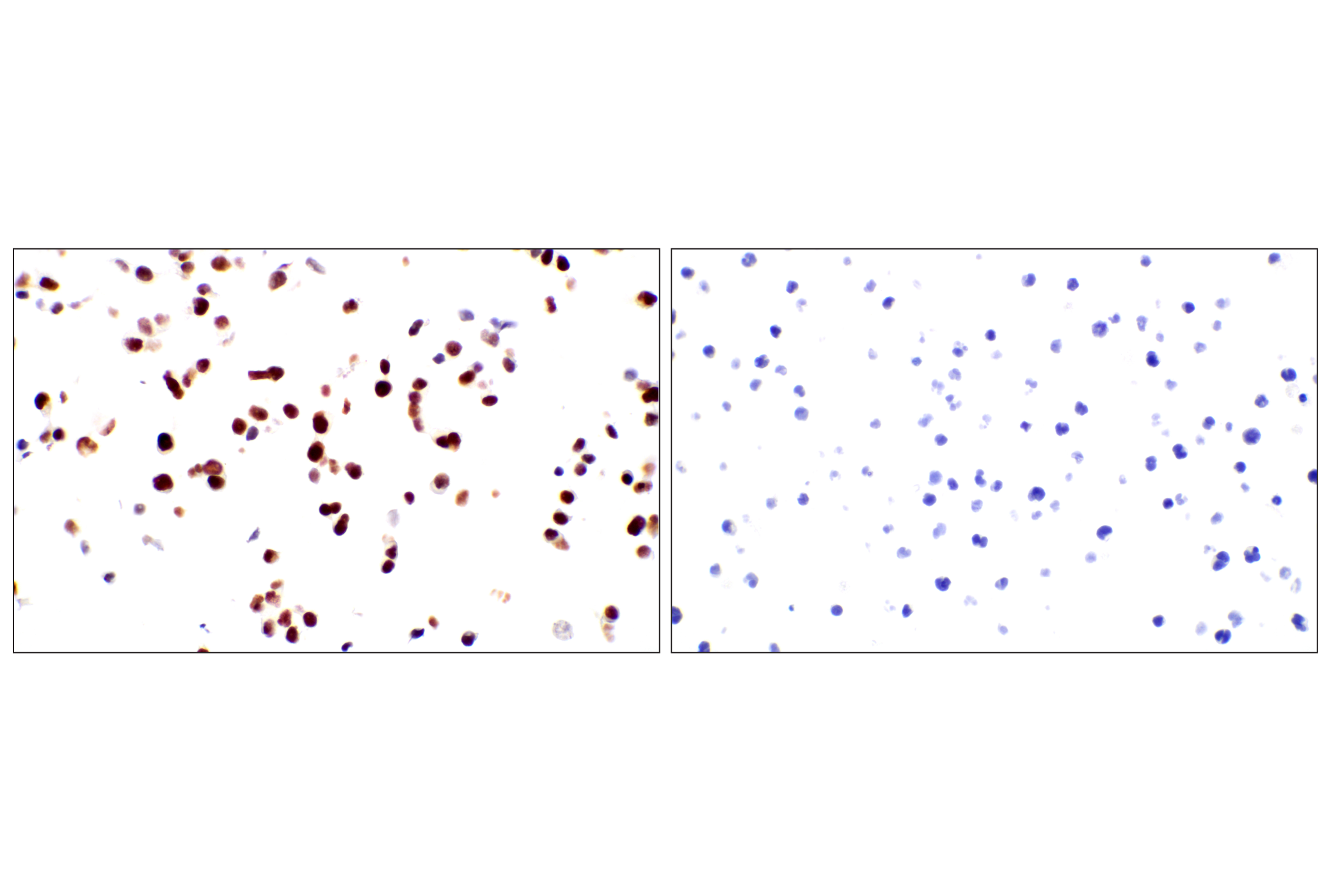
Monoclonal antibodies are produced by immunizing animals with synthetic peptides corresponding to residues surrounding Glu13 of human GATA-1, near the amino terminus of human GATA-3, GATA-4, and GATA-6 protein, and with recombinant protein specific to the amino terminus of human GATA-2 protein.
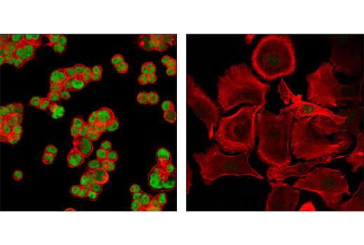
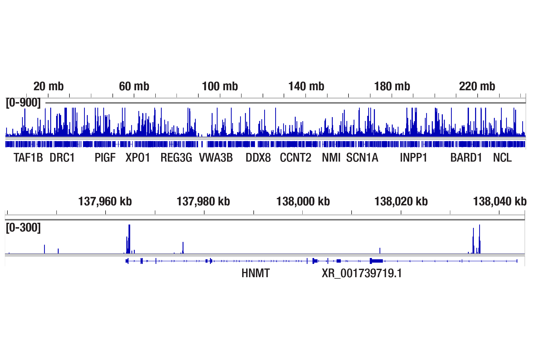
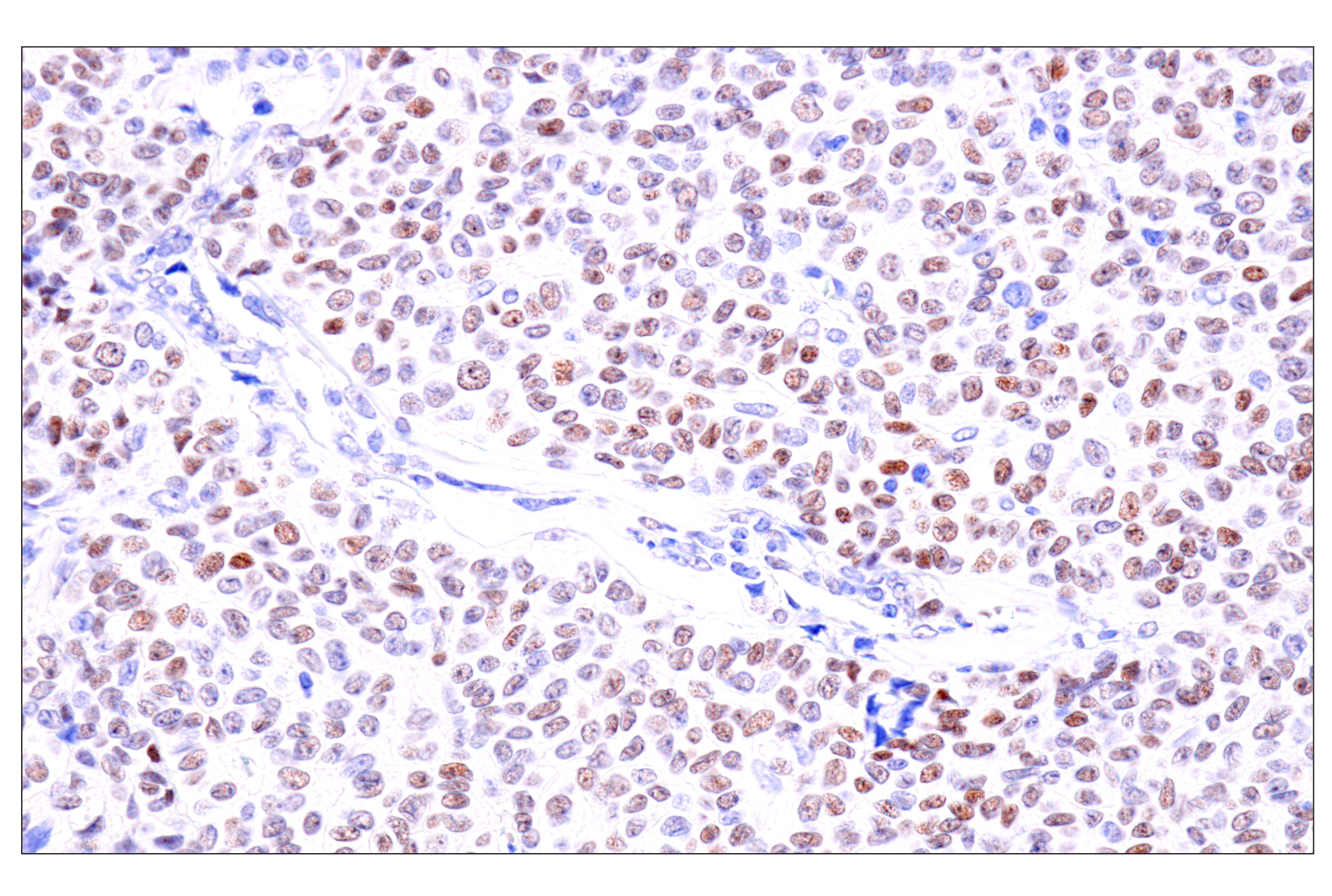
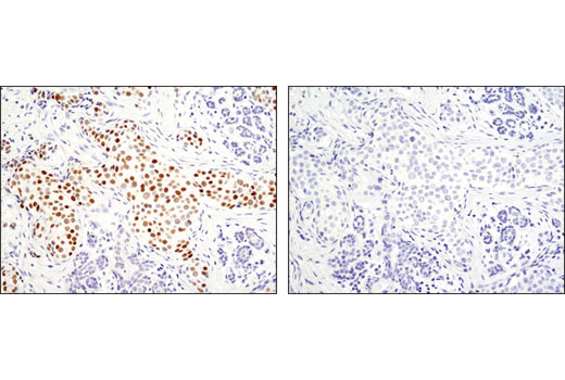
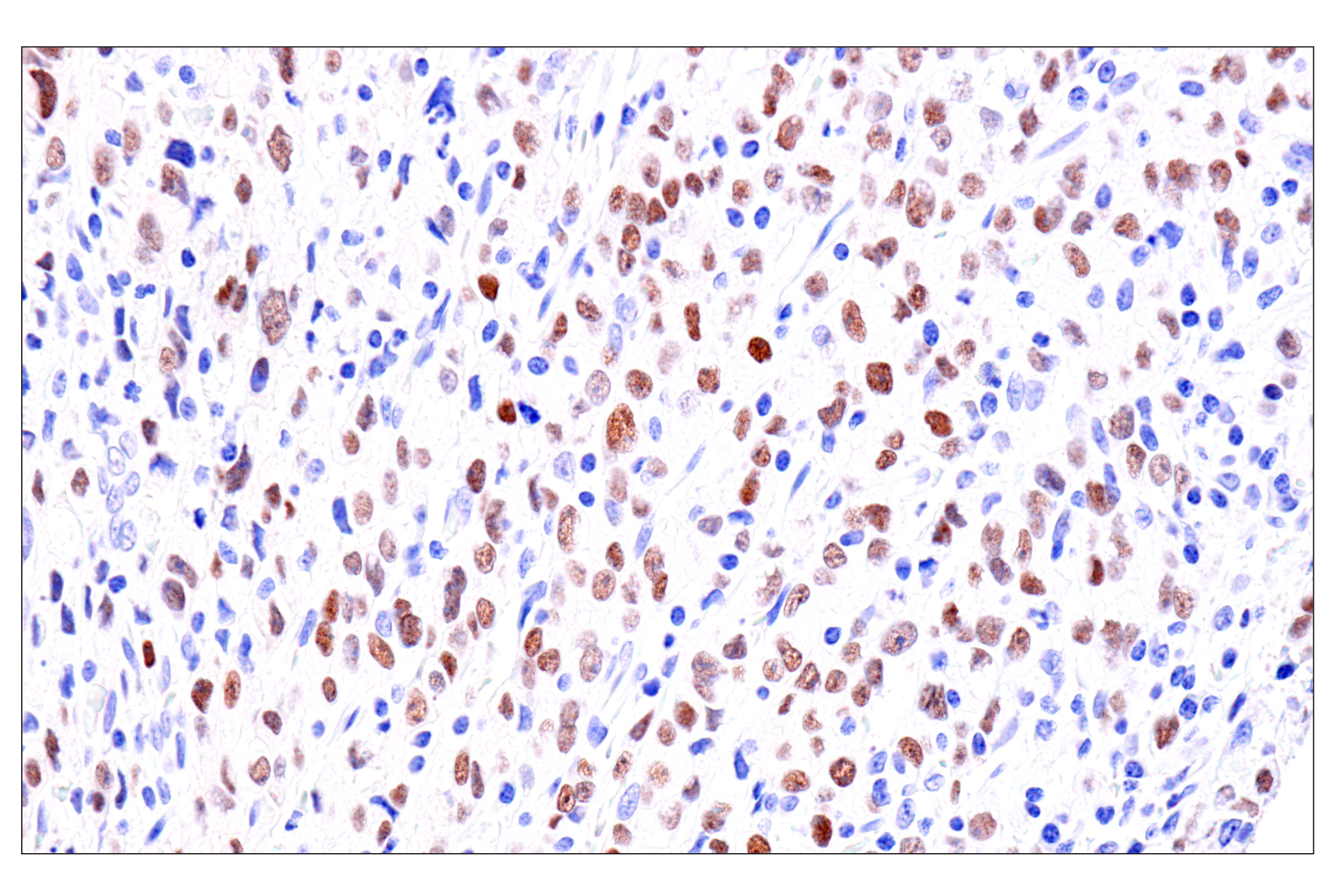
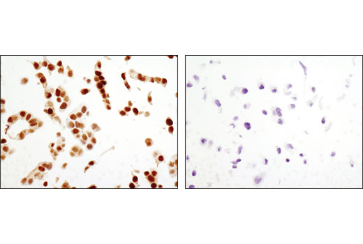
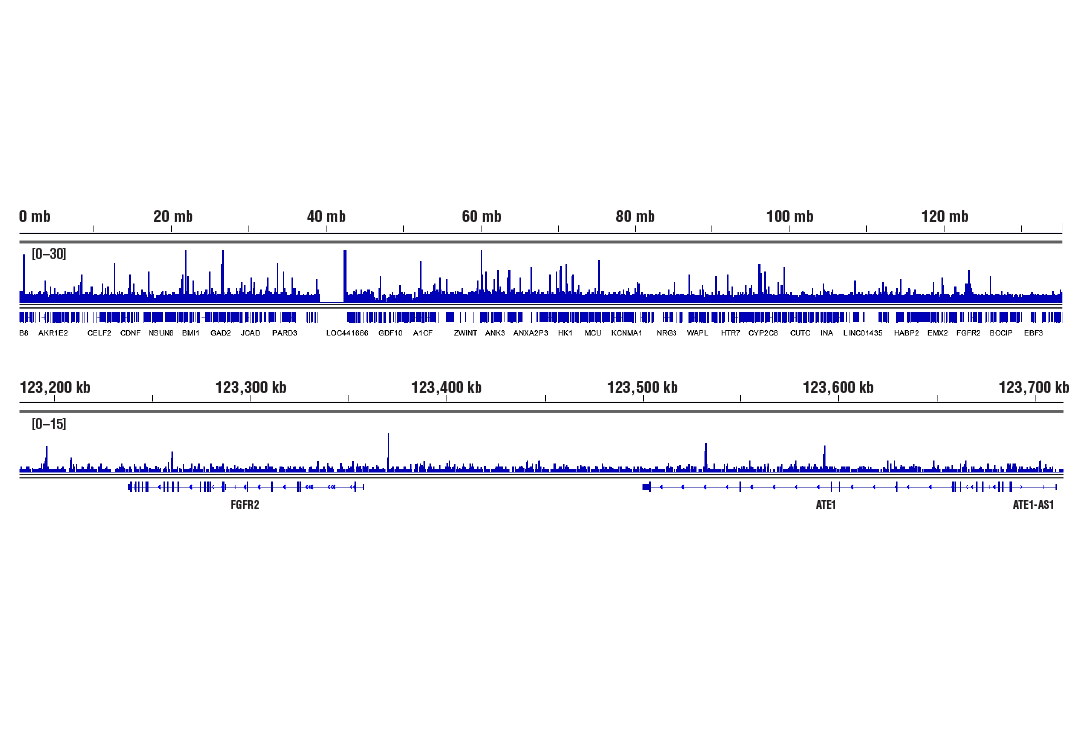
GATA proteins comprise a group of transcription factors that are related by the presence of conserved zinc finger DNA-binding domains, which bind directly to the nucleotide sequence core element GATA (1-3). There are six vertebrate GATA proteins, designated GATA-1 to GATA-6 (3). Although they are commonly divided as hematopoietic (GATA-1-3) or cardiac (GATA-4-6) factors, GATA proteins are expressed in a wide variety of tissue and play critical roles in embryonic development and organ differentiation (4). GATA-1 is the founding member of the GATA family and is required for erythroid and megakaryocytic cell development (5,6). Mutations in the corresponding GATA-1 gene are linked to many human diseases, including acute megakaryoblastic leukemia in Down Syndrome children (DS-AMKL), X-linked thrombocytopenia, and gray platelet syndrome (7-10). GATA-2 is widely expressed and plays an essential role in many developmental processes (11). Studies on GATA-2 knockout mice indicate that this protein is required in hematopoiesis (12). GATA-2 also inhibits the differentiation of white and brown adipocytes and has been shown to suppress the proliferation of neuronal progenitor cells (13-15). GATA-3 is a critical regulator of development and is expressed in both hematopoietic and non-hematopoietic tissues, including the kidney, skin, mammary gland, and central nervous system (16-19). GATA-3 knockout mouse embryos die between E11 and E12 due to growth retardation and deformities in the brain and spinal cord (20). The function of GATA-3 has also been extensively studied in T cell development and has been shown to be a downstream target of Notch in Notch-mediated differentiation of TH2 cells (21,22). GATA-4 is crucial for cardiomyocyte differentiation, and not surprisingly, mutations in the GATA-4 gene are implicated in many cardiac diseases, such as tetralogy of Fallot, familial and sporadic dilated cardiomyopathy, and atrial septal defect (23-27). GATA-4 and GATA-6 together maintain intestinal epithelial structure by regulating enterocyte gene expression (28). They also have overlapping roles in steroidogenesis and genital ridge formation during gonadal development (29). GATA-6 plays a critical role in endoderm development and is essential for the development of the heart, gut, and other organs (30-32). Knockout of GATA-6 is embryonic lethal due to defects in the formation of the heart tube and a failure to develop extraembryonic endoderm (30).
Except as otherwise expressly agreed in a writing signed by a legally authorized representative of CST, the following terms apply to Products provided by CST, its affiliates or its distributors. Any Customer's terms and conditions that are in addition to, or different from, those contained herein, unless separately accepted in writing by a legally authorized representative of CST, are rejected and are of no force or effect.
Products are labeled with For Research Use Only or a similar labeling statement and have not been approved, cleared, or licensed by the FDA or other regulatory foreign or domestic entity, for any purpose. Customer shall not use any Product for any diagnostic or therapeutic purpose, or otherwise in any manner that conflicts with its labeling statement. Products sold or licensed by CST are provided for Customer as the end-user and solely for research and development uses. Any use of Product for diagnostic, prophylactic or therapeutic purposes, or any purchase of Product for resale (alone or as a component) or other commercial purpose, requires a separate license from CST. Customer shall (a) not sell, license, loan, donate or otherwise transfer or make available any Product to any third party, whether alone or in combination with other materials, or use the Products to manufacture any commercial products, (b) not copy, modify, reverse engineer, decompile, disassemble or otherwise attempt to discover the underlying structure or technology of the Products, or use the Products for the purpose of developing any products or services that would compete with CST products or services, (c) not alter or remove from the Products any trademarks, trade names, logos, patent or copyright notices or markings, (d) use the Products solely in accordance with CST Product Terms of Sale and any applicable documentation, and (e) comply with any license, terms of service or similar agreement with respect to any third party products or services used by Customer in connection with the Products.