Revision 3
#69219
Store at -20C
Glycolysis/TCA Cycle Molecular Checkpoint Antibody Sampler Kit
1 Kit
(5 x 20 microliters)
877-616-CELL (2355)
877-678-TECH (8324)
3 Trask Lane | Danvers | Massachusetts | 01923 | USA
For Research Use Only. Not for Use in Diagnostic Procedures.
| Product Includes | Product # | Quantity | Mol. Wt | Isotype/Source |
|---|---|---|---|---|
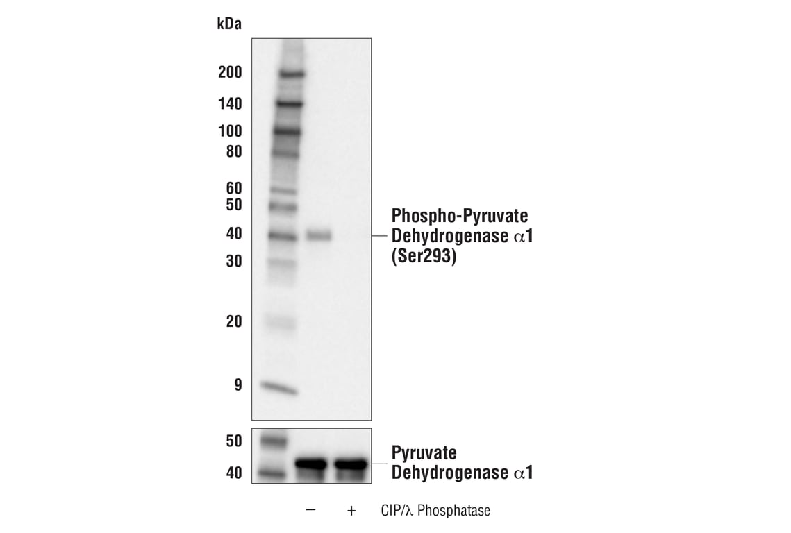
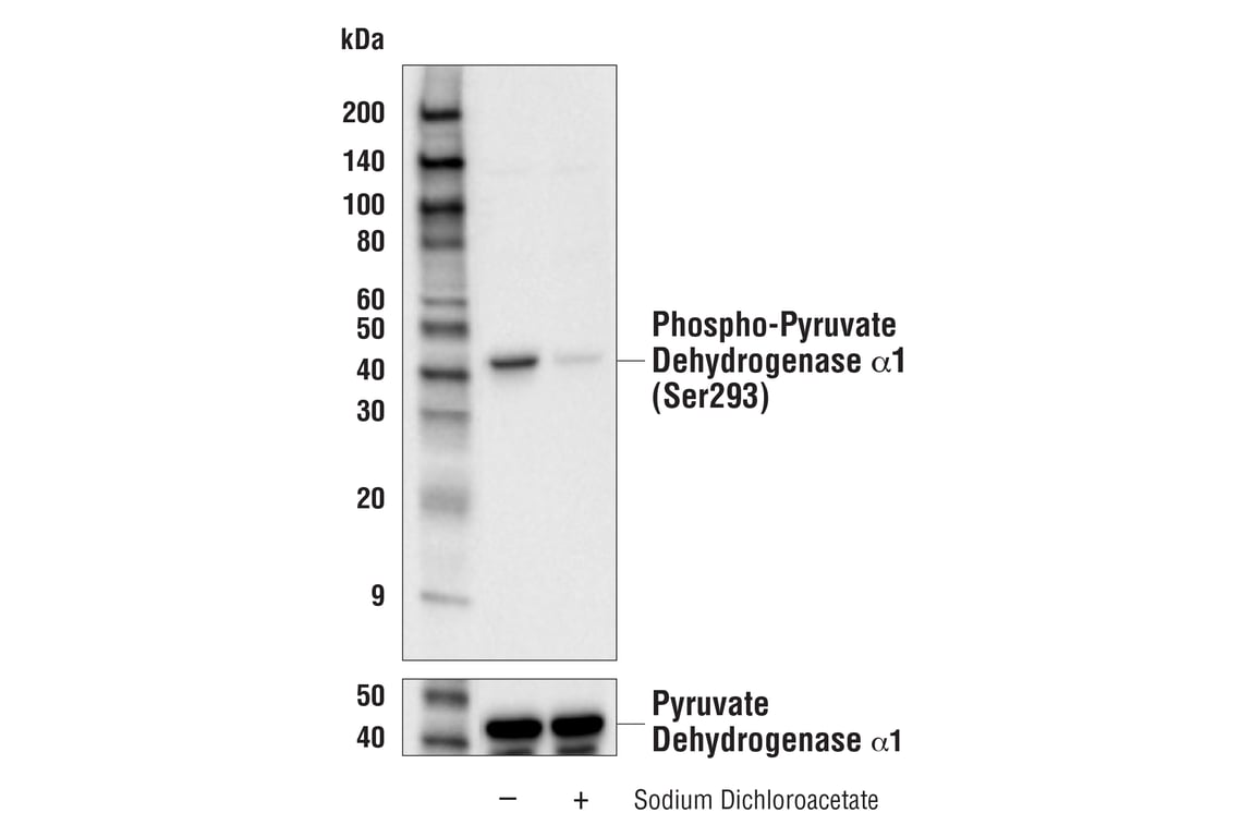
| Phospho-Pyruvate Dehydrogenase alpha1 (Ser293) (E4V9L) Rabbit Monoclonal Antibody | 37115 | 20 µl | 43 kDa | Rabbit IgG |
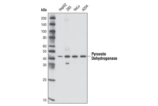
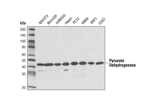
| Pyruvate Dehydrogenase (C54G1) Rabbit Monoclonal Antibody | 3205 | 20 µl | 43 kDa | Rabbit IgG |
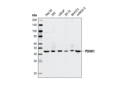
| PDHK1 (C47H1) Rabbit Monoclonal Antibody | 3820 | 20 µl | 47 kDa | Rabbit IgG |
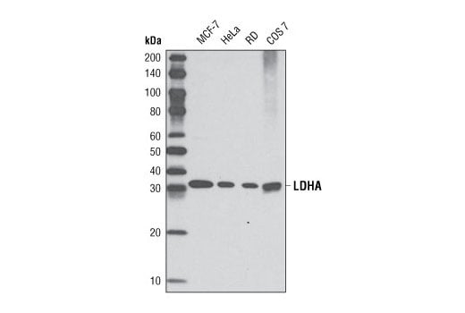
| LDHA (C4B5) Rabbit Monoclonal Antibody | 3582 | 20 µl | 37 kDa | Rabbit IgG |
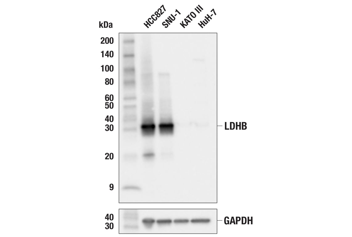
| LDHB (E8J8T) Rabbit Monoclonal Antibody | 56298 | 20 µl | 37 kDa | Rabbit IgG |
| Anti-rabbit IgG, HRP-linked Antibody | 7074 | 100 µl | Goat |
Please visit cellsignal.com for individual component applications, species cross-reactivity, dilutions, protocols, and additional product information.
Description
Storage
Background
Pyruvate dehydrogenase kinase 1 (PDHK1) phosphorylates pyruvate dehydrogenase (E1) α1 subunit at Ser293 to inactivate its activity (3,4). This phosphorylation contributes to the tumor metabolic reprogramming toward glycolysis in hypoxia by inhibiting the citric acid cycle (TCA cycle) (4).
Lactate dehydrogenase (LDH) catalyzes the reversible conversion between pyruvate and lactate. LDH is a tetramer composed of various combinations of LDHA subunit and LDHB subunit to form five different isozymes. LDHA has a higher affinity for pyruvate and preferentially catalyzes the conversion of pyruvate to lactate. LDHA levels are upregulated in many cancers. On the other hand, LDHB has a higher affinity for lactate and preferentially catalyzes the conversion of lactate to pyruvate, enabling cells to use lactate as a nutrient (5-7). Studies show that LDHA/LDHB deficiency suppresses glycolysis and ATP production, inhibiting STING signaling and antitumor immune responses mediated by dendritic cells (8). In addition, acetylation of LDHB inhibits its activity, reduces hepatic lactate clearance, and promotes the progression of non-alcoholic fatty liver disease (NAFLD) (9).
Background References
- Strumiło, S. (2005) Acta Biochim Pol 52, 759-64.
- Stacpoole, P.W. et al. (2003) Curr Gene Ther 3, 239-45.
- Fan, J. et al. (2014) J Biol Chem 289, 26533-26541.
- Chae, Y.C. et al. (2016) Cancer Cell 30, 257-272.
- Doherty, J.R. and Cleveland, J.L. (2013) J Clin Invest 123, 3685-92.
- Hong, S.M. et al. (2019) J Biol Chem 294, 7810-7820.
- Urbańska, K. and Orzechowski, A. (2019) Int J Mol Sci 20, 2085. doi: 10.3390/ijms20092085.
- Hu, Z. et al. (2023) J Clin Invest 133, e166031. doi: 10.1172/JCI166031.
- Wang, T. et al. (2021) J Hepatol 74, 1038-1052.
Trademarks and Patents
Cell Signaling Technology is a trademark of Cell Signaling Technology, Inc.
U.S. Patent No. 7,429,487, foreign equivalents, and child patents deriving therefrom.
All other trademarks are the property of their respective owners. Visit cellsignal.com/trademarks for more information.
Limited Uses
Except as otherwise expressly agreed in a writing signed by a legally authorized representative of CST, the following terms apply to Products provided by CST, its affiliates or its distributors. Any Customer's terms and conditions that are in addition to, or different from, those contained herein, unless separately accepted in writing by a legally authorized representative of CST, are rejected and are of no force or effect.
Products are labeled with For Research Use Only or a similar labeling statement and have not been approved, cleared, or licensed by the FDA or other regulatory foreign or domestic entity, for any purpose. Customer shall not use any Product for any diagnostic or therapeutic purpose, or otherwise in any manner that conflicts with its labeling statement. Products sold or licensed by CST are provided for Customer as the end-user and solely for research and development uses. Any use of Product for diagnostic, prophylactic or therapeutic purposes, or any purchase of Product for resale (alone or as a component) or other commercial purpose, requires a separate license from CST. Customer shall (a) not sell, license, loan, donate or otherwise transfer or make available any Product to any third party, whether alone or in combination with other materials, or use the Products to manufacture any commercial products, (b) not copy, modify, reverse engineer, decompile, disassemble or otherwise attempt to discover the underlying structure or technology of the Products, or use the Products for the purpose of developing any products or services that would compete with CST products or services, (c) not alter or remove from the Products any trademarks, trade names, logos, patent or copyright notices or markings, (d) use the Products solely in accordance with CST Product Terms of Sale and any applicable documentation, and (e) comply with any license, terms of service or similar agreement with respect to any third party products or services used by Customer in connection with the Products.
Revision 3
Revision 3

Revision 3

Revision 3


Revision 3


Revision 3


