Revision 6
#38313
Store at -20C
877-616-CELL (2355)
877-678-TECH (8324)
3 Trask Lane | Danvers | Massachusetts | 01923 | USA
For Research Use Only. Not for Use in Diagnostic Procedures.
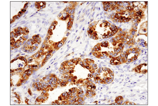
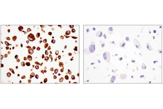
Applications:
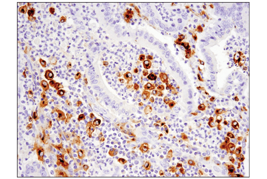
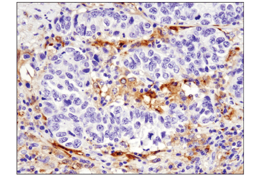
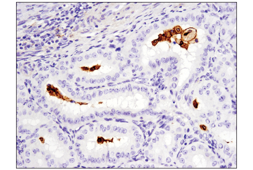
W, IP, IHC-P
Reactivity:
H
Sensitivity:
Endogenous
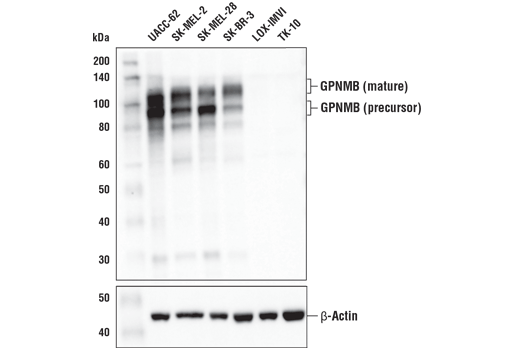
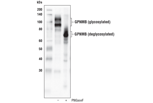
MW (kDa):
95, 120
Source/Isotype:
Rabbit IgG
UniProt ID:
#Q14956
Entrez-Gene Id:
10457
Product Usage Information
| Application | Dilution |
|---|---|
| Western Blotting | 1:1000 |
| Immunoprecipitation | 1:50 |
| Immunohistochemistry (Paraffin) | 1:250 - 1:1000 |
Storage
For a carrier free (BSA and azide free) version of this product see product #79846.
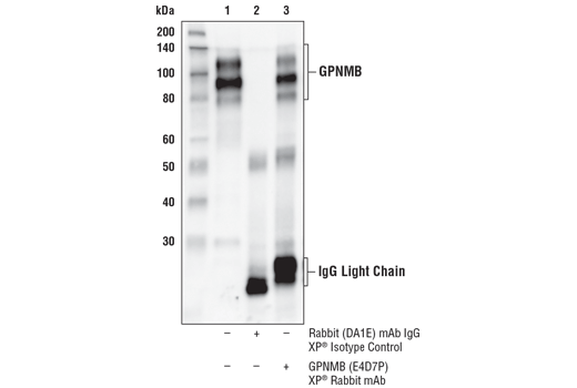
Specificity/Sensitivity
Source / Purification
Background
While typical GPNMB expression is seen in tissues including skin, heart, kidney, lung, liver, and skeletal muscle (3,6), research studies show elevated GPNMB expression often contributes to the metastatic phenotype in numerous cancers (reviewed in 7). GPNMB is typically localized to intracellular compartments in normal cells (1,8), but investigators found it primarily on the cell surface of tumor cells (9,10). Differential localization and expression, and the role of GPNMB as a tumor promoter in many cancer types make this protein a viable therapeutic target (11).
The GPNMB ectodomain can be cleaved by matrix metalloproteinases and shed from the cell surface (12). Research studies identify the sheddase ADAM10 as one peptidase responsible for cleavage of the GPNMB ectodomain at the surface of breast cancer cells. Shedded GPNMB ectodomains may promote angiogenesis by inducing endothelial cell migration (13).
Background References
- Tomihari, M. et al. (2009) Exp Dermatol 18, 586-95.
- Sheng, M.H. et al. (2012) PLoS One 7, e35280.
- Shikano, S. et al. (2001) J Biol Chem 276, 8125-34.
- Li, B. et al. (2010) FASEB J 24, 4767-81.
- Patel-Chamberlin, M. et al. (2011) Kidney Int 79, 1138-48.
- Bandari, P.S. et al. (2003) Regul Pept 111, 169-78.
- Maric, G. et al. (2013) Onco Targets Ther 6, 839-52.
- Ripoll, V.M. et al. (2007) J Immunol 178, 6557-66.
- Tse, K.F. et al. (2006) Clin Cancer Res 12, 1373-82.
- Rose, A.A. et al. (2010) Clin Cancer Res 16, 2147-56.
- Keir, C.H. and Vahdat, L.T. (2012) Expert Opin Biol Ther 12, 259-63.
- Furochi, H. et al. (2007) FEBS Lett 581, 5743-50.
- Rose, A.A. et al. (2010) PLoS One 5, e12093.
Species Reactivity
Species reactivity is determined by testing in at least one approved application (e.g., western blot).
Western Blot Buffer
IMPORTANT: For western blots, incubate membrane with diluted primary antibody in 5% w/v BSA, 1X TBS, 0.1% Tween® 20 at 4°C with gentle shaking, overnight.
Applications Key
W: Western Blotting IP: Immunoprecipitation IHC-P: Immunohistochemistry (Paraffin)
Cross-Reactivity Key
H: Human
Trademarks and Patents
Cell Signaling Technology is a trademark of Cell Signaling Technology, Inc.
SignalStain is a registered trademark of Cell Signaling Technology, Inc.
All other trademarks are the property of their respective owners. Visit cellsignal.com/trademarks for more information.
Limited Uses
Except as otherwise expressly agreed in a writing signed by a legally authorized representative of CST, the following terms apply to Products provided by CST, its affiliates or its distributors. Any Customer's terms and conditions that are in addition to, or different from, those contained herein, unless separately accepted in writing by a legally authorized representative of CST, are rejected and are of no force or effect.
Products are labeled with For Research Use Only or a similar labeling statement and have not been approved, cleared, or licensed by the FDA or other regulatory foreign or domestic entity, for any purpose. Customer shall not use any Product for any diagnostic or therapeutic purpose, or otherwise in any manner that conflicts with its labeling statement. Products sold or licensed by CST are provided for Customer as the end-user and solely for research and development uses. Any use of Product for diagnostic, prophylactic or therapeutic purposes, or any purchase of Product for resale (alone or as a component) or other commercial purpose, requires a separate license from CST. Customer shall (a) not sell, license, loan, donate or otherwise transfer or make available any Product to any third party, whether alone or in combination with other materials, or use the Products to manufacture any commercial products, (b) not copy, modify, reverse engineer, decompile, disassemble or otherwise attempt to discover the underlying structure or technology of the Products, or use the Products for the purpose of developing any products or services that would compete with CST products or services, (c) not alter or remove from the Products any trademarks, trade names, logos, patent or copyright notices or markings, (d) use the Products solely in accordance with CST Product Terms of Sale and any applicable documentation, and (e) comply with any license, terms of service or similar agreement with respect to any third party products or services used by Customer in connection with the Products.
Revision 6
Revision 6
Revision 6