Revision 2
#8339
Store at -20C
HER/ErbB Family Antibody Sampler Kit
1 Kit
(8 x 20 microliters)
877-616-CELL (2355)
877-678-TECH (8324)
3 Trask Lane | Danvers | Massachusetts | 01923 | USA
For Research Use Only. Not for Use in Diagnostic Procedures.
| Product Includes | Product # | Quantity | Mol. Wt | Isotype/Source |
|---|---|---|---|---|
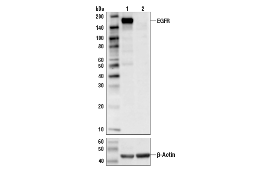
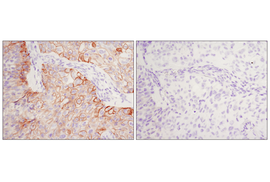
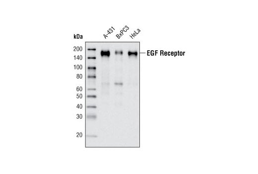
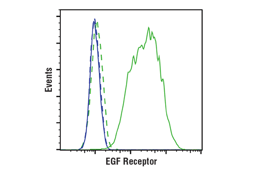
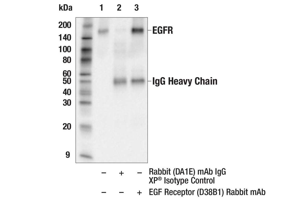
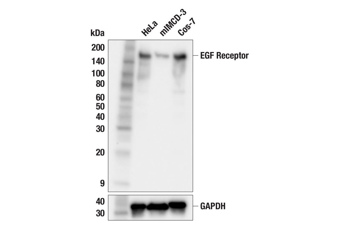
| EGF Receptor (D38B1) Rabbit Monoclonal Antibody | 4267 | 20 µl | 175 kDa | Rabbit IgG |
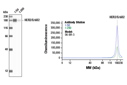
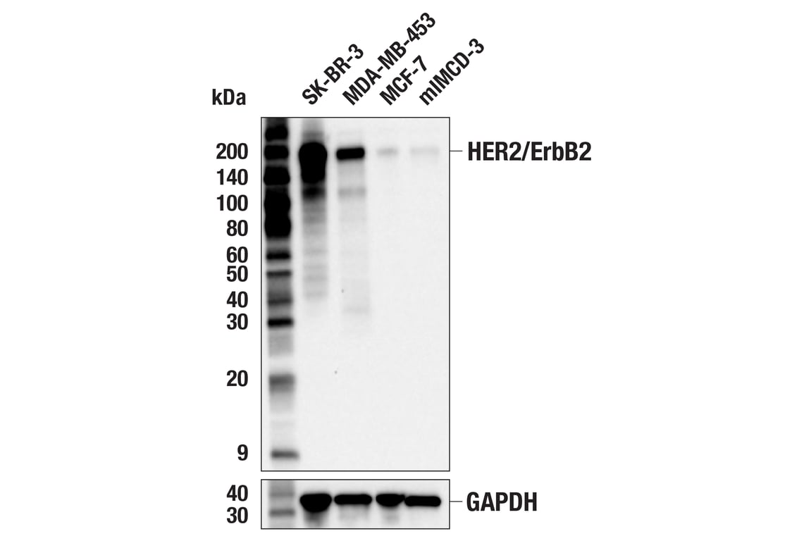
| HER2/ErbB2 (D8F12) Rabbit Monoclonal Antibody | 4290 | 20 µl | 185 kDa | Rabbit IgG |
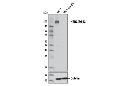
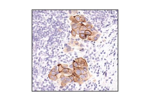
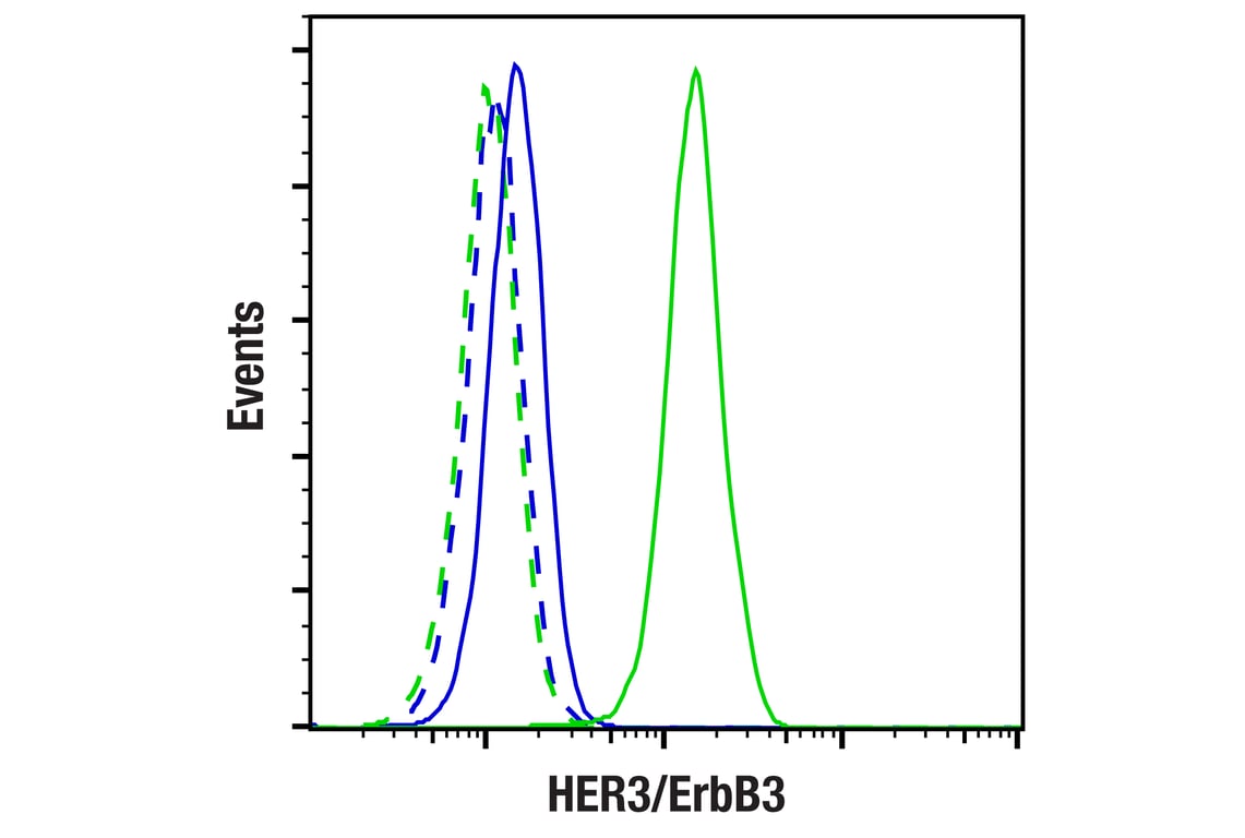
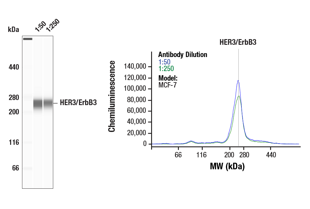
| HER3/ErbB3 (D22C5) Rabbit Monoclonal Antibody | 12708 | 20 µl | 185 kDa | Rabbit IgG |
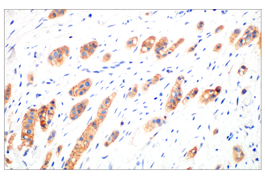
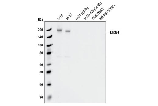
| HER4/ErbB4 (111B2) Rabbit Monoclonal Antibody | 4795 | 20 µl | 180 kDa | Rabbit IgG |
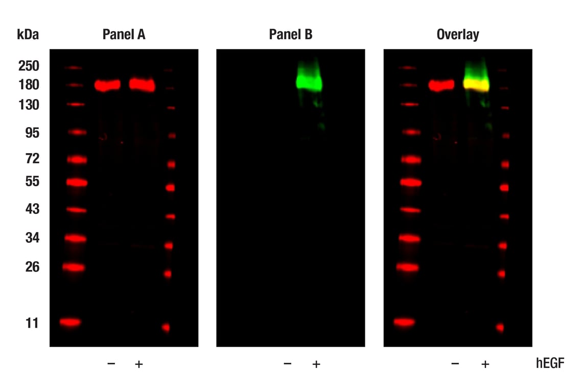
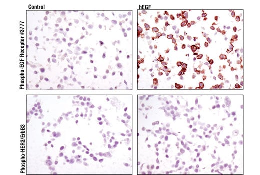
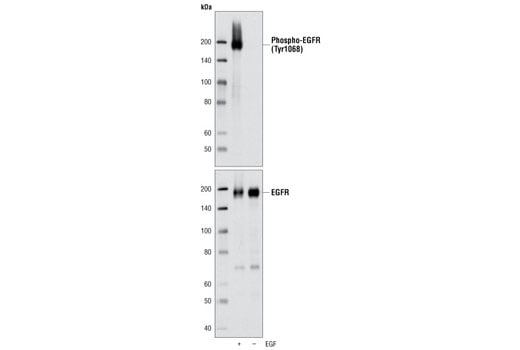
| Phospho-EGF Receptor (Tyr1068) (D7A5) Rabbit Monoclonal Antibody | 3777 | 20 µl | 175 kDa | Rabbit IgG |
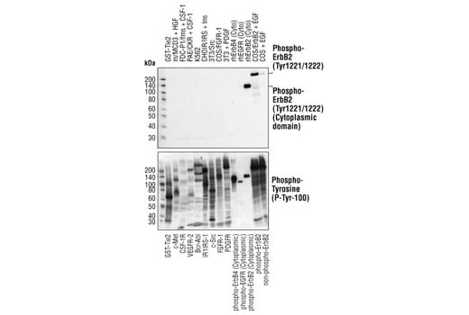
| Phospho-HER2/ErbB2 (Tyr1221/1222) (6B12) Rabbit Monoclonal Antibody | 2243 | 20 µl | 185 kDa | Rabbit IgG |
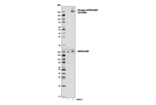
| Phospho-HER3/ErbB3 (Tyr1289) (D1B5) Rabbit Monoclonal Antibody | 2842 | 20 µl | 185 kDa | Rabbit IgG |
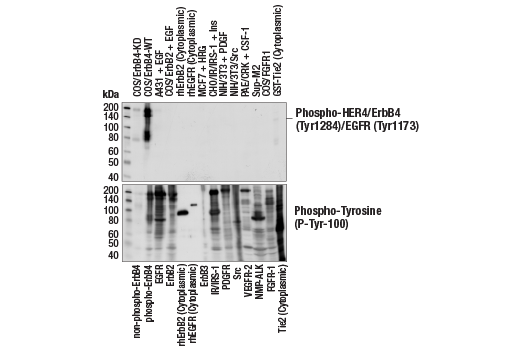
| Phospho-HER4/ErbB4 (Tyr1284)/EGFR (Tyr1173) (21A9) Rabbit Monoclonal Antibody | 4757 | 20 µl | 180 kDa | Rabbit |
| Anti-rabbit IgG, HRP-linked Antibody | 7074 | 100 µl | Goat |
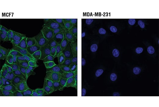
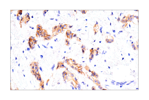
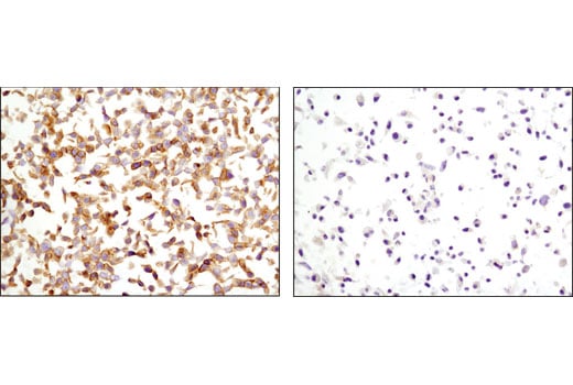
Please visit cellsignal.com for individual component applications, species cross-reactivity, dilutions, protocols, and additional product information.
Description
Storage
Background
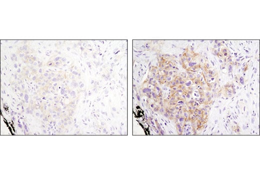
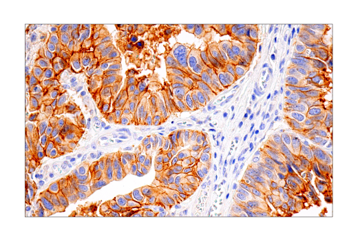
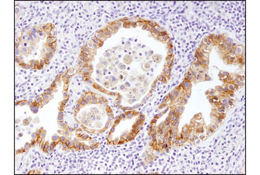
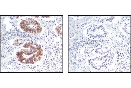
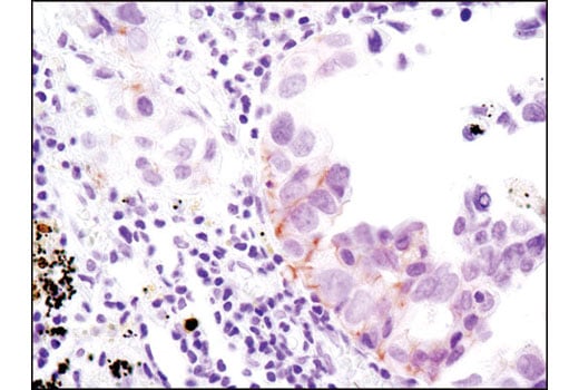
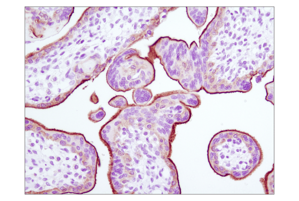
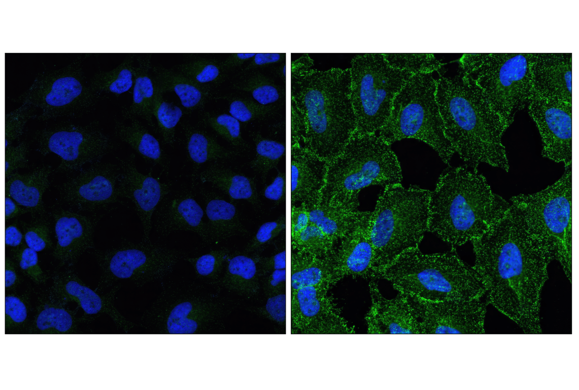
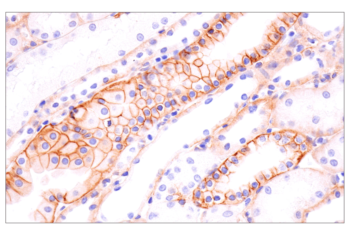
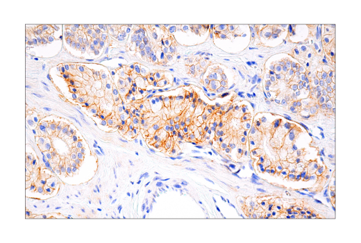
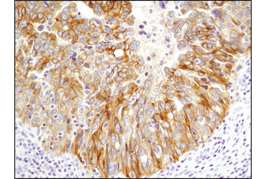
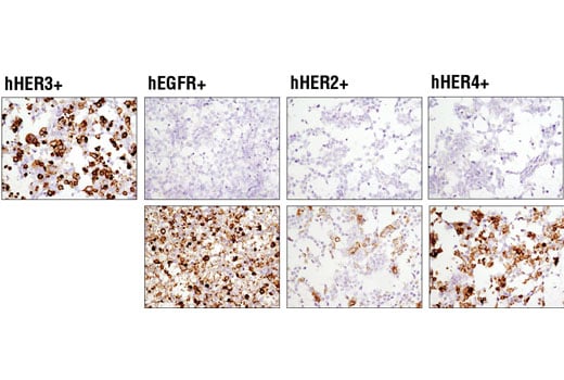
The ErbB2 (HER2) proto-oncogene encodes a 185 kDa transmembrane, receptor-like glycoprotein with intrinsic tyrosine kinase activity (4). While ErbB2 lacks an identified ligand, ErbB2 kinase activity can be activated in the absence of a ligand when overexpressed and through heteromeric associations with other ErbB family members (5). Amplification of the ErbB2 gene and overexpression of its product are detected in almost 40% of human breast cancers (6). The major autophosphorylation sites in ErbB2 are Tyr1248 and Tyr1221/1222; phosphorylation of these sites couples ErbB2 to the Ras-Raf-MAP kinase signal transduction pathway (4,7).
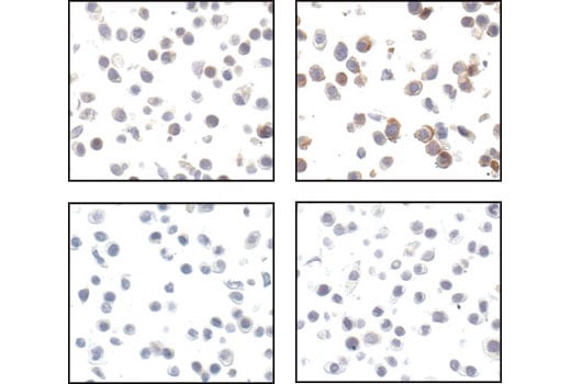
HER3/ErbB3 is a member of the ErbB receptor protein tyrosine kinase family, but lacks tyrosine kinase activity. Tyrosine phosphorylation of ErbB3 depends on its association with other ErbB tyrosine kinases. Upon ligand binding, heterodimers form between ErbB3 and other ErbB proteins, and ErbB3 is phosphorylated on tyrosine residues by the activated ErbB kinase (8,9). There are at least 9 potential tyrosine phosphorylation sites in the carboxy-terminal tail of ErbB3. These sites serve as consensus binding sites for signal transducing proteins, including Src family members, GRB2, and the p85 subunit of PI3 kinase, which mediate ErbB-downstream signaling (10). Both Tyr1222 and Tyr1289 of ErbB3 reside within a YXXM motif and participate in signaling to PI3 kinase (11).
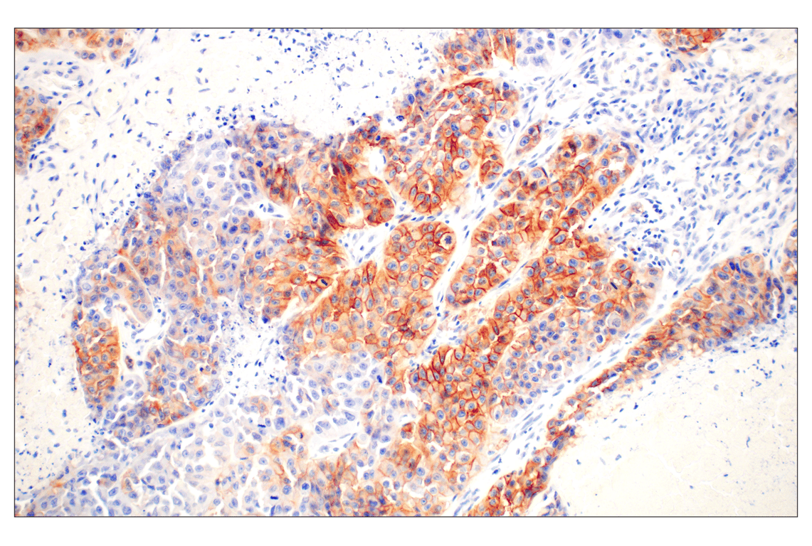
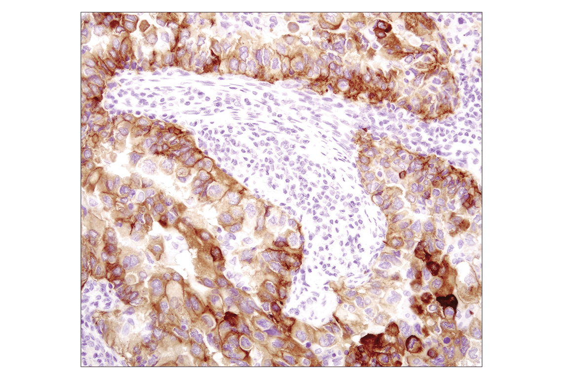
HER4/ErbB4, like other family members, has four ectodomains, a single transmembrane domain, and a cytoplasmic tail containing the active tyrosine kinase domain (12). By binding to neuregulins and/or EGF family ligands, ErbB4 forms either a homodimer or heterodimer with other ErbB family members, which results in receptor activation and signaling (12). ErbB4 is ubiquitously expressed with the highest expression occurring in the brain and heart. The expression of ErbB4 in breast cancer, pediatric brain cancer, and other types of carcinomas has been reported, suggesting that ErbB4 expression is involved in both normal tissue development and carcinogenesis (12).
Background References
- Hackel, P.O. et al. (1999) Curr Opin Cell Biol 11, 184-9.
- Zwick, E. et al. (1999) Trends Pharmacol Sci 20, 408-12.
- Rojas, M. et al. (1996) J Biol Chem 271, 27456-61.
- Muthuswamy, S.K. et al. (1999) Mol Cell Biol 19, 6845-57.
- Qian, X. et al. (1994) Proc Natl Acad Sci U S A 91, 1500-4.
- Dittadi, R. and Gion, M. (2000) J Natl Cancer Inst 92, 1443-4.
- Kwon, Y.K. et al. (1997) J Neurosci 17, 8293-9.
- Yarden, Y. and Sliwkowski, M.X. (2001) Nat Rev Mol Cell Biol 2, 127-37.
- Guy, P.M. et al. (1994) Proc Natl Acad Sci U S A 91, 8132-6.
- Songyang, Z. et al. (1993) Cell 72, 767-78.
- Kim, H.H. et al. (1994) J Biol Chem 269, 24747-55.
- Carpenter, G. (2003) Exp Cell Res 284, 66-77.
Trademarks and Patents
Cell Signaling Technology is a trademark of Cell Signaling Technology, Inc.
All other trademarks are the property of their respective owners. Visit cellsignal.com/trademarks for more information.
Limited Uses
Except as otherwise expressly agreed in a writing signed by a legally authorized representative of CST, the following terms apply to Products provided by CST, its affiliates or its distributors. Any Customer's terms and conditions that are in addition to, or different from, those contained herein, unless separately accepted in writing by a legally authorized representative of CST, are rejected and are of no force or effect.
Products are labeled with For Research Use Only or a similar labeling statement and have not been approved, cleared, or licensed by the FDA or other regulatory foreign or domestic entity, for any purpose. Customer shall not use any Product for any diagnostic or therapeutic purpose, or otherwise in any manner that conflicts with its labeling statement. Products sold or licensed by CST are provided for Customer as the end-user and solely for research and development uses. Any use of Product for diagnostic, prophylactic or therapeutic purposes, or any purchase of Product for resale (alone or as a component) or other commercial purpose, requires a separate license from CST. Customer shall (a) not sell, license, loan, donate or otherwise transfer or make available any Product to any third party, whether alone or in combination with other materials, or use the Products to manufacture any commercial products, (b) not copy, modify, reverse engineer, decompile, disassemble or otherwise attempt to discover the underlying structure or technology of the Products, or use the Products for the purpose of developing any products or services that would compete with CST products or services, (c) not alter or remove from the Products any trademarks, trade names, logos, patent or copyright notices or markings, (d) use the Products solely in accordance with CST Product Terms of Sale and any applicable documentation, and (e) comply with any license, terms of service or similar agreement with respect to any third party products or services used by Customer in connection with the Products.
Revision 2
Revision 2
Revision 2
Revision 2

Revision 2
Revision 2
Revision 2
Revision 2


Revision 2
Revision 2
Revision 2
Revision 2
Revision 2
Revision 2
Revision 2
Revision 2
Revision 2
Revision 2

Revision 2