Product Information
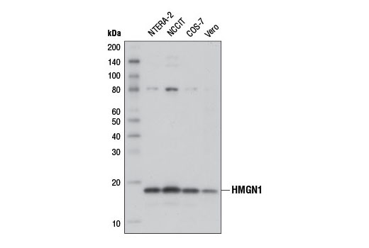
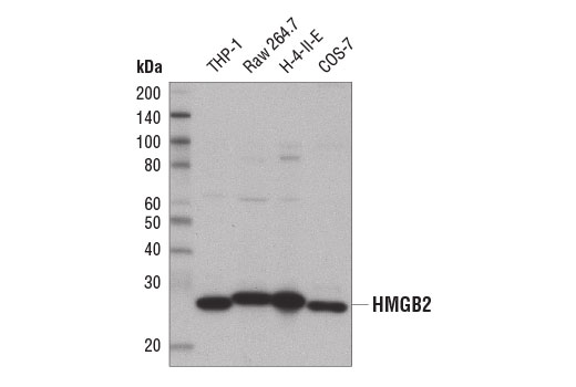
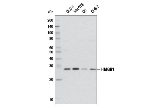
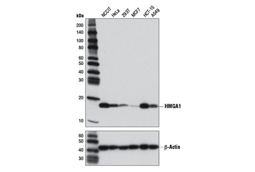
Monoclonal antibody is produced by immunizing animals with synthetic peptides corresponding to residues surrounding Gly68 of human HMGA1, Ala137 of Human HMGB1, Glu169 of human HMGB2, Val32 of human HMGN1, or Asp74 of human HMGN2 protein.
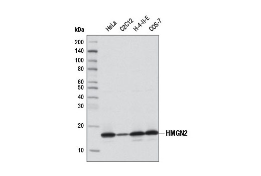
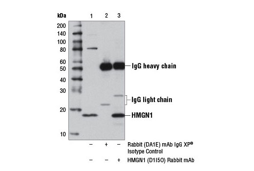
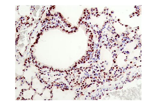
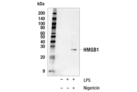
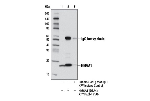
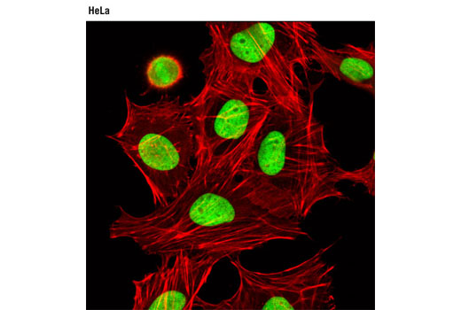
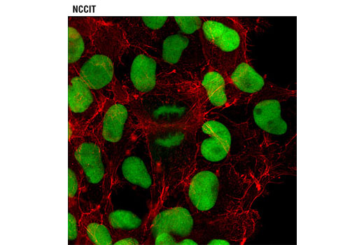
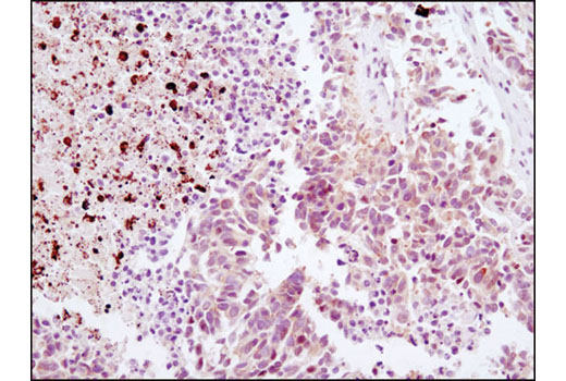
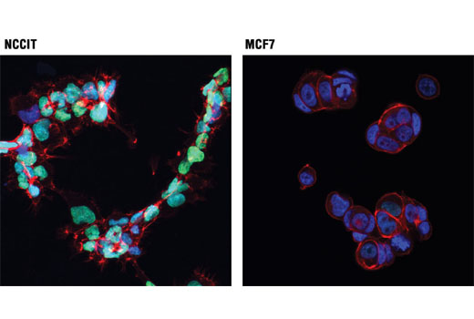
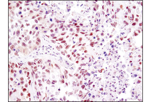
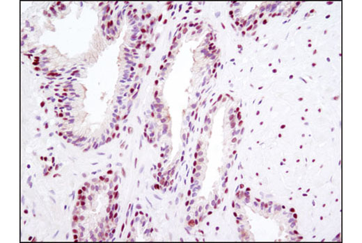
High mobility group (HMG) proteins are a superfamily of abundant and ubiquitous nuclear proteins that bind DNA without sequence specificity and induce structural changes to the chromatin fiber to regulate access to the underlying DNA (1). HMGA1, formerly known as HMG-I/Y, belongs to a family of high mobility group proteins known as HMGA. HMGA proteins are considered architectural transcription factors; they do not have direct transcriptional activation capacity, but instead regulate gene expression by changing DNA conformation through binding to AT-rich regions in the DNA and/or direct interaction with other transcription factors (2). HMGA1 is highly expressed during embryogenesis and in embryonic stem cells, but not in fully differentiated adult tissues (3,4). High mobility group protein B1 (HMGB1) and high mobility group protein B2 (HMGB2) belong to a family of highly conserved proteins that contain HMG box domains (5). HMGB1 is a widely expressed and highly abundant protein (6). HMGB2 is widely expressed during embryonic development, but it is restricted to lymphoid organs and testis in adult animals (7). While expression varies, the biochemical properties of the different family members may be indistinguishable. HMGB proteins are recruited by and help facilitate the assembly of site-specific DNA binding proteins to their cognate binding sites in chromatin. For example, HMGB1 and HMGB2 facilitate the binding of Hox proteins, Oct proteins, p53, Rel proteins, and steroid hormone receptor proteins to their target gene promoters (5,6). In addition to their functions in the nucleus, HMGB proteins play a significant role in extracellular signaling associated with inflammation. HMGB1 is massively released into the extracellular environment during cell necrosis, but not apoptosis. Extracellular HMGB1 "alarms" the innate immune system by acting as a chemoattractant for inflammatory cells triggering activation of T cells and dendritic cells. In addition, activated monocytes, macrophages, and dendritic cells also secrete HMGB1 (6). HMGB2 is secreted by myeloid cells and promotes proliferation and migration of endothelial cells by binding to the receptor for advanced glycation end products (RAGE) (8). The HMGN family of proteins, which includes five members (HMGN1-5) (1) function in transcriptional regulation and are recruited to gene promoters by transcription factors, such as estrogen receptor α (ERα), serum responsive factor (SRF), and PITX2, where they can facilitate either gene activation or repression (9-11). The expression of HMGN1 (also known as HMG14) and HMGN2 (also known as HMG17) is tightly linked to cellular differentiation. HMGN1 and HMNG2 are ubiquitous and highly expressed in all embryonic tissues. During mouse embryogenesis, expression is down-regulated throughout the embryo, except in committed but continuously renewing cell types undergoing active differentiation, such as the basal layer of the epithelium and kidney cells undergoing mesenchyme to epithelium transition (12,13).
Explore pathways related to this product.
STRING - Known and Predicted Protein-Protein Interactions.
Except as otherwise expressly agreed in a writing signed by a legally authorized representative of CST, the following terms apply to Products provided by CST, its affiliates or its distributors. Any Customer's terms and conditions that are in addition to, or different from, those contained herein, unless separately accepted in writing by a legally authorized representative of CST, are rejected and are of no force or effect.
Products are labeled with For Research Use Only or a similar labeling statement and have not been approved, cleared, or licensed by the FDA or other regulatory foreign or domestic entity, for any purpose. Customer shall not use any Product for any diagnostic or therapeutic purpose, or otherwise in any manner that conflicts with its labeling statement. Products sold or licensed by CST are provided for Customer as the end-user and solely for research and development uses. Any use of Product for diagnostic, prophylactic or therapeutic purposes, or any purchase of Product for resale (alone or as a component) or other commercial purpose, requires a separate license from CST. Customer shall (a) not sell, license, loan, donate or otherwise transfer or make available any Product to any third party, whether alone or in combination with other materials, or use the Products to manufacture any commercial products, (b) not copy, modify, reverse engineer, decompile, disassemble or otherwise attempt to discover the underlying structure or technology of the Products, or use the Products for the purpose of developing any products or services that would compete with CST products or services, (c) not alter or remove from the Products any trademarks, trade names, logos, patent or copyright notices or markings, (d) use the Products solely in accordance with CST Product Terms of Sale and any applicable documentation, and (e) comply with any license, terms of service or similar agreement with respect to any third party products or services used by Customer in connection with the Products.