Revision 5
#5692
Store at -20C
877-616-CELL (2355)
877-678-TECH (8324)
3 Trask Lane | Danvers | Massachusetts | 01923 | USA
For Research Use Only. Not for Use in Diagnostic Procedures.
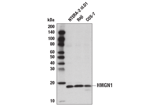
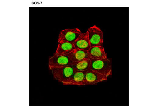
Applications:
W, IF-IC
Reactivity:
H Mk
Sensitivity:
Endogenous
MW (kDa):
18
Source/Isotype:
Rabbit
UniProt ID:
#P05114
Entrez-Gene Id:
3150
Product Usage Information
| Application | Dilution |
|---|---|
| Western Blotting | 1:1000 |
| Immunofluorescence (Immunocytochemistry) | 1:800 - 1:1600 |
Storage
Specificity/Sensitivity
Species predicted to react based on 100% sequence homology
Source / Purification
Background
HMGN1 (also known as HMG14) expression is tightly linked to cellular differentiation. HMGN1 is ubiquitous and highly expressed in all embryonic tissues. During mouse embryogenesis, expression is down-regulated throughout the embryo, except in committed but continuously renewing cell types undergoing active differentiation, such as the basal layer of the epithelium and kidney cells undergoing mesenchyme to epithelium transition (11,12). HMGN1 expression is also down-regulated during myogenesis, erythropoiesis, and osteogenesis (11). Over-expression of HMGN1 inhibits myotube formation in C2C12 myoblast cells and chondrocyte differentiation in primary limb bud mesenchymal cells, suggesting a role in blocking cellular differentiation (11,13). HGMN1-/- mice appear normal, most likely due to partial redundancy with other family members such as HMGN2. However, these mice are hypersensitive to various stress conditions, including exposure to UV light and ionizing radiation (IR) (14,15). Further studies have shown that HMGN1 is required for efficient transcription-coupled repair (TCR) following UV treatment, and proper activation of ATM following IR treatment, both of which require HMGN1 chromatin binding activity, suggesting a direct role for HMGN1 in chromatin remodeling during DNA repair (14-17).
Background References
- Hock, R. et al. (2007) Trends Cell Biol 17, 72-9.
- Gerlitz, G. Biochim Biophys Acta 1799, 80-5.
- Zhu, N. and Hansen, U. (2007) Mol Cell Biol 27, 8859-73.
- Amen, M. et al. (2008) Nucleic Acids Res 36, 462-76.
- Belova, G.I. et al. (2008) J Biol Chem 283, 8080-8.
- Catez, F. et al. (2002) EMBO Rep 3, 760-6.
- Lim, J.H. et al. (2005) EMBO J 24, 3038-48.
- Lim, J.H. et al. (2004) Mol Cell 15, 573-84.
- Postnikov, Y.V. et al. (2006) Biochemistry 45, 15092-9.
- Rattner, B.P. et al. (2009) Mol Cell 34, 620-6.
- Furusawa, T. et al. (2006) Mol Cell Biol 26, 592-604.
- Lehtonen, S. and Lehtonen, E. (2001) Differentiation 67, 154-63.
- Pash, J.M. et al. (1993) J Biol Chem 268, 13632-8.
- Birger, Y. et al. (2003) EMBO J 22, 1665-75.
- Birger, Y. et al. (2005) Cancer Res 65, 6711-8.
- Fousteri, M. et al. (2006) Mol Cell 23, 471-82.
- Kim, Y.C. et al. (2009) Nat Cell Biol 11, 92-6.
Species Reactivity
Species reactivity is determined by testing in at least one approved application (e.g., western blot).
Western Blot Buffer
IMPORTANT: For western blots, incubate membrane with diluted primary antibody in 5% w/v BSA, 1X TBS, 0.1% Tween® 20 at 4°C with gentle shaking, overnight.
Applications Key
W: Western Blotting IF-IC: Immunofluorescence (Immunocytochemistry)
Cross-Reactivity Key
H: Human Mk: Monkey
Trademarks and Patents
Cell Signaling Technology is a trademark of Cell Signaling Technology, Inc.
All other trademarks are the property of their respective owners. Visit cellsignal.com/trademarks for more information.
Limited Uses
Except as otherwise expressly agreed in a writing signed by a legally authorized representative of CST, the following terms apply to Products provided by CST, its affiliates or its distributors. Any Customer's terms and conditions that are in addition to, or different from, those contained herein, unless separately accepted in writing by a legally authorized representative of CST, are rejected and are of no force or effect.
Products are labeled with For Research Use Only or a similar labeling statement and have not been approved, cleared, or licensed by the FDA or other regulatory foreign or domestic entity, for any purpose. Customer shall not use any Product for any diagnostic or therapeutic purpose, or otherwise in any manner that conflicts with its labeling statement. Products sold or licensed by CST are provided for Customer as the end-user and solely for research and development uses. Any use of Product for diagnostic, prophylactic or therapeutic purposes, or any purchase of Product for resale (alone or as a component) or other commercial purpose, requires a separate license from CST. Customer shall (a) not sell, license, loan, donate or otherwise transfer or make available any Product to any third party, whether alone or in combination with other materials, or use the Products to manufacture any commercial products, (b) not copy, modify, reverse engineer, decompile, disassemble or otherwise attempt to discover the underlying structure or technology of the Products, or use the Products for the purpose of developing any products or services that would compete with CST products or services, (c) not alter or remove from the Products any trademarks, trade names, logos, patent or copyright notices or markings, (d) use the Products solely in accordance with CST Product Terms of Sale and any applicable documentation, and (e) comply with any license, terms of service or similar agreement with respect to any third party products or services used by Customer in connection with the Products.
Revision 5
#5692
HMGN1 Antibody