Revision 1
#43049
Store at -20C
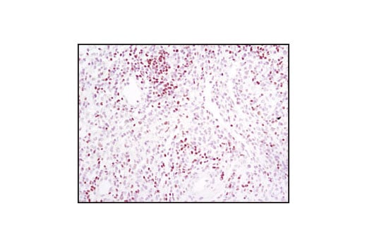
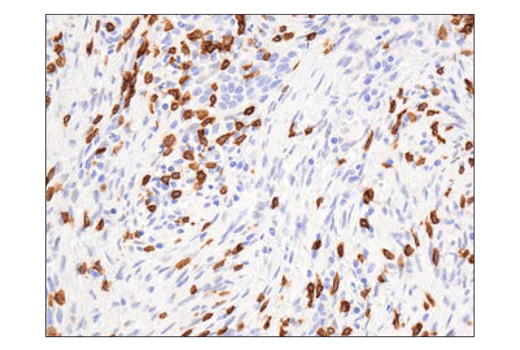
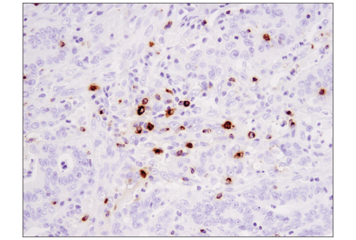
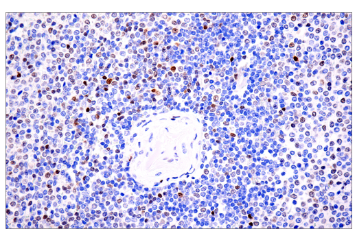
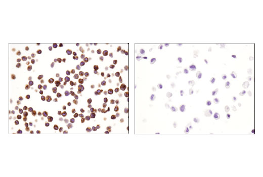
Human Exhausted CD8+ T Cell IHC Antibody Sampler Kit
1 Kit
(9 x 20 microliters)
877-616-CELL (2355)
877-678-TECH (8324)
3 Trask Lane | Danvers | Massachusetts | 01923 | USA
For Research Use Only. Not for Use in Diagnostic Procedures.
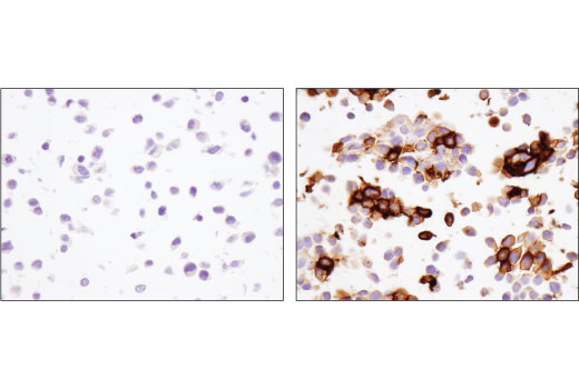
| Product Includes | Product # | Quantity | Mol. Wt | Isotype/Source |
|---|---|---|---|---|
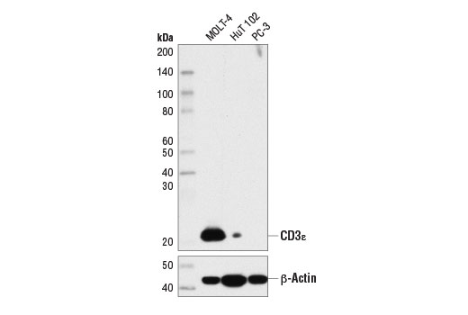
| CD3 epsilon (D7A6E) Rabbit Monoclonal Antibody | 85061 | 20 µl | 23 kDa | Rabbit IgG |
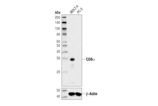
| CD8 alpha (D8A8Y) Rabbit Monoclonal Antibody | 85336 | 20 µl | 29 kDa | Rabbit IgG |
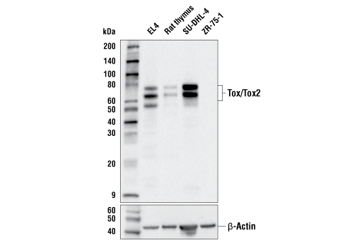
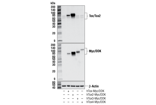
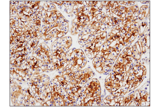
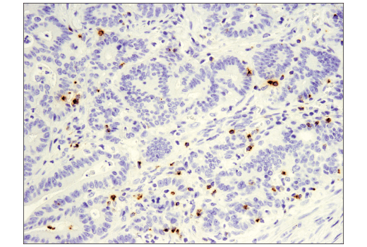
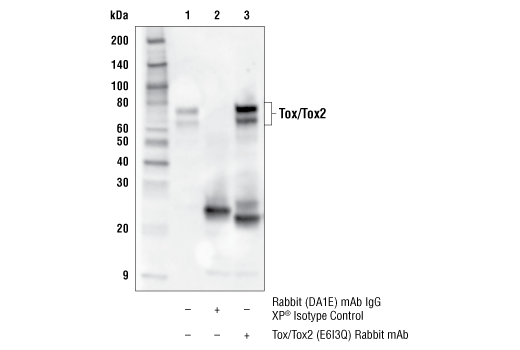
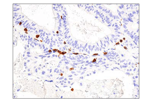
| Tox/Tox2 (E6I3Q) Rabbit Monoclonal Antibody | 73758 | 20 µl | 60-80 kDa | Rabbit IgG |
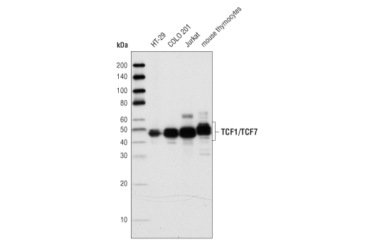
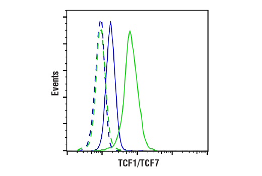
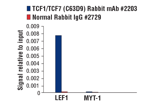
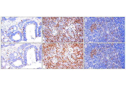
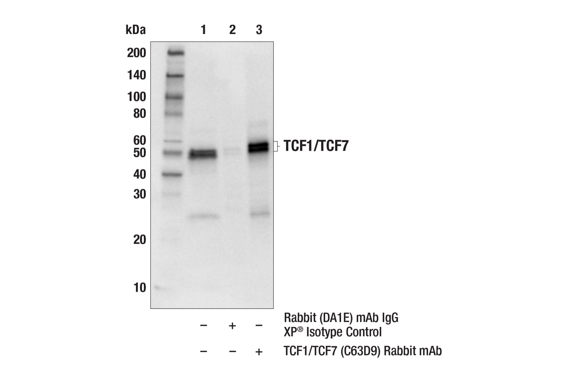
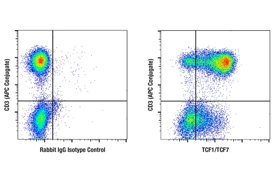
| TCF1/TCF7 (C63D9) Rabbit Monoclonal Antibody | 2203 | 20 µl | 48, 50 kDa | Rabbit IgG |
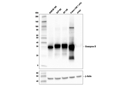
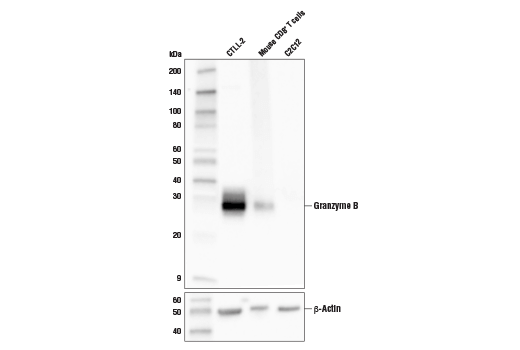
| Granzyme B (D6E9W) Rabbit Monoclonal Antibody | 46890 | 20 µl | 30 kDa | Rabbit IgG |
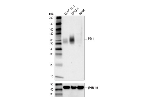
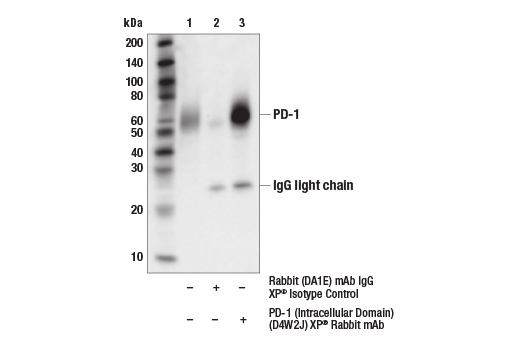
| PD-1 (Intracellular Domain) (D4W2J) Rabbit Monoclonal Antibody | 86163 | 20 µl | 52-65 kDa | Rabbit IgG |
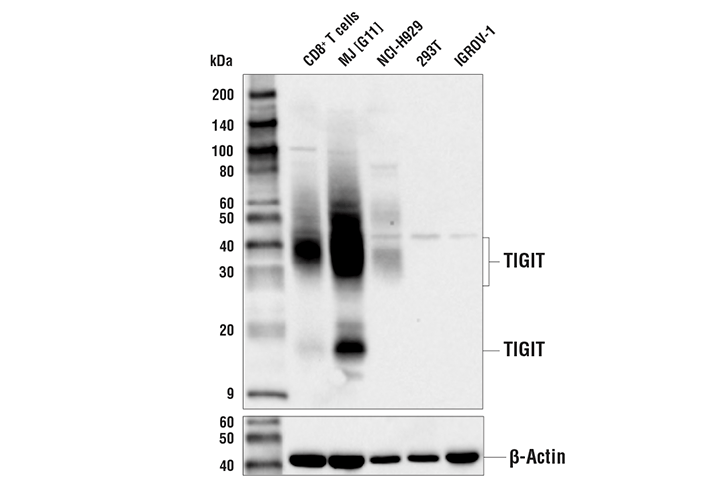
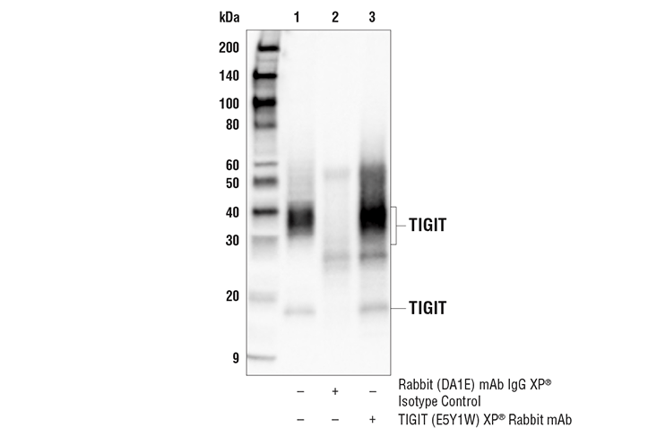
| TIGIT (E5Y1W) Rabbit Monoclonal Antibody | 99567 | 20 µl | 18, 30-40 kDa | Rabbit IgG |
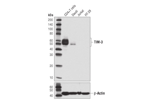
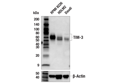
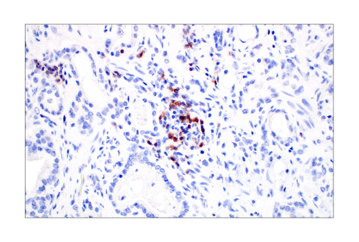
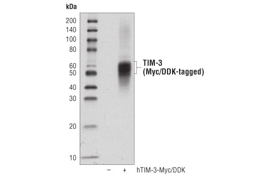
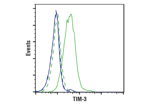
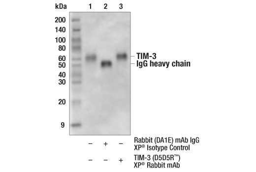
| TIM-3 (D5D5R) Rabbit Monoclonal Antibody | 45208 | 20 µl | 45-70 kDa | Rabbit IgG |
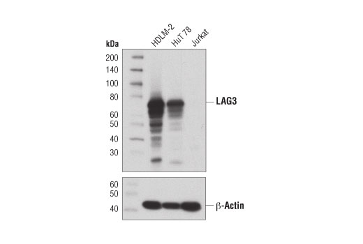
| LAG3 (D2G4O) Rabbit Monoclonal Antibody | 15372 | 20 µl | 60-80 kDa | Rabbit IgG |
| Anti-rabbit IgG, HRP-linked Antibody | 7074 | 100 µl | Goat |
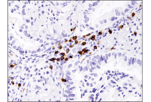
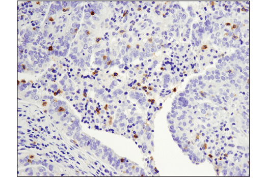
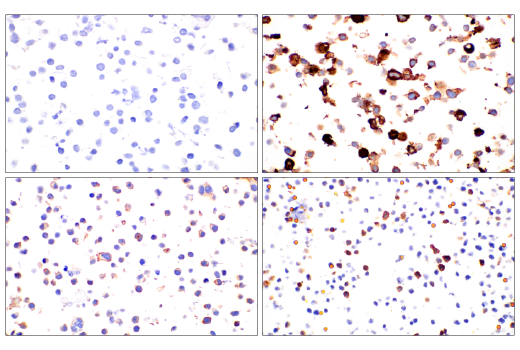
Please visit cellsignal.com for individual component applications, species cross-reactivity, dilutions, protocols, and additional product information.
Description
Storage
Background
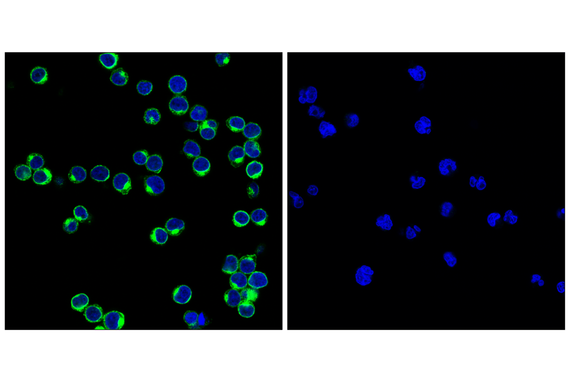
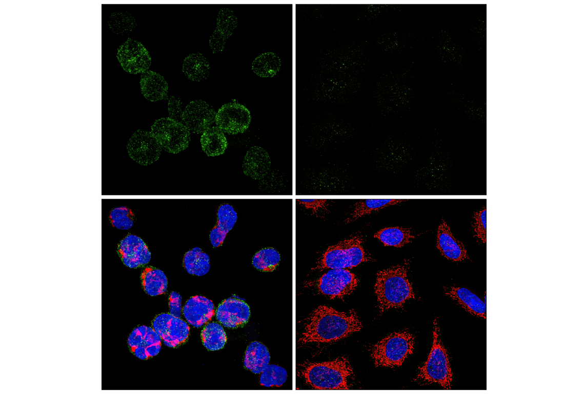
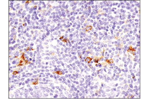
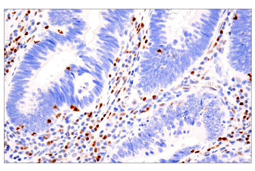
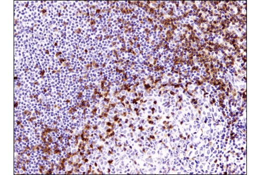
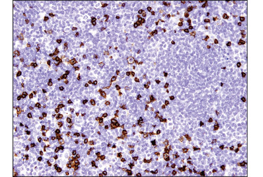
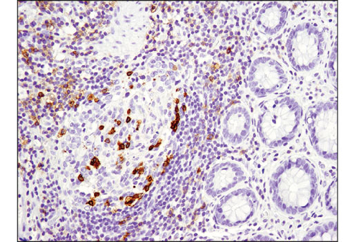
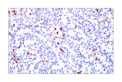
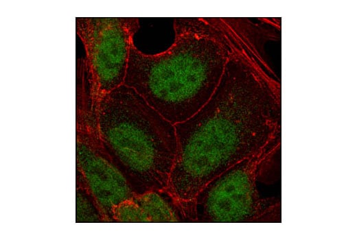
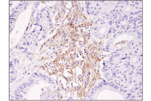
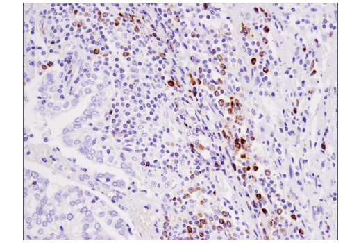
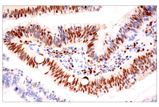
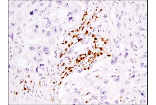
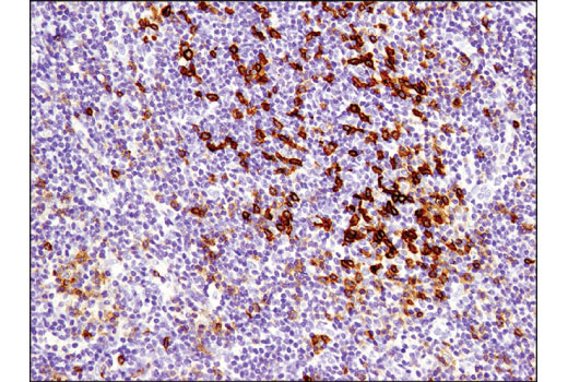
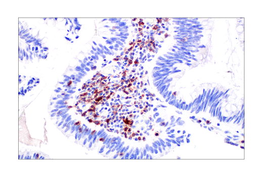
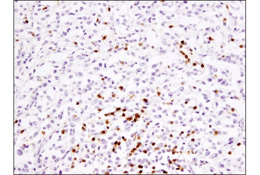
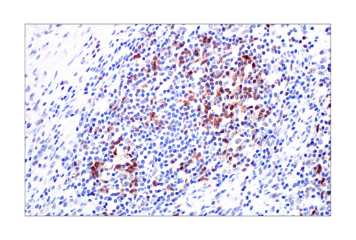
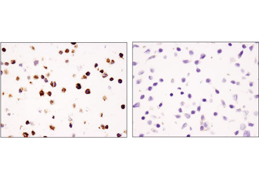
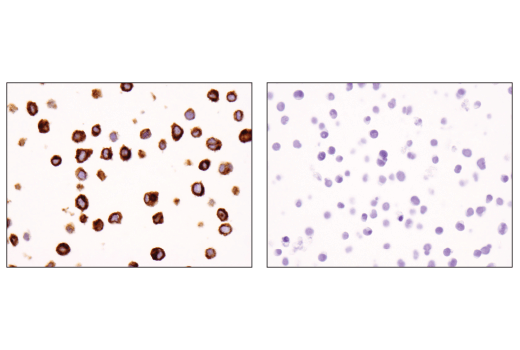
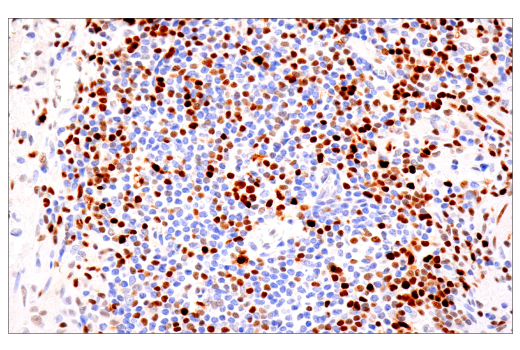
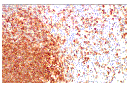
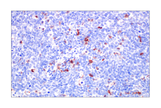
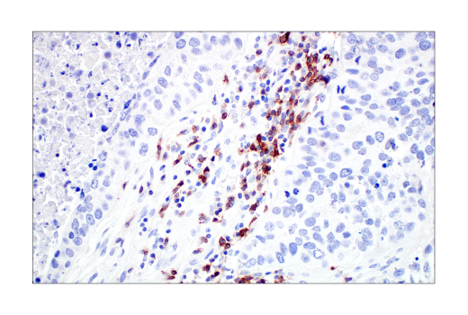
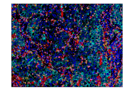
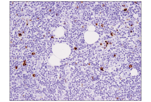
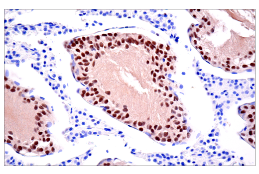
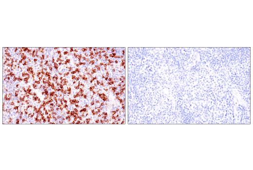
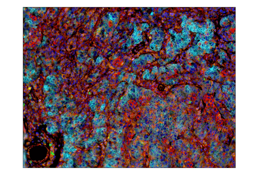
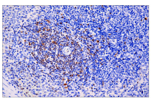
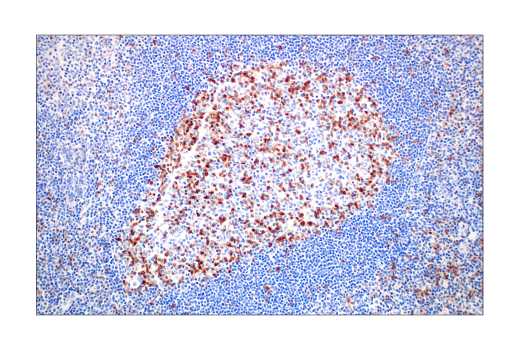
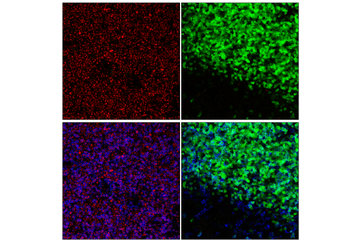
Tox, Tox2, and TCF1/TCF7 play key roles in T cell development. Tox is also induced by high antigen stimulation during chronic viral infection or cancer, regulating T cell persistence and exhaustion. TCF1/TCF7 preserves the effector function of exhausted T cells during viral infection or cancer. EOMES is a key transcription factor for memory T cells and for full effector differentiation of CD8+ T cells. The dynamic expression of these transcription factors help characterize the extent to which a T cell is exhausted and will respond to antigen stimulation (4-8). Granzyme B is a serine protease expressed by cytotoxic T lymphocytes and natural killer (NK) cells and is a key component of immune responses to pathogens and transformed cells (9).
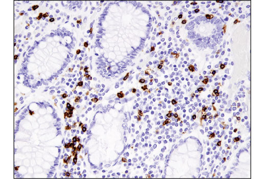
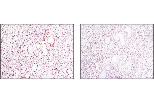
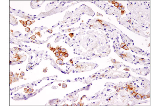
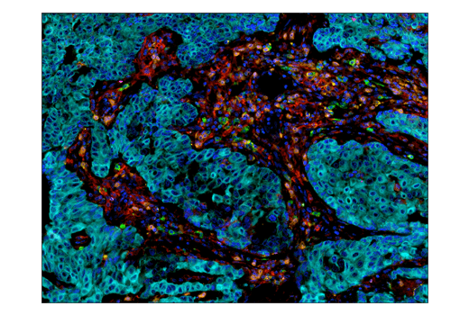
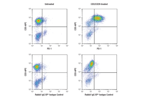
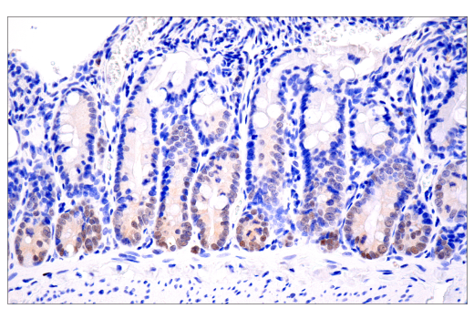
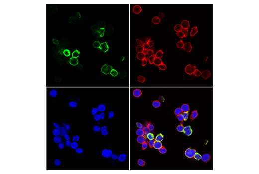
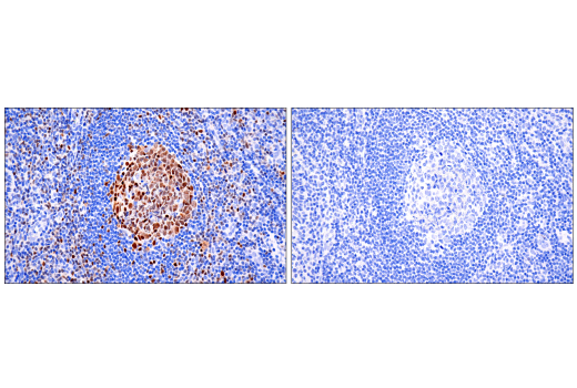
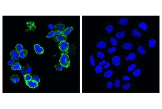
PD-1 (PDCD1, CD279), TIGIT (VSIG9, VSTM3), TIM-3 (HAVCR2), and LAG3 (CD223) are immune cell co-inhibitory receptors (also known as immune checkpoints) that negatively regulate T cell function and dampen the immune response to pathogens and cancer (10-15). In addition to activated T cells, PD-1 is expressed by activated B cells and monocytes. Following interaction with its ligands, PD-L1 and PD-L2, PD-1 is phosphorylated at ITIM and ITSM motifs leading to recruitment of protein tyrosine phosphatases SHP-1 and SHP-2 and suppression of TCR signaling. TIGIT is expressed at low levels on subsets of T cells and NK cells, and is upregulated at the protein level following activation of these cells. TIGIT marks exhausted T cells in the tumor microenvironment and during human immunodeficiency virus (HIV) infection. TIM-3 is expressed by exhausted T cells in the settings of chronic infection and cancer. Tumor-infiltrating macrophages and dendritic cells also express TIM-3. LAG3 is primarily expressed by activated CD4+ T cells, CD8+ T cells, FoxP3+ T regulatory cells (Tregs), and natural killer (NK) cells. Co-expression of multiple immune checkpoints help characterize the extent to which a T cell is exhausted and will respond to antigen stimulation. Therapeutic blockade of several of these immune checkpoint receptors is a promising strategy for neoplastic intervention by enabling anti-tumor immune responses (10-15).
Background References
- Kuhns, M.S. et al. (2006) Immunity 24, 133-9.
- Zamoyska, R. (1994) Immunity 1, 243-6.
- Shortman, K. and Heath, W.R. (2010) Immunol Rev 234, 18-31.
- Aliahmad, P. et al. (2012) Curr Opin Immunol 24, 173-7.
- Yao, C. et al. (2019) Nat Immunol 20, 890-901.
- Alfei, F. et al. (2019) Nature 571, 265-269.
- Seo, H. et al. (2019) Proc Natl Acad Sci U S A 116, 12410-12415.
- Wang, Y. et al. (2019) Front Immunol 10, 169.
- Trapani, J.A. (2001) Genome Biol 2, REVIEWS3014.
- Schildberg, F.A. et al. (2016) Immunity 44, 955-72.
- Anderson, A.C. et al. (2016) Immunity 44, 989-1004.
- Callahan, M.K. et al. (2016) Immunity 44, 1069-78.
- Chen, L. and Flies, D.B. (2013) Nat Rev Immunol 13, 227-42.
- Chauvin, J.M. et al. (2015) J Clin Invest 125, 2046-58.
- Chew, G.M. et al. (2016) PLoS Pathog 12, e1005349.
Trademarks and Patents
Cell Signaling Technology is a trademark of Cell Signaling Technology, Inc.
U.S. Patent No. 7,429,487, foreign equivalents, and child patents deriving therefrom.
All other trademarks are the property of their respective owners. Visit cellsignal.com/trademarks for more information.
Limited Uses
Except as otherwise expressly agreed in a writing signed by a legally authorized representative of CST, the following terms apply to Products provided by CST, its affiliates or its distributors. Any Customer's terms and conditions that are in addition to, or different from, those contained herein, unless separately accepted in writing by a legally authorized representative of CST, are rejected and are of no force or effect.
Products are labeled with For Research Use Only or a similar labeling statement and have not been approved, cleared, or licensed by the FDA or other regulatory foreign or domestic entity, for any purpose. Customer shall not use any Product for any diagnostic or therapeutic purpose, or otherwise in any manner that conflicts with its labeling statement. Products sold or licensed by CST are provided for Customer as the end-user and solely for research and development uses. Any use of Product for diagnostic, prophylactic or therapeutic purposes, or any purchase of Product for resale (alone or as a component) or other commercial purpose, requires a separate license from CST. Customer shall (a) not sell, license, loan, donate or otherwise transfer or make available any Product to any third party, whether alone or in combination with other materials, or use the Products to manufacture any commercial products, (b) not copy, modify, reverse engineer, decompile, disassemble or otherwise attempt to discover the underlying structure or technology of the Products, or use the Products for the purpose of developing any products or services that would compete with CST products or services, (c) not alter or remove from the Products any trademarks, trade names, logos, patent or copyright notices or markings, (d) use the Products solely in accordance with CST Product Terms of Sale and any applicable documentation, and (e) comply with any license, terms of service or similar agreement with respect to any third party products or services used by Customer in connection with the Products.
Revision 1
Revision 1

Revision 1
Revision 1

Revision 1
Revision 1
Revision 1

Revision 1
Revision 1
Revision 1

Revision 1
Revision 1
Revision 1

Revision 1
Revision 1

Revision 1

Revision 1


Revision 1

Revision 1
Revision 1
Revision 1


Revision 1
Revision 1

Revision 1
Revision 1
Revision 1
Revision 1

Revision 1
Revision 1
Revision 1
Revision 1