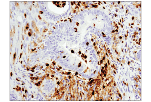
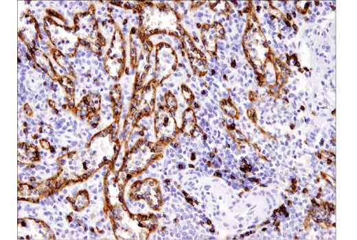
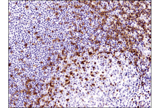
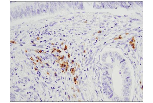
| Cat. # | Size | Qty. | Price |
|---|---|---|---|
| 62714T | 1 Kit (9 x 20 microliters) |
|
| Product Includes | Quantity | Applications | Reactivity | MW(kDa) | Isotype |
|---|---|---|---|---|---|
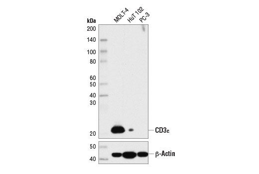
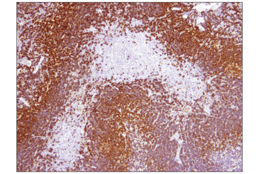
| CD3ε (D7A6E™) XP® Rabbit mAb 85061 | 20 µl |
|
H Mk | 23 | Rabbit IgG |
| CD8α (C8/144B) Mouse mAb 70306 | 20 µl |
|
H | Mouse IgG1 | |
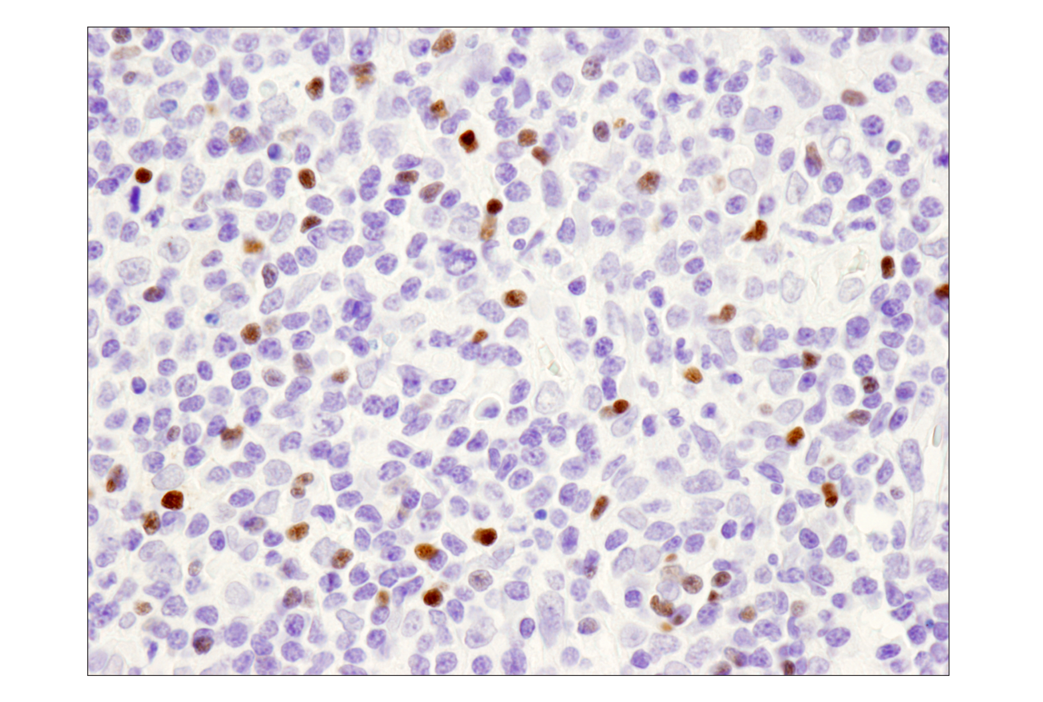
| FoxP3 (D2W8E™) Rabbit mAb 98377 | 20 µl |
|
H Mk | 45 | Rabbit IgG |
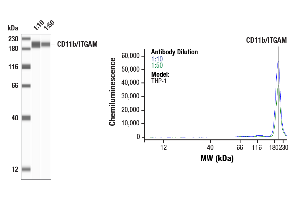
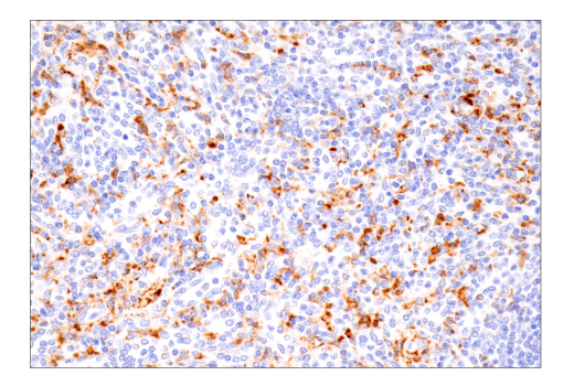
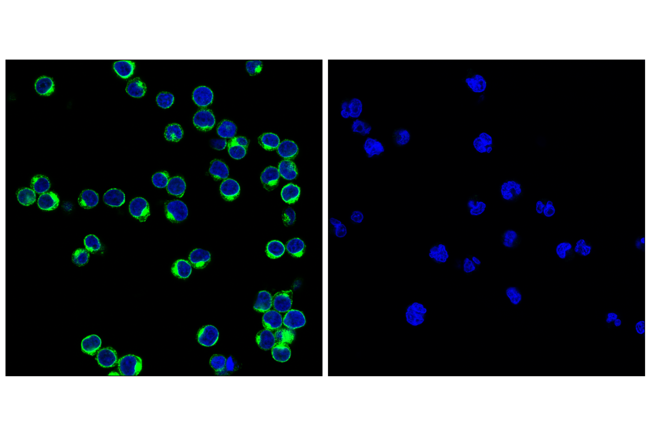
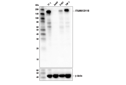
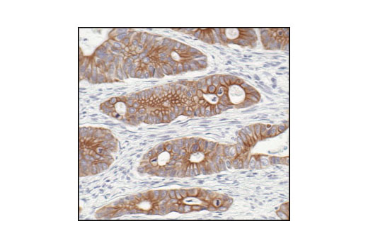
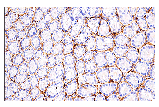
| CD11b/ITGAM (D6X1N) Rabbit mAb 49420 | 20 µl |
|
H Mk | 170 | Rabbit IgG |
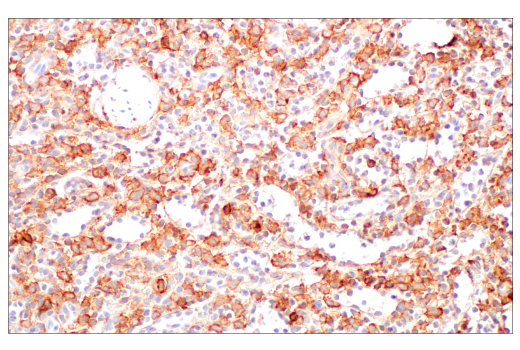
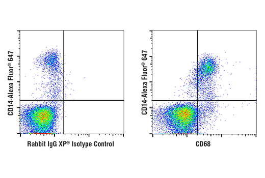
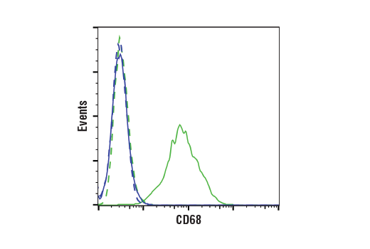
| CD68 (D4B9C) XP® Rabbit mAb 76437 | 20 µl |
|
H Mk | Rabbit IgG | |
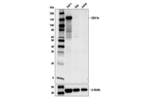
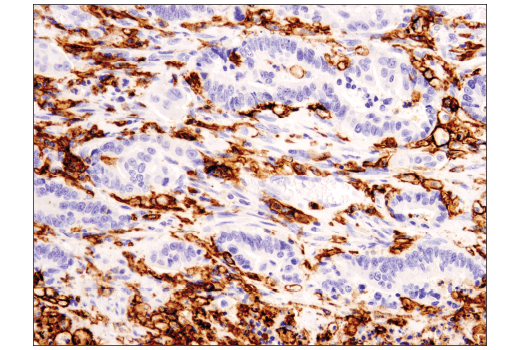
| CD11c (D3V1E) XP® Rabbit mAb 45581 | 20 µl |
|
H | 145 | Rabbit IgG |
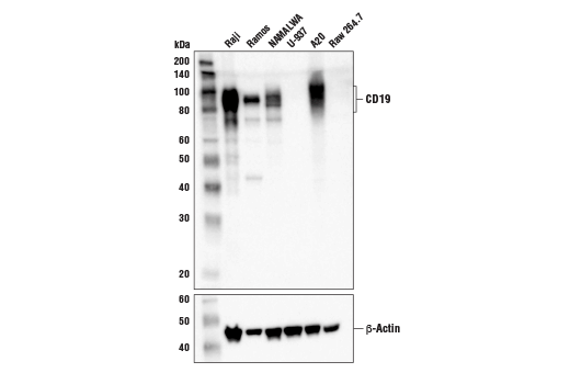
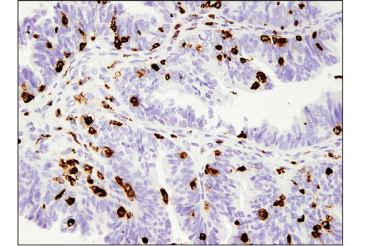
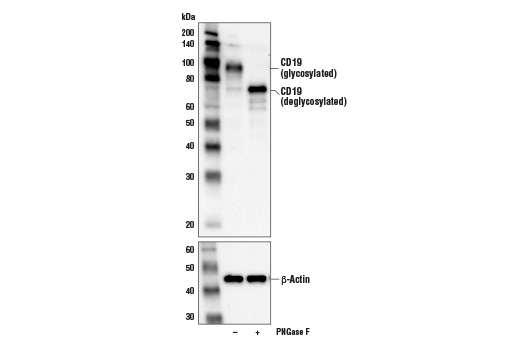
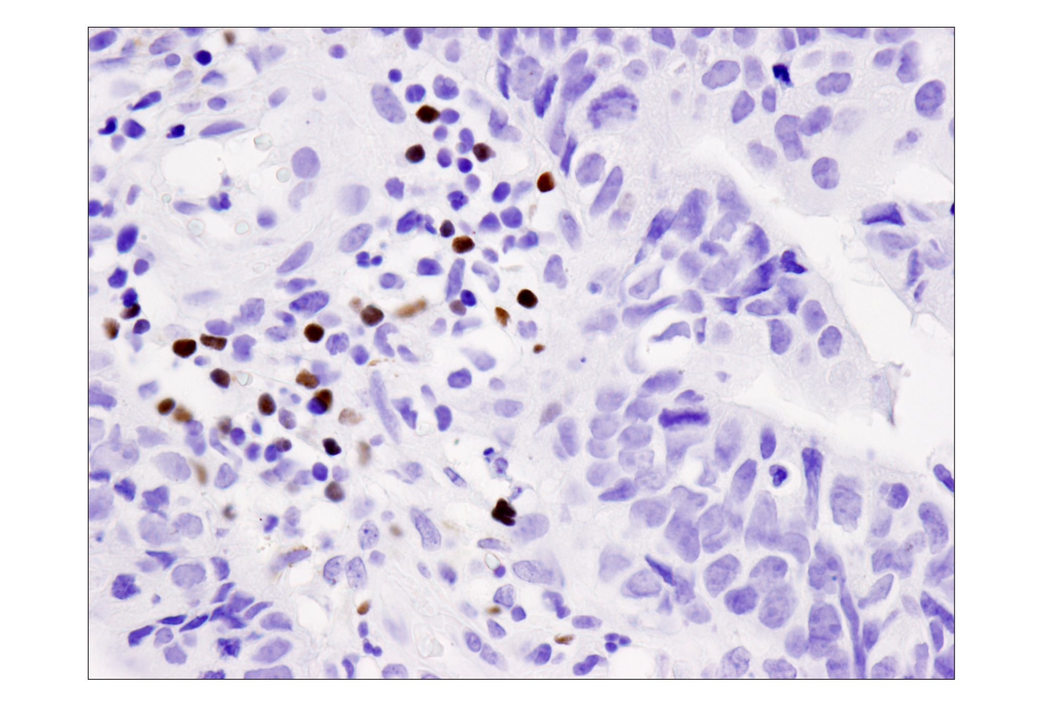
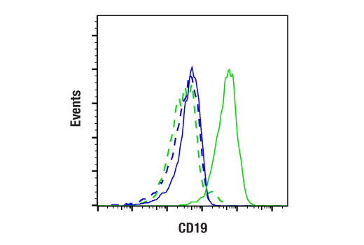
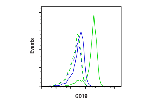
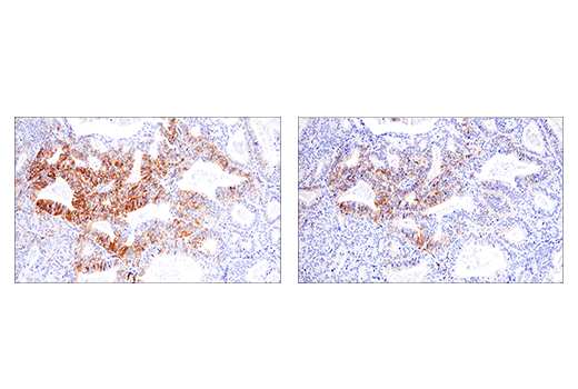
| CD19 (Intracellular Domain) (D4V4B) XP® Rabbit mAb 90176 | 20 µl |
|
H M Mk | 95 | Rabbit IgG |
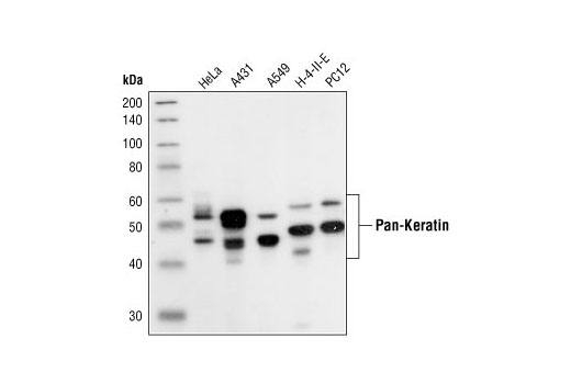
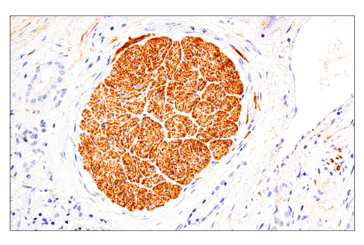
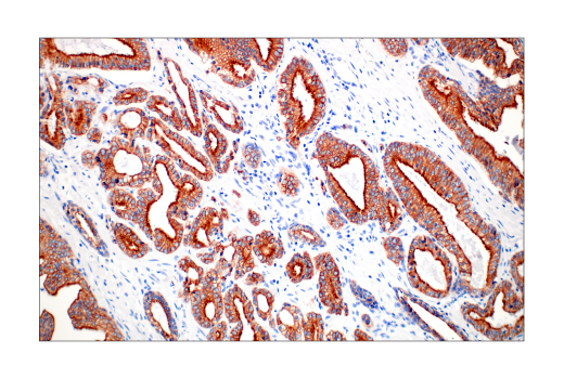
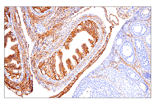
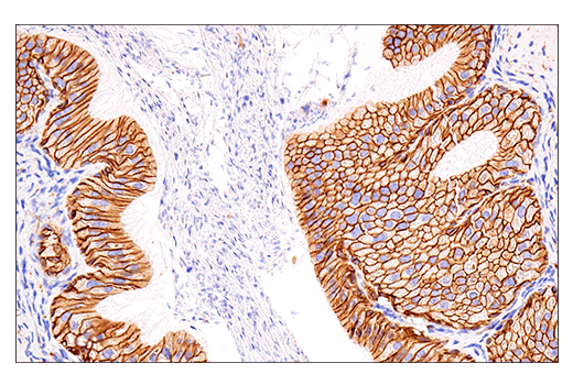
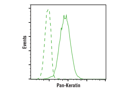
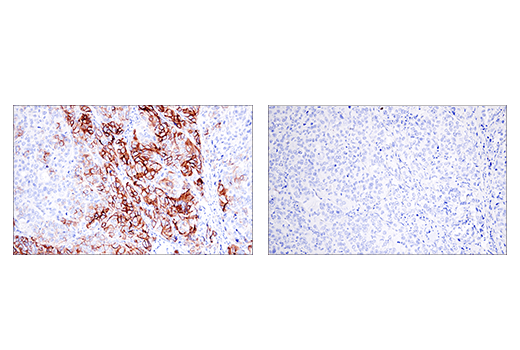
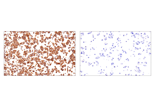
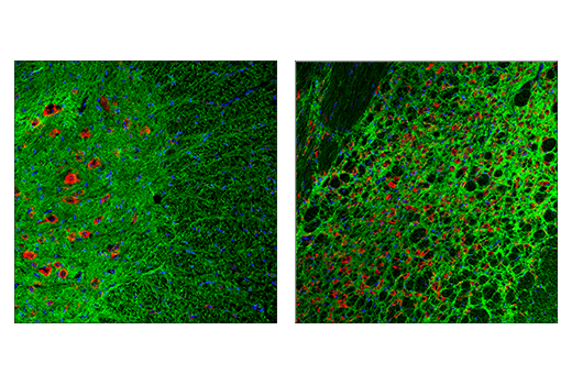
| Pan-Keratin (C11) Mouse mAb 4545 | 20 µl |
|
H M R Mk | 46-58 | Mouse IgG1 |
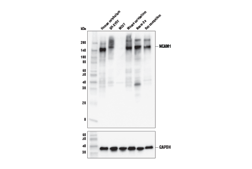
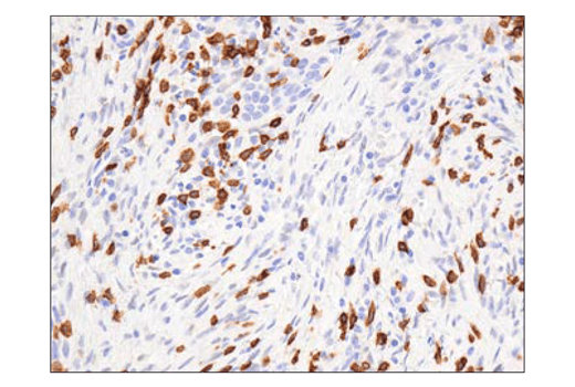
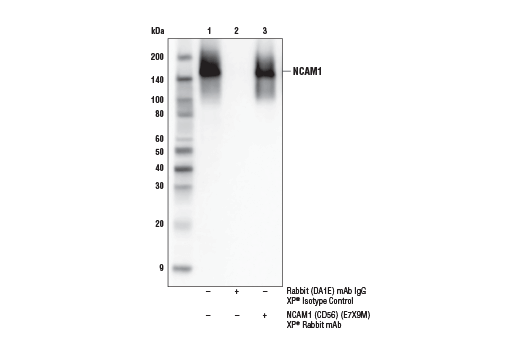
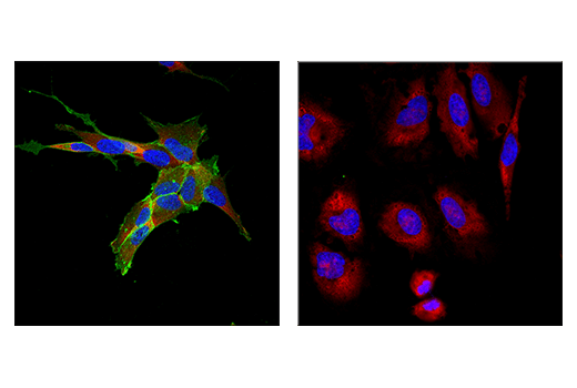
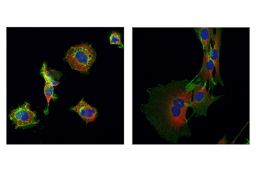
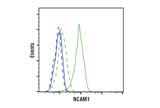
| NCAM1 (CD56) (E7X9M) XP® Rabbit mAb 99746 | 20 µl |
|
H M R Mk | 120 to 220 | Rabbit IgG |
Product Information
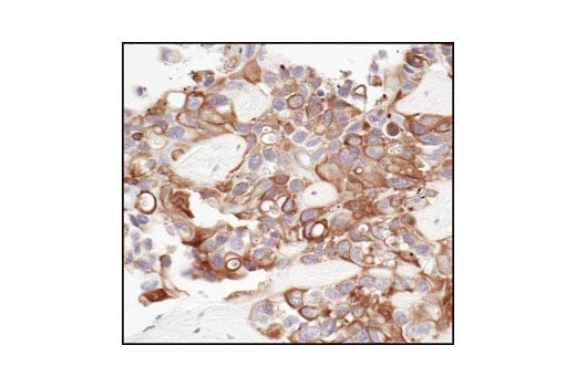
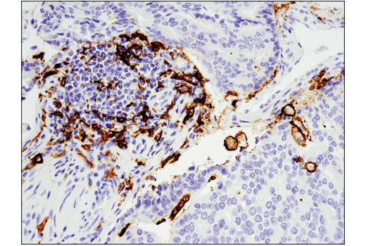
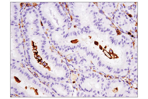
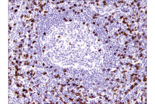
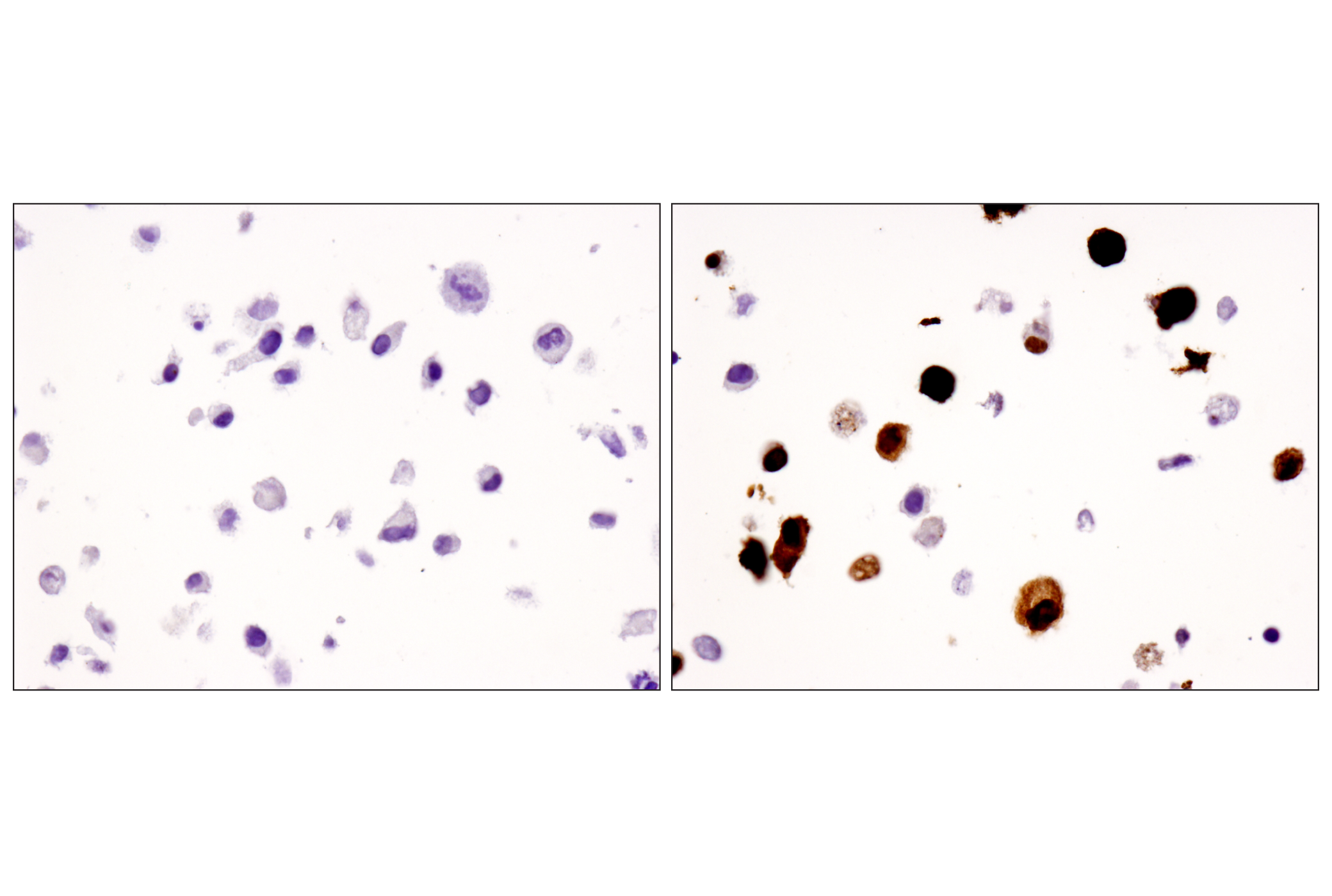
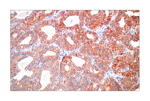
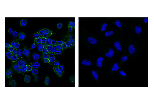
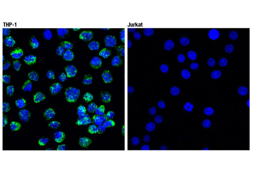
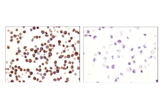
Monoclonal antibodies are produced by immunizing animals with synthetic peptides corresponding to residues surrounding Glu178 of human CD3ε protein, Leu427 of human CD19 protein, Pro799 of human NCAM1/CD56 protein, near the carboxy terminus of human CD8 protein, or with recombinant protein specific to human FoxP3, human CD68, human CD11c, or the amino terminus of human CD11b/ITGAM protein. Pan-Keratin (C11) Mouse mAb (isotype: IgG1) is produced by immunizing a BALB/c mouse with a cytoskeleton preparation from A431 cells.
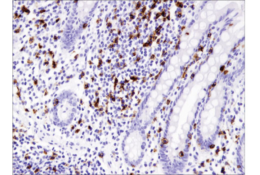
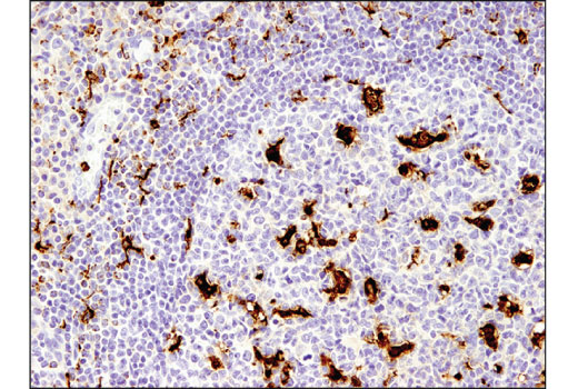
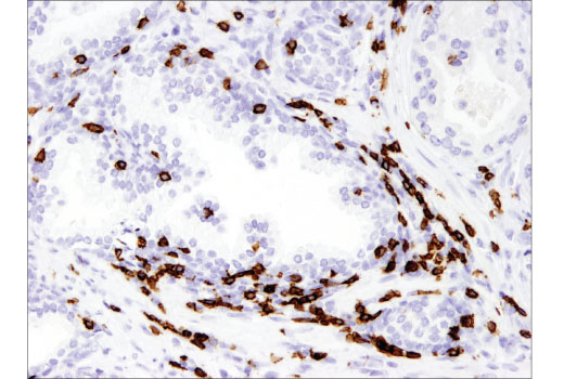
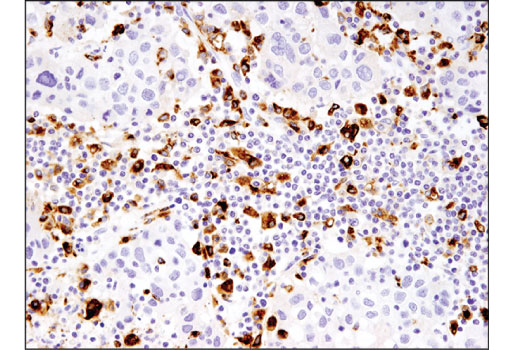
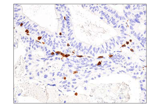
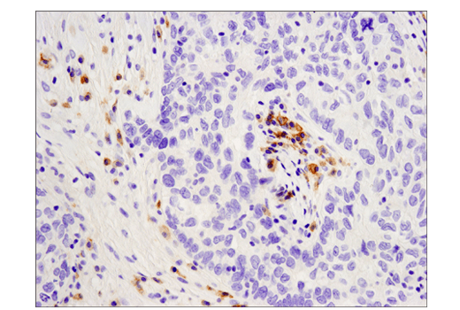
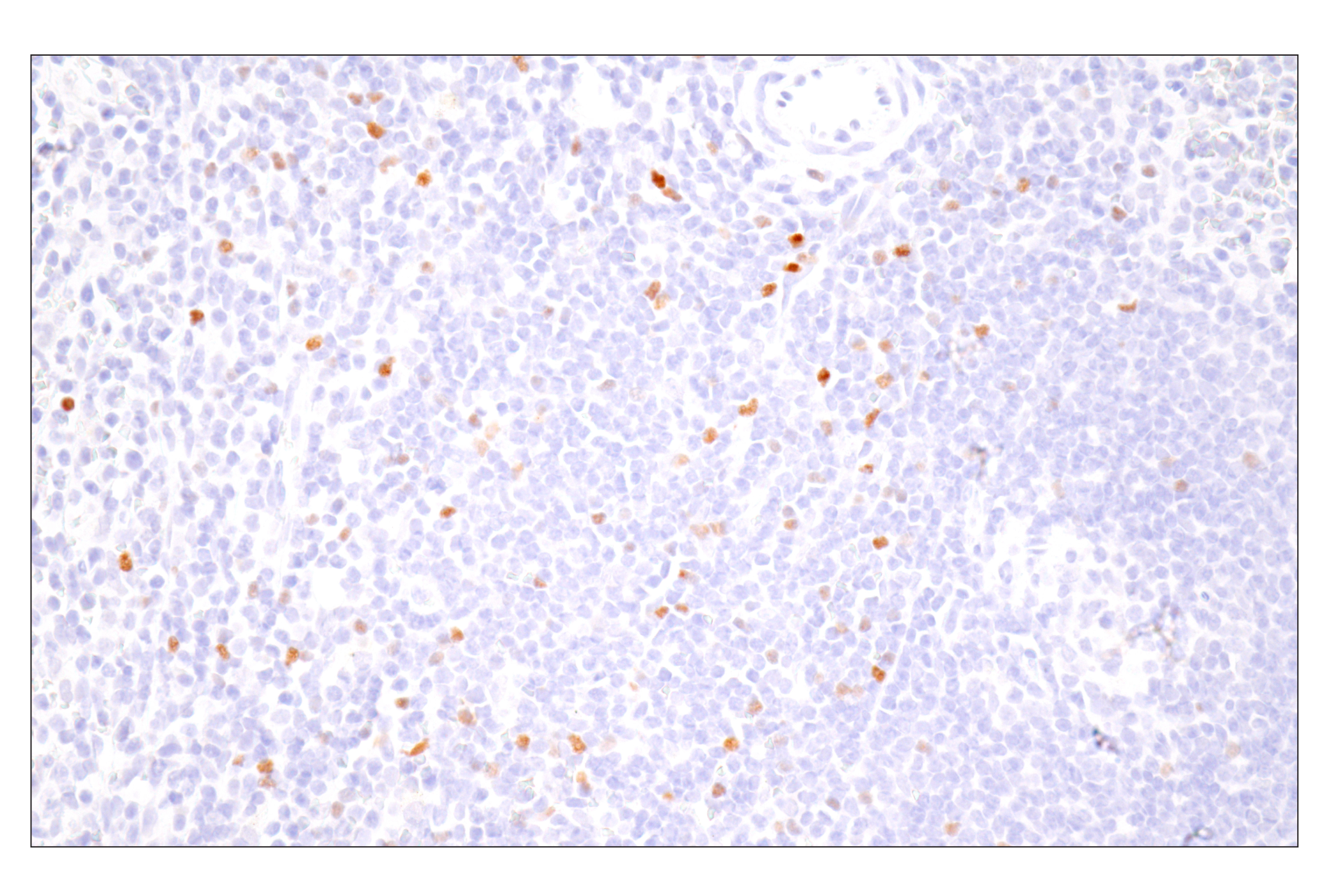
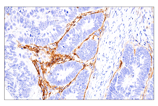
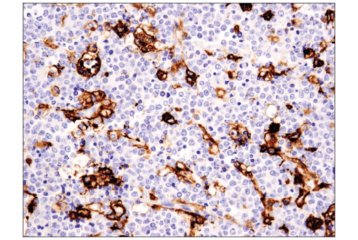
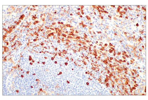
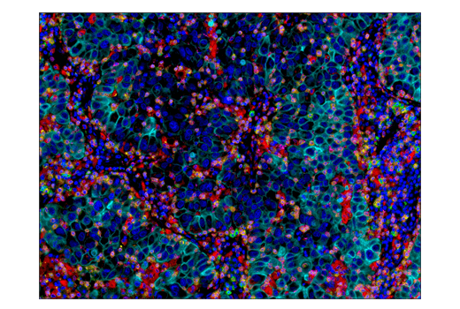
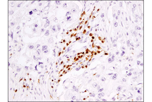
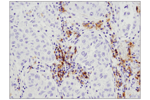
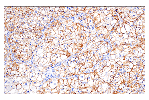
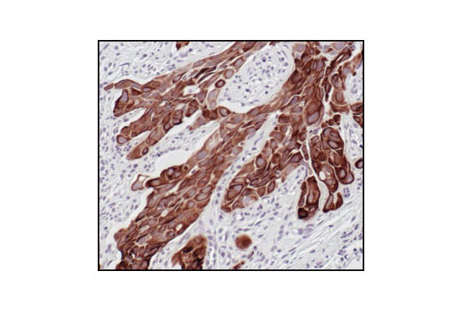
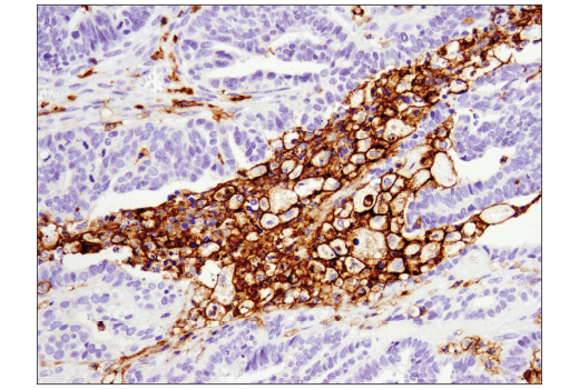
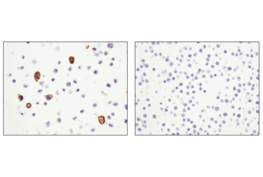
Cluster of Differentiation 3 (CD3) is a multiunit protein complex expressed on the surface of T cells that directly associates with the T cell receptor (TCR). CD3 is composed of four polypeptides: ζ, γ, ε and δ. Engagement of the TCR complex with antigens presented in Major Histocompatibility Complexes (MHC) induces tyrosine phosphorylation in the immunoreceptor tyrosine-based activation motif (ITAM) of CD3 proteins. CD3 phosphorylation is required for downstream signaling through ZAP-70 and p85 subunit of PI-3 kinase, leading to T cell activation, proliferation, and effector functions (1). CD8 is a transmembrane glycoprotein expressed primarily on cytotoxic T cells, but has also been described on a subset of dendritic cells in mice (2,3). On T cells, CD8 is a co-receptor for the TCR, and these two distinct structures are required to recognize antigen bound to MHC Class I. CD8 ensures specificity of the TCR–antigen interaction, prolongs the contact between the T cell and the antigen presenting cell, and recruits the tyrosine kinase Lck, which is essential for T cell activation (2). Forkhead box P3 (FoxP3) is crucial for the development of T cells with immunosuppressive regulatory properties and is a well-established marker for CD4+ T regulatory cells (Tregs) (4). Cluster of differentiation molecule 11b (CD11b)/Integrin alpha M (ITGAM) is a transmembrane protein forming heterodimers that are composed of α and β subunits (5). CD11b is expressed by, and commonly used as a marker for, myeloid lineage cells, including neutrophils, monocytes, macrophages, and microglia (6). CD68 (macrosialin) is a heavily glycosylated transmembrane protein that is expressed by and commonly used as a marker for monocytes and macrophages (7,8). It is found on the plasma membrane, as well as endosomal and lysosomal membranes (7-9). CD11c (integrin αX, ITGAX) is a transmembrane glycoprotein highly expressed by dendritic cells, and has also been observed on activated NK cells, subsets of B and T cells, monocytes, granulocytes, and some B cell malignancies including hairy cell leukemia (10,11). CD19 is a co-receptor expressed on B cells that amplifies the signaling cascade initiated by the B cell receptor (BCR) to induce activation. It is a biomarker of B lymphocyte development, lymphoma diagnosis, and can be utilized as a target for leukemia immunotherapies (12,13). NCAM (neural cell adhesion molecule, CD56) is an adhesion glycoprotein with five extracellular immunoglobulin-like domains followed by two fibronectin type III repeats (14). CD56 and CD16 are commonly used to identify NK cells although some cells with the T cell markers CD3 and CD4 also express CD56 (15). Keratins (cytokeratins) are intermediate filament proteins that are mainly expressed in epithelial cells. Keratin heterodimers composed of an acidic keratin (or type I keratin, keratins 9 to 23) and a basic keratin (or type II keratin, keratins 1 to 8) assemble to form filaments (16,17). Keratin isoforms demonstrate tissue- and differentiation-specific profiles that make them useful as research biomarkers (16).
Explore pathways related to this product.
STRING - Known and Predicted Protein-Protein Interactions.
UniProt ID: P15391 , P07766 , P48668 , P13645 , P04259 , P05787 , P13646 , P11215 , P02538 , P20702 , P05783 , P13647 , P19013 , P01732 , P13591 , Q9BZS1 , P34810
Entrez-Gene Id: 930 , 916 , 286887 , 3858 , 3854 , 3856 , 3860 , 3684 , 3853 , 3687 , 3875 , 3852 , 3851 , 925 , 4684 , 50943 , 968
Except as otherwise expressly agreed in a writing signed by a legally authorized representative of CST, the following terms apply to Products provided by CST, its affiliates or its distributors. Any Customer's terms and conditions that are in addition to, or different from, those contained herein, unless separately accepted in writing by a legally authorized representative of CST, are rejected and are of no force or effect.
Products are labeled with For Research Use Only or a similar labeling statement and have not been approved, cleared, or licensed by the FDA or other regulatory foreign or domestic entity, for any purpose. Customer shall not use any Product for any diagnostic or therapeutic purpose, or otherwise in any manner that conflicts with its labeling statement. Products sold or licensed by CST are provided for Customer as the end-user and solely for research and development uses. Any use of Product for diagnostic, prophylactic or therapeutic purposes, or any purchase of Product for resale (alone or as a component) or other commercial purpose, requires a separate license from CST. Customer shall (a) not sell, license, loan, donate or otherwise transfer or make available any Product to any third party, whether alone or in combination with other materials, or use the Products to manufacture any commercial products, (b) not copy, modify, reverse engineer, decompile, disassemble or otherwise attempt to discover the underlying structure or technology of the Products, or use the Products for the purpose of developing any products or services that would compete with CST products or services, (c) not alter or remove from the Products any trademarks, trade names, logos, patent or copyright notices or markings, (d) use the Products solely in accordance with CST Product Terms of Sale and any applicable documentation, and (e) comply with any license, terms of service or similar agreement with respect to any third party products or services used by Customer in connection with the Products.