Revision 1
#92114
Store at -20C
Human TREM2 Activity Antibody Sampler Kit
1 Kit
(6 x 20 microliters)
877-616-CELL (2355)
877-678-TECH (8324)
3 Trask Lane | Danvers | Massachusetts | 01923 | USA
For Research Use Only. Not for Use in Diagnostic Procedures.
| Product Includes | Product # | Quantity | Mol. Wt | Isotype/Source |
|---|---|---|---|---|
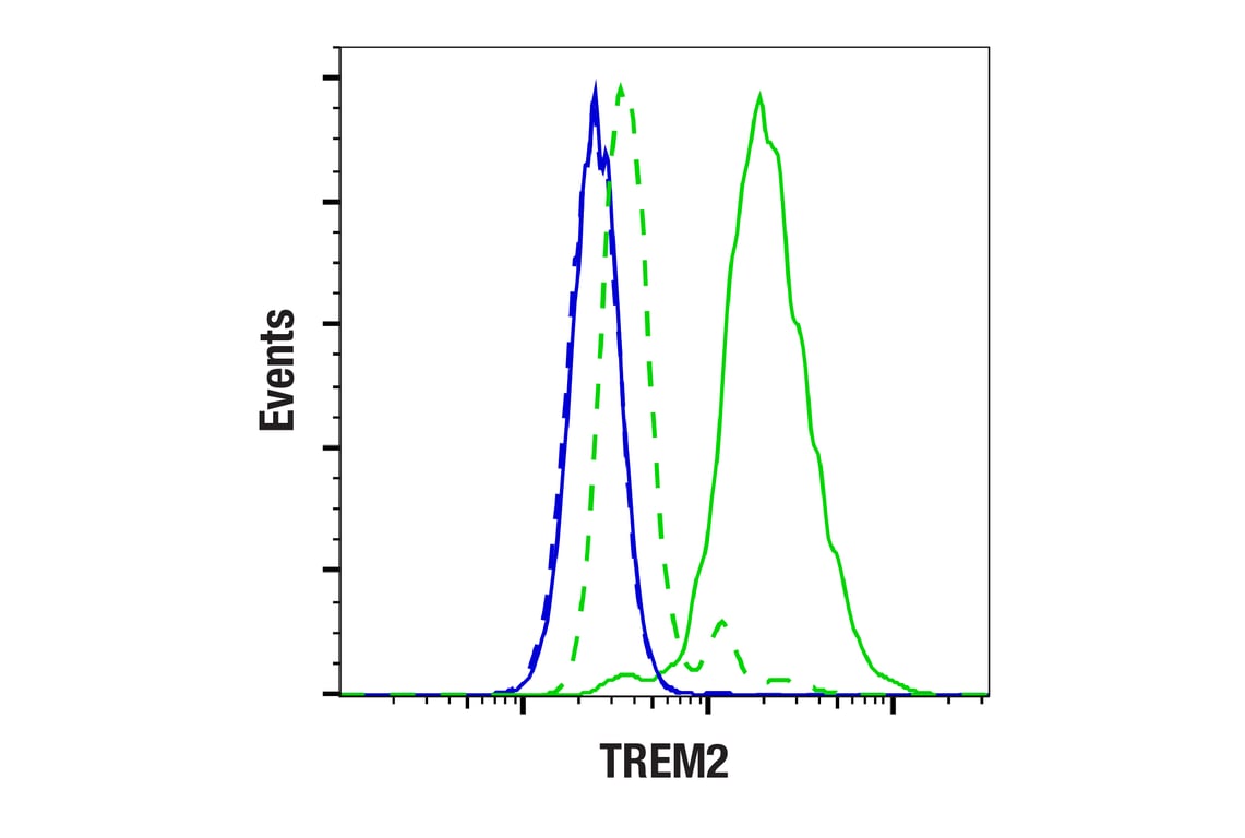
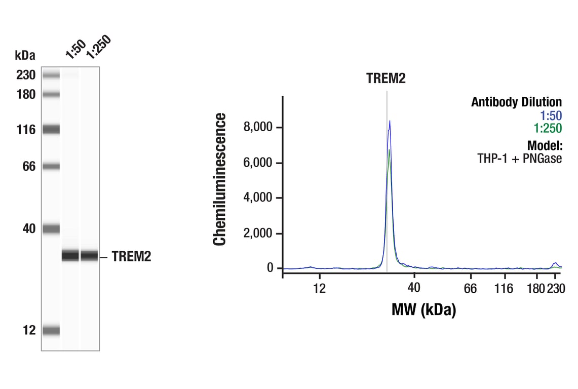
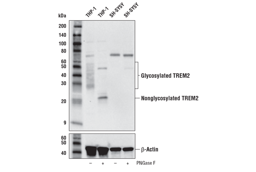
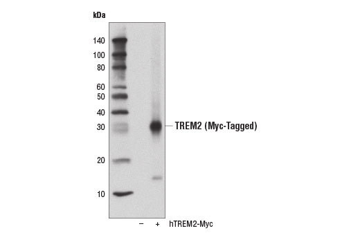
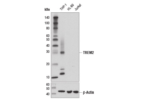
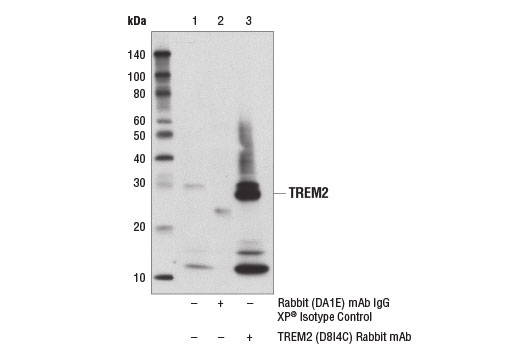
| TREM2 (D8I4C) Rabbit Monoclonal Antibody | 91068 | 20 µl | 28 kDa | Rabbit IgG |
| TREM2 (E9U8L) Rabbit Monoclonal Antibody (Amino-terminal Antigen) | 70551 | 20 µl | 28 kDa | Rabbit IgG |
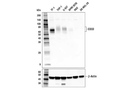
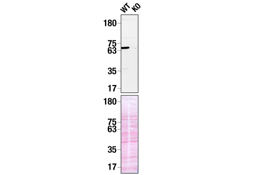
| CD33 Antibody | 77576 | 20 µl | 70-80 kDa | Rabbit |
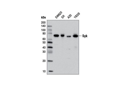
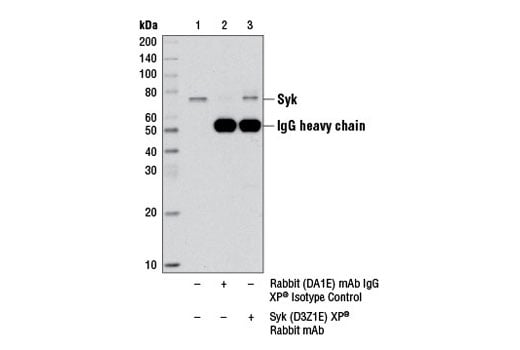
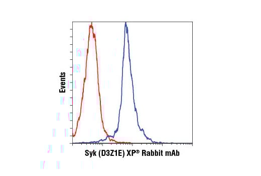
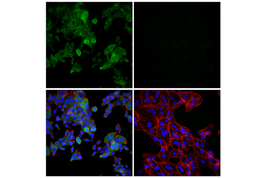
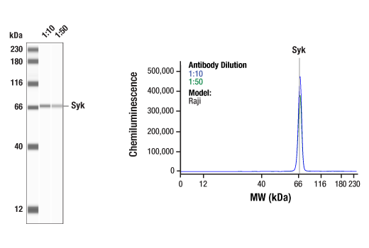
| Syk (D3Z1E) Rabbit Monoclonal Antibody | 13198 | 20 µl | 72 kDa | Rabbit IgG |
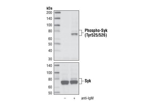
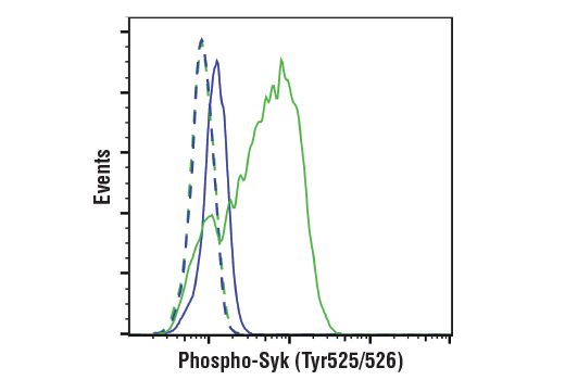
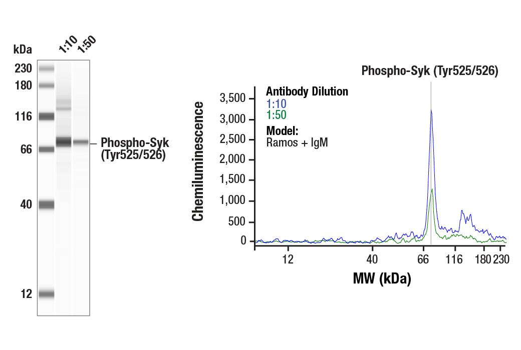
| Phospho-Syk (Tyr525/526) (C87C1) Rabbit Monoclonal Antibody | 2710 | 20 µl | 72 kDa | Rabbit IgG |
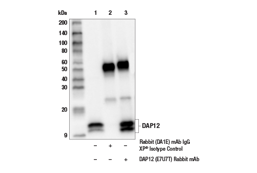
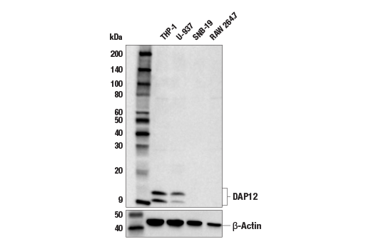
| DAP12 (E7U7T) Rabbit Monoclonal Antibody | 97415 | 20 µl | 10, 12 kDa | Rabbit IgG |
| Anti-rabbit IgG, HRP-linked Antibody | 7074 | 100 µl | Goat |
Please visit cellsignal.com for individual component applications, species cross-reactivity, dilutions, protocols, and additional product information.
Description
Storage
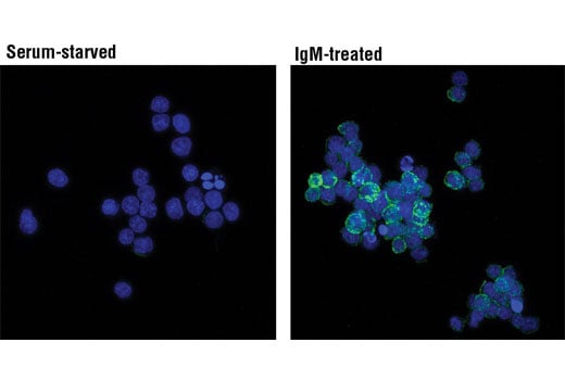
Background
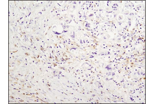
There is also evidence that these processes may be regulated via crosstalk between TREM2 and the cell surface receptor CD33, a sialic acid-binding Ig-like lectin (Siglec-3) type I transmembrane protein. Much like TREM2, CD33 has been identified as a risk gene in AD. CD33 binds preferentially to alpha-2, 6-linked sialic acid, which can be found in sialylated gangliosides in the brain. Activation of CD33 has been shown to be inhibitory to a variety of cellular processes. Evidence suggests that TREM2 may act downstream of CD33 and that TREM2-dependent microglial signaling in AD may be directly inhibited by CD33 activation (14-17).
Background References
- Nguyen, A.T. et al. (2020) Acta Neuropathol 140, 477-493.
- Gratuze, M. et al. (2018) Mol Neurodegener 13, 66.
- Jonsson, T. et al. (2013) N Engl J Med 368, 107-16.
- Jay, T.R. et al. (2017) Mol Neurodegener 12, 56.
- McQuade, A. et al. (2020) Nat Commun 11, 5370.
- Schlepckow, K. et al. (2020) EMBO Mol Med 12, e11227.
- Zhao, Y. et al. (2018) Neuron 97, 1023-1031.e7.
- Colonna, M. (2003) Nat Rev Immunol 3, 445-53.
- Lanier, L.L. et al. (1998) Nature 391, 703-7.
- Zhang, J. et al. (2000) J Biol Chem 275, 35442-7.
- Mansueto, M.S. et al. (2019) J Biol Chem 294, 7658-7668.
- Grädler, U. et al. (2013) J Mol Biol 425, 309-33.
- Turner, M. et al. (2000) Immunol Today 21, 148-54.
- Karch, C.M. et al. (2012) PLoS One 7, e50976.
- Griciuc, A. et al. (2013) Neuron 78, 631-43.
- Griciuc, A. et al. (2019) Neuron 103, 820-835.e7.
- Salminen, A. et al. (2021) Neurochem Int 150, 105186.
Trademarks and Patents
Cell Signaling Technology is a trademark of Cell Signaling Technology, Inc.
U.S. Patent No. 7,429,487, foreign equivalents, and child patents deriving therefrom.
All other trademarks are the property of their respective owners. Visit cellsignal.com/trademarks for more information.
Limited Uses
Except as otherwise expressly agreed in a writing signed by a legally authorized representative of CST, the following terms apply to Products provided by CST, its affiliates or its distributors. Any Customer's terms and conditions that are in addition to, or different from, those contained herein, unless separately accepted in writing by a legally authorized representative of CST, are rejected and are of no force or effect.
Products are labeled with For Research Use Only or a similar labeling statement and have not been approved, cleared, or licensed by the FDA or other regulatory foreign or domestic entity, for any purpose. Customer shall not use any Product for any diagnostic or therapeutic purpose, or otherwise in any manner that conflicts with its labeling statement. Products sold or licensed by CST are provided for Customer as the end-user and solely for research and development uses. Any use of Product for diagnostic, prophylactic or therapeutic purposes, or any purchase of Product for resale (alone or as a component) or other commercial purpose, requires a separate license from CST. Customer shall (a) not sell, license, loan, donate or otherwise transfer or make available any Product to any third party, whether alone or in combination with other materials, or use the Products to manufacture any commercial products, (b) not copy, modify, reverse engineer, decompile, disassemble or otherwise attempt to discover the underlying structure or technology of the Products, or use the Products for the purpose of developing any products or services that would compete with CST products or services, (c) not alter or remove from the Products any trademarks, trade names, logos, patent or copyright notices or markings, (d) use the Products solely in accordance with CST Product Terms of Sale and any applicable documentation, and (e) comply with any license, terms of service or similar agreement with respect to any third party products or services used by Customer in connection with the Products.
Revision 1
Revision 1

Revision 1
Revision 1
Revision 1

Revision 1
Revision 1
Revision 1
Revision 1