| Cat. # | Size | Qty. | Price |
|---|---|---|---|
| 92114T | 1 Kit (6 x 20 microliters) |
|
| Product Includes | Quantity | Applications | Reactivity | MW(kDa) | Isotype |
|---|---|---|---|---|---|
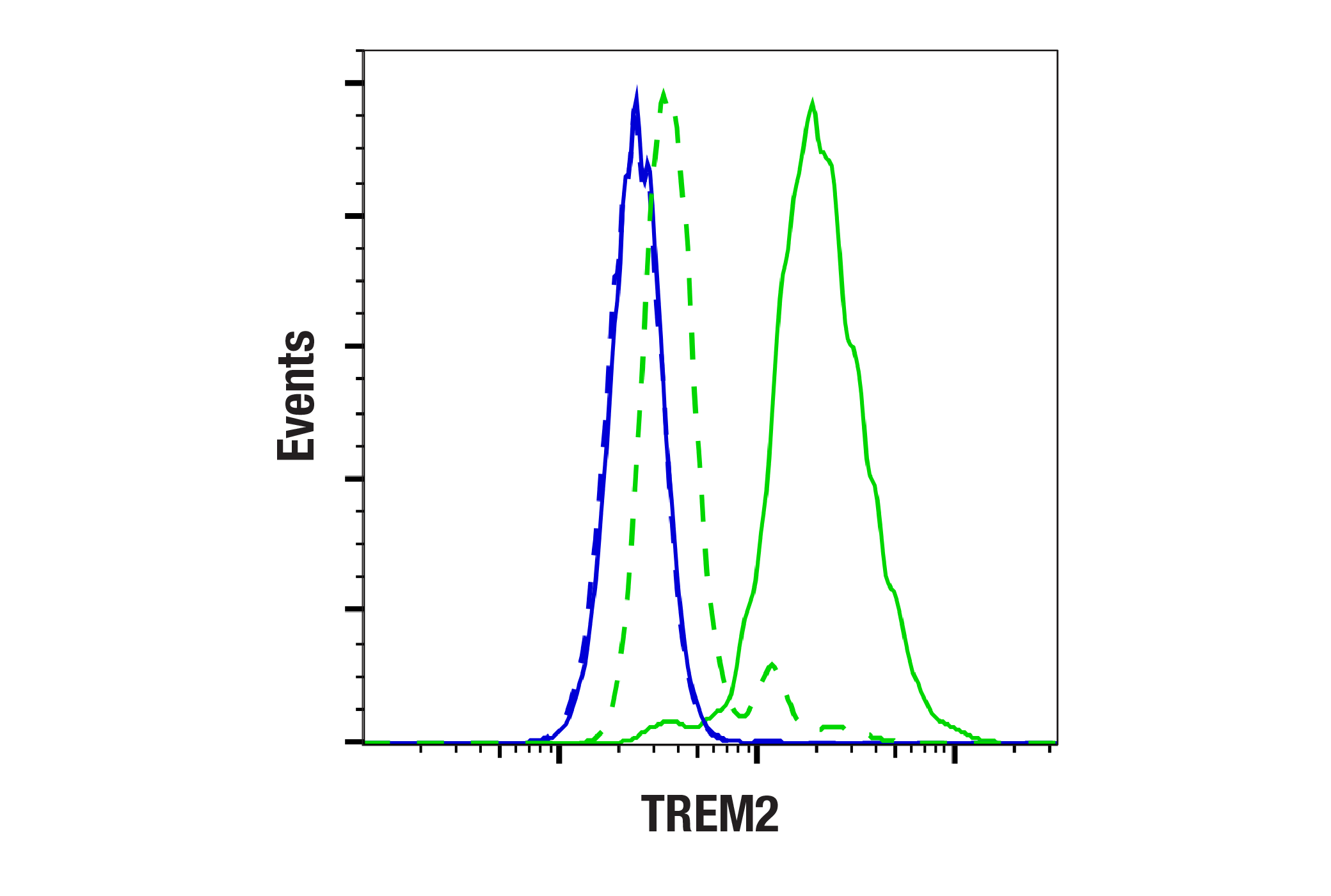
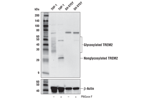
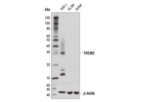
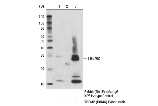
| TREM2 (D8I4C) Rabbit mAb 91068 | 20 µl |
|
H | 28 | Rabbit IgG |
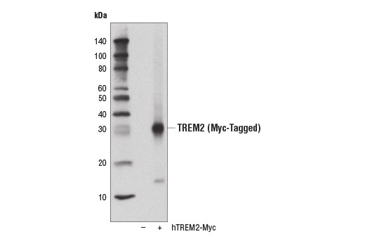
| TREM2 (E9U8L) Rabbit mAb (Amino-terminal Antigen) 70551 | 20 µl |
|
H | 28 | Rabbit IgG |
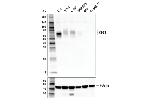
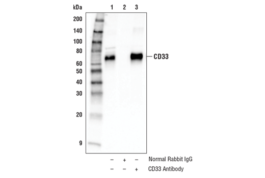
| CD33 Antibody 77576 | 20 µl |
|
H | 70-80 | Rabbit |
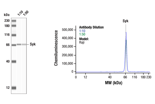
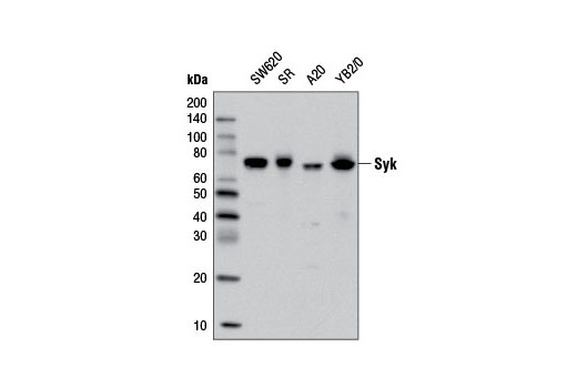
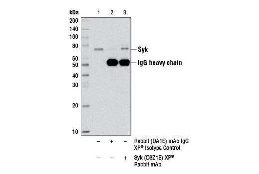
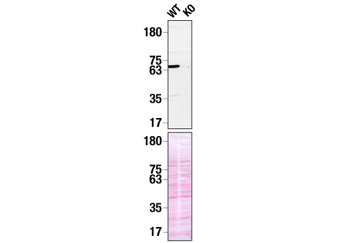
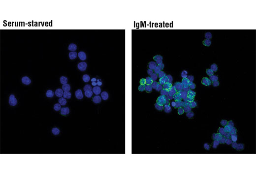
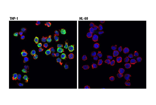
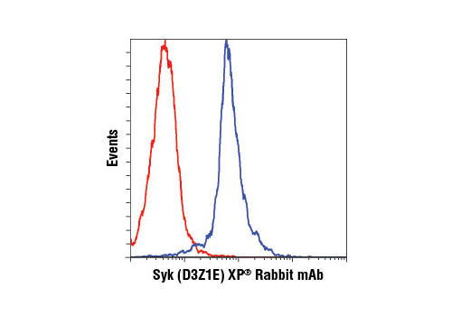
| Syk (D3Z1E) XP® Rabbit mAb 13198 | 20 µl |
|
H M R | 72 | Rabbit IgG |
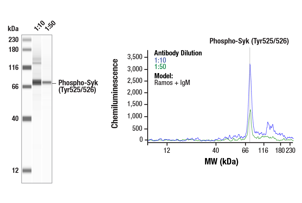
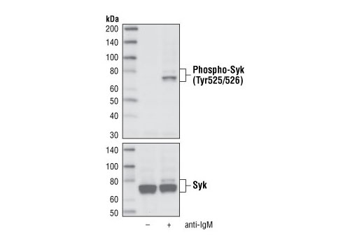
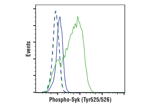
| Phospho-Syk (Tyr525/526) (C87C1) Rabbit mAb 2710 | 20 µl |
|
H | 72 | Rabbit IgG |
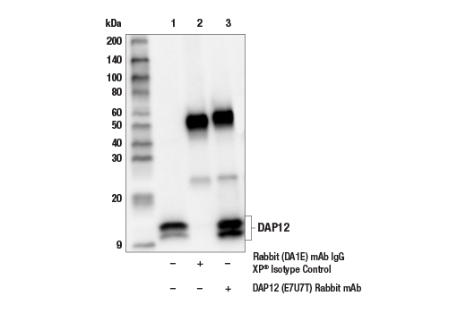
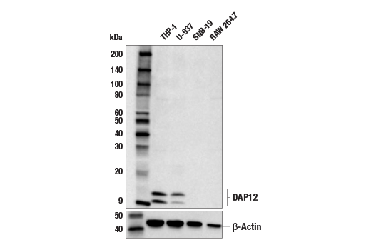
| DAP12 (E7U7T) Rabbit mAb 97415 | 20 µl |
|
H | 10, 12 | Rabbit IgG |
| Anti-rabbit IgG, HRP-linked Antibody 7074 | 100 µl |
|
Goat |
Product Information
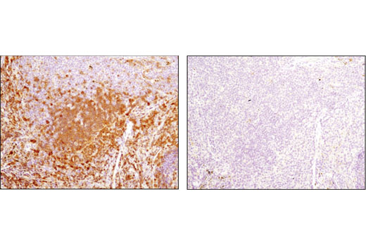
Monoclonal antibodies are produced by immunizing animals with synthetic peptides corresponding to residues surrounding Leu221 of human TREM2 protein, Pro110 of human DAP12 protein, Asn463 of human Syk protein, a synthetic phosphopeptide corresponding to residues surrounding Tyr525/526 of human Syk, and a recombinant protein specific to the amino terminus of human TREM2 protein. Polyclonal antibodies are produced by immunizing animals with a synthetic peptide corresponding to residues near the carboxy terminus of human CD33 protein. Antibodies are purified by peptide affinity chromatography.
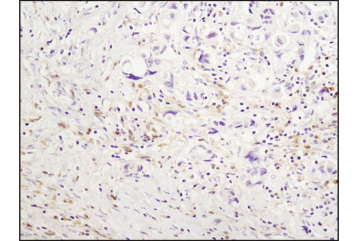
Alzheimer's Disease (AD) is one of the most common neurodegenerative diseases worldwide. Clinically, it is characterized by the presence of extracellular amyloid plaques and intracellular neurofibrillary tangles, resulting in neuronal dysfunction and cell death. Triggering receptor expressed on myeloid cells 2 (TREM2), a protein localized at the membrane of innate immune cells, including microglia in the brain, has been genetically linked to AD, with specific variants increasing disease risk by as much as threefold (1,2). The TREM2 receptor is a single-pass type I membrane glycoprotein that consists of an extracellular immunoglobulin-like domain, a transmembrane domain, and a cytoplasmic tail. Upon activation, TREM2 interacts with the tyrosine kinase-binding protein DNAX-activating protein 12 (DAP12, TYROBP) to form a receptor-signaling complex. The DAP12 protein structure consists of a short extracellular domain, a transmembrane domain, and a cytoplasmic immunoreceptor tyrosine-based activation motif (ITAM) (2-9). ITAMs function as a binding site for tyrosine kinases, including spleen tyrosine kinase (Syk). Syk is comprised of two tandem amino-terminal Src homology (SH) 2 domains separated by an SH2-kinase linker, and a C-terminal tyrosine kinase domain, separated from the SH2 domains by an inter-domain linker. When Syk binds to an ITAM, it changes conformation, allowing for residues within the inter-domain linker region, including Tyr352, to become phosphorylated. Residues within the activation loop subsequently become phosphorylated, leading to full Syk activation. Tyr525 and Tyr526 are located in the activation loop of the Syk kinase domain and phosphorylation at these residues (equivalent to Tyr519/520 of mouse Syk) is essential for Syk function (10-12). This activation can lead to the mediation of a variety of cellular responses, including proliferation, differentiation, inflammation, and phagocytosis. Evidence suggests that TREM2 and DAP12 may act in a Syk-dependent manner to drive microglial cellular responses in AD (2,4-8,13).
There is also evidence that these processes may be regulated via crosstalk between TREM2 and the cell surface receptor CD33, a sialic acid-binding Ig-like lectin (Siglec-3) type I transmembrane protein. Much like TREM2, CD33 has been identified as a risk gene in AD. CD33 binds preferentially to alpha-2, 6-linked sialic acid, which can be found in sialylated gangliosides in the brain. Activation of CD33 has been shown to be inhibitory to a variety of cellular processes. Evidence suggests that TREM2 may act downstream of CD33 and that TREM2-dependent microglial signaling in AD may be directly inhibited by CD33 activation (14-17).
Explore pathways related to this product.
STRING - Known and Predicted Protein-Protein Interactions.
Except as otherwise expressly agreed in a writing signed by a legally authorized representative of CST, the following terms apply to Products provided by CST, its affiliates or its distributors. Any Customer's terms and conditions that are in addition to, or different from, those contained herein, unless separately accepted in writing by a legally authorized representative of CST, are rejected and are of no force or effect.
Products are labeled with For Research Use Only or a similar labeling statement and have not been approved, cleared, or licensed by the FDA or other regulatory foreign or domestic entity, for any purpose. Customer shall not use any Product for any diagnostic or therapeutic purpose, or otherwise in any manner that conflicts with its labeling statement. Products sold or licensed by CST are provided for Customer as the end-user and solely for research and development uses. Any use of Product for diagnostic, prophylactic or therapeutic purposes, or any purchase of Product for resale (alone or as a component) or other commercial purpose, requires a separate license from CST. Customer shall (a) not sell, license, loan, donate or otherwise transfer or make available any Product to any third party, whether alone or in combination with other materials, or use the Products to manufacture any commercial products, (b) not copy, modify, reverse engineer, decompile, disassemble or otherwise attempt to discover the underlying structure or technology of the Products, or use the Products for the purpose of developing any products or services that would compete with CST products or services, (c) not alter or remove from the Products any trademarks, trade names, logos, patent or copyright notices or markings, (d) use the Products solely in accordance with CST Product Terms of Sale and any applicable documentation, and (e) comply with any license, terms of service or similar agreement with respect to any third party products or services used by Customer in connection with the Products.