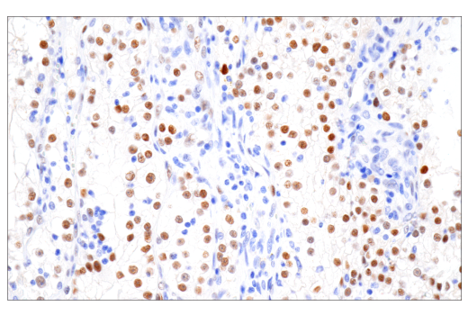
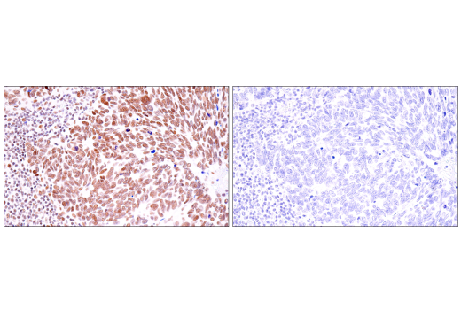
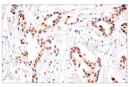
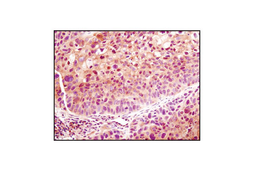
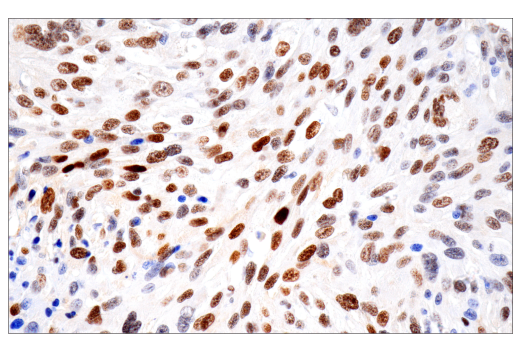
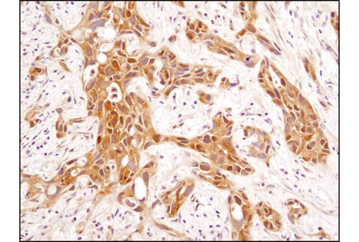
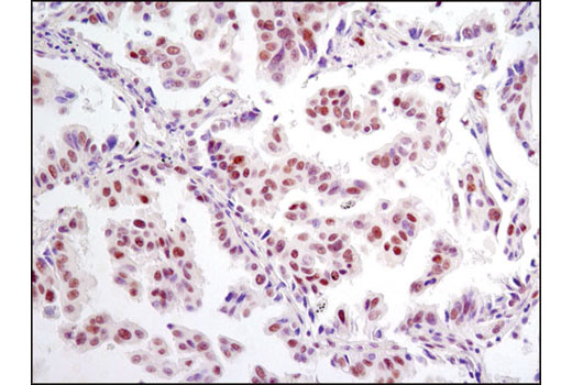
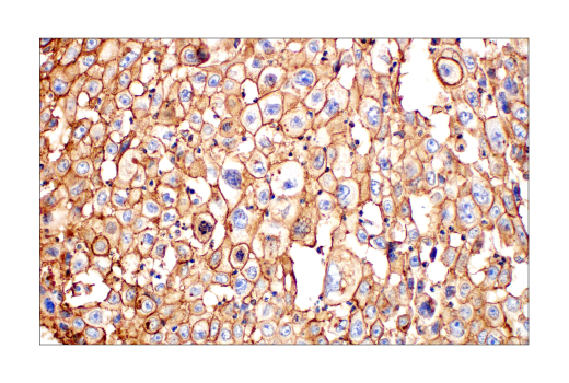
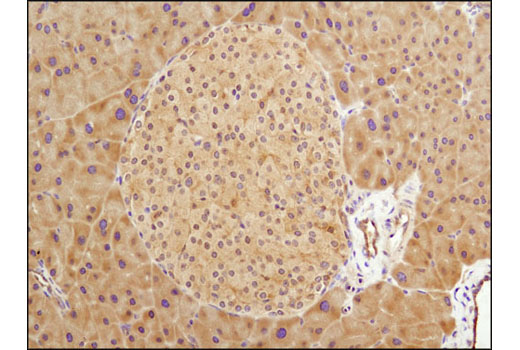
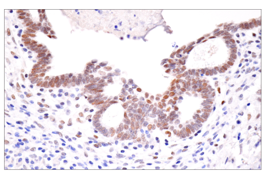
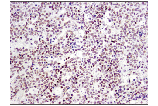
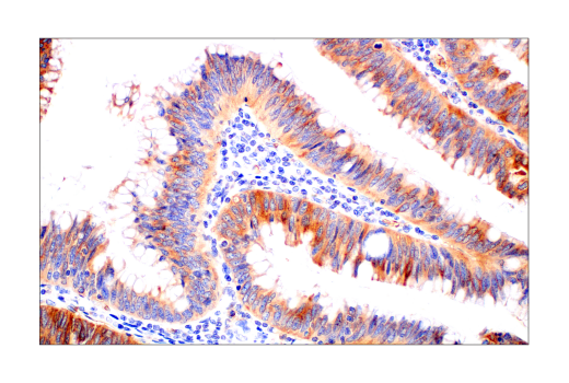
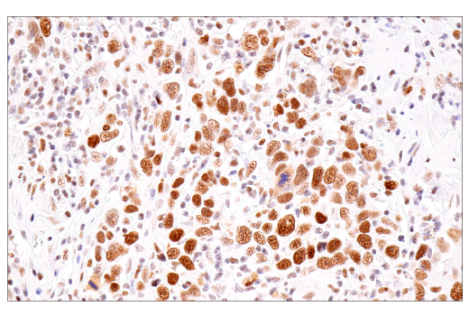
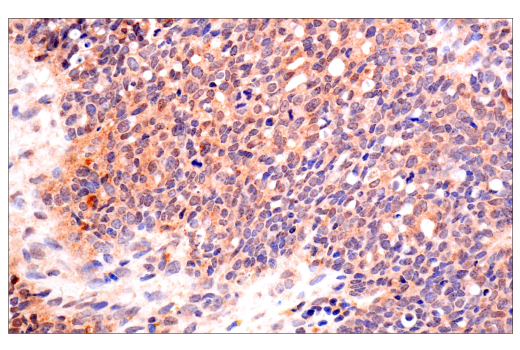
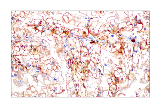
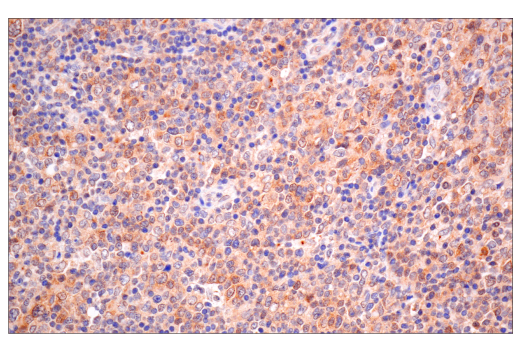
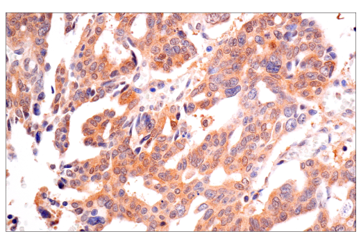
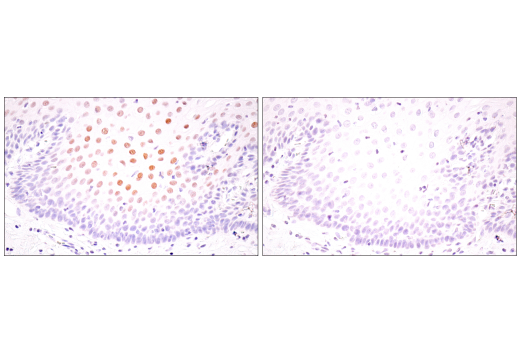
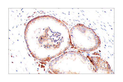
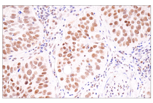
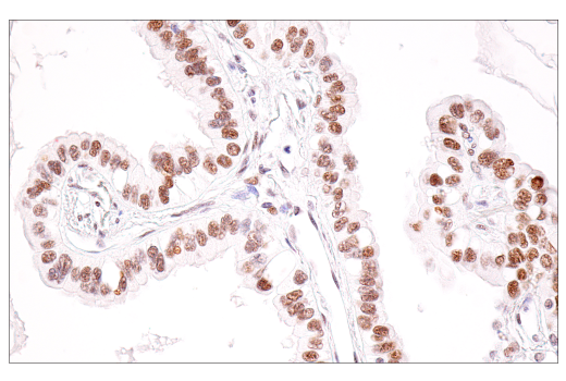
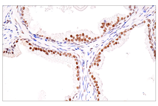
Immunohistochemical analysis of paraffin-embedded human ductal breast carcinoma using VHL (E3X9K) Rabbit mAb.
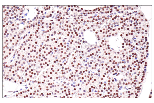
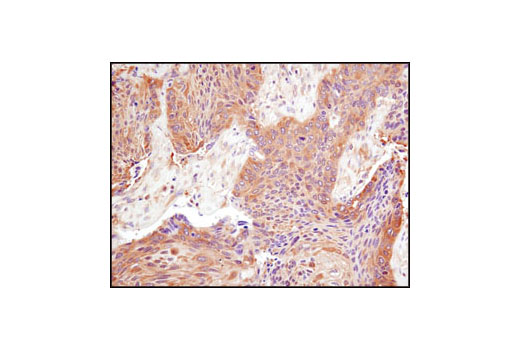
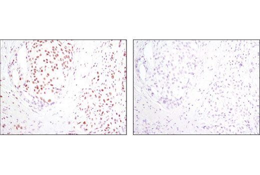
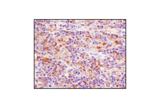
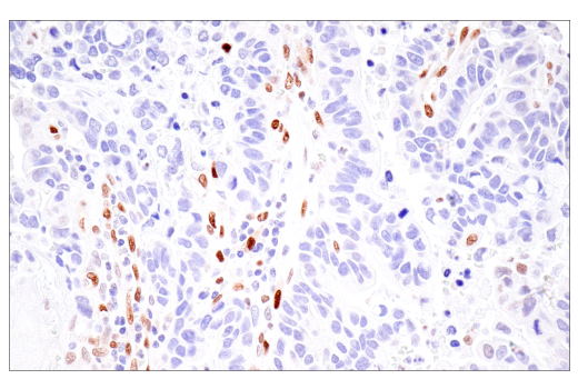
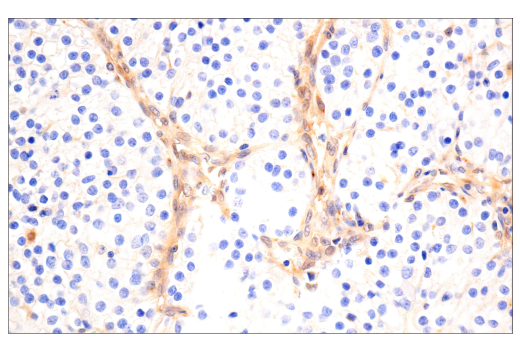
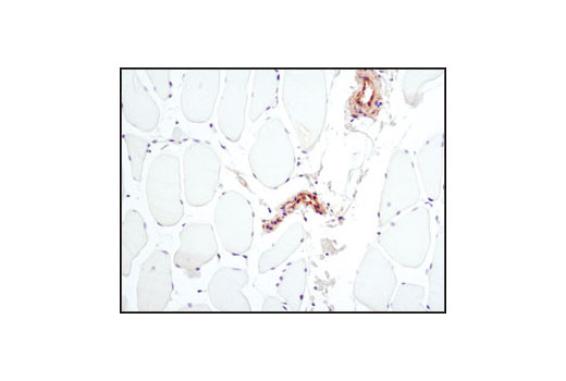
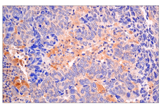
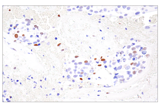
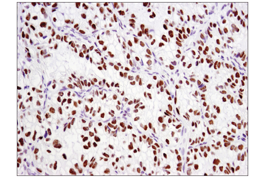
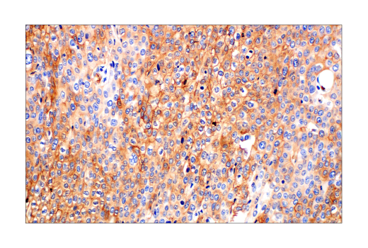
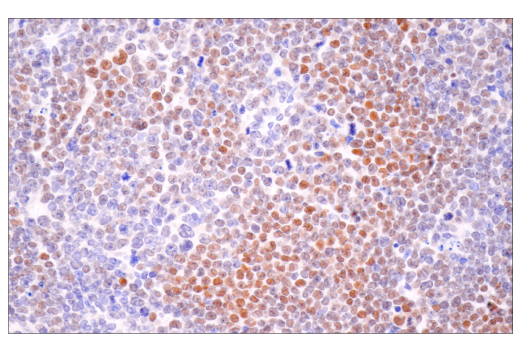
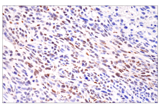
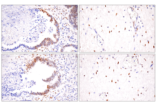
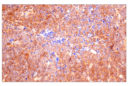
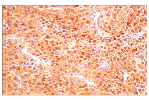
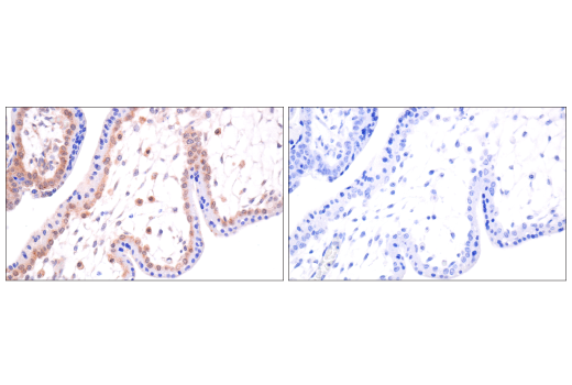
Immunohistochemical analysis of paraffin-embedded Renca syngeneic tumor using VHL (E3X9K) Rabbit mAb.
| Cat. # | Size | Qty. | Price |
|---|---|---|---|
| 43065T | 1 Kit (6 x 20 microliters) |
|
| Product Includes | Quantity | Applications | Reactivity | MW(kDa) | Isotype |
|---|---|---|---|---|---|
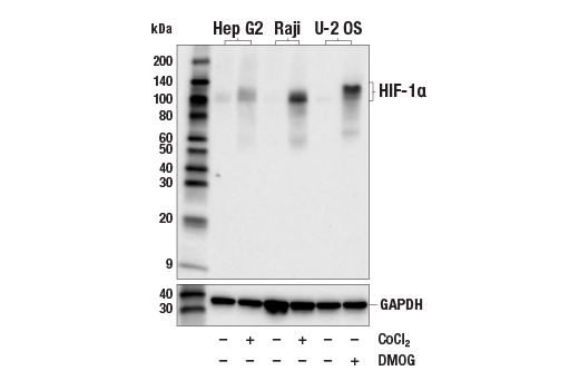
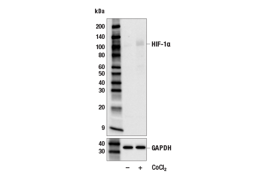
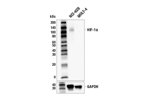
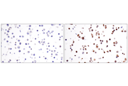
| HIF-1α (E1V6A) Rabbit mAb 48085 | 20 µl |
|
H M | 120 | Rabbit IgG |
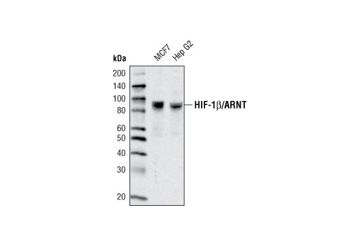
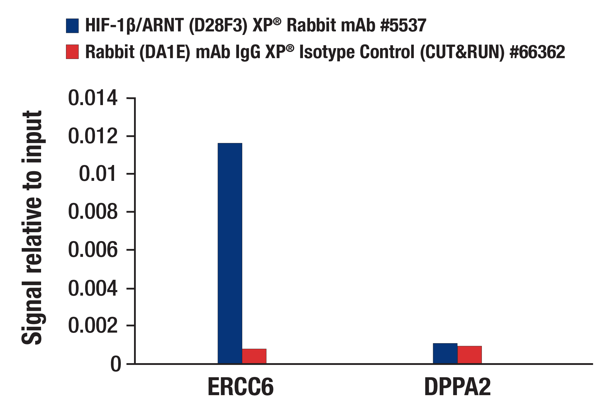
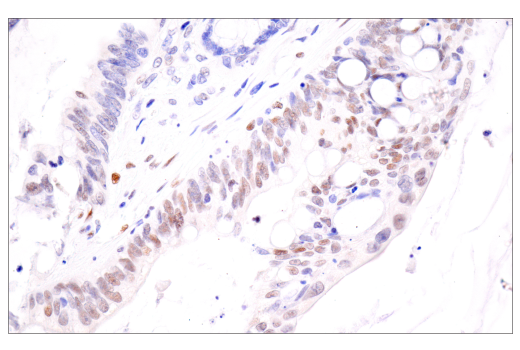
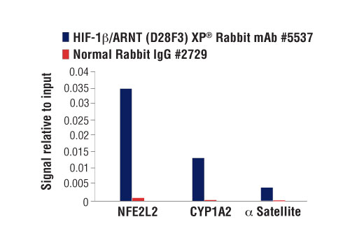
| HIF-1β/ARNT (D28F3) XP® Rabbit mAb 5537 | 20 µl |
|
H M R Mk | 87 | Rabbit IgG |
| VHL (E3X9K) Rabbit mAb 81292 | 20 µl |
|
H M | Rabbit IgG | |
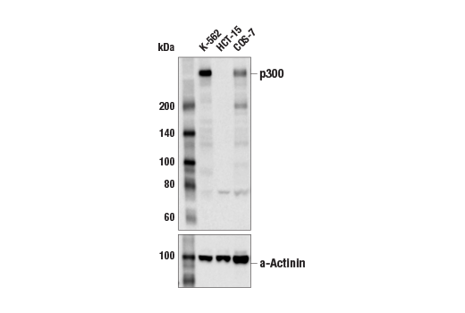
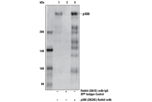
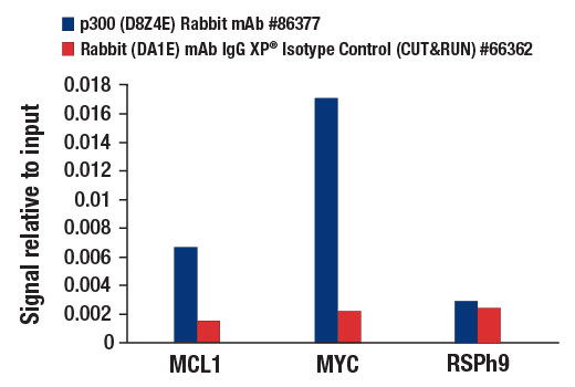
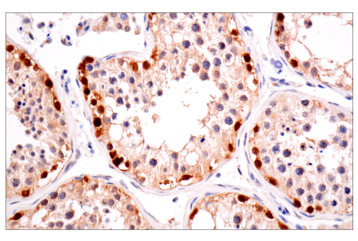
| p300 (D8Z4E) Rabbit mAb 86377 | 20 µl |
|
H Mk | 300 | Rabbit IgG |
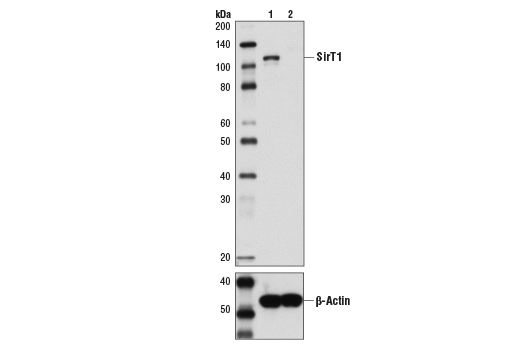
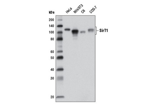
| SirT1 (1F3) Mouse mAb 8469 | 20 µl |
|
H M R Mk | 120 | Mouse IgG1 |
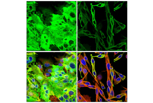
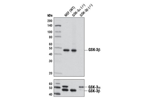
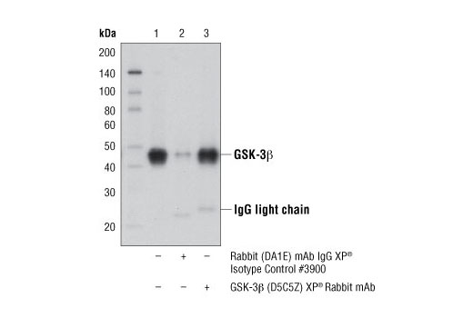
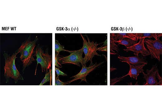
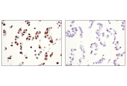
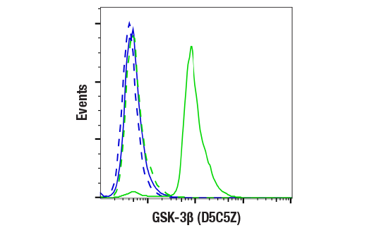
| GSK-3β (D5C5Z) XP® Rabbit mAb 12456 | 20 µl |
|
H M R Mk | 46 | Rabbit IgG |
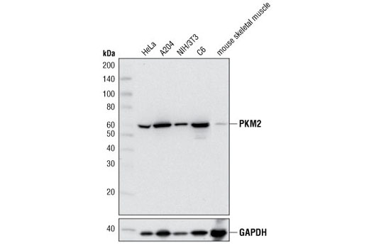
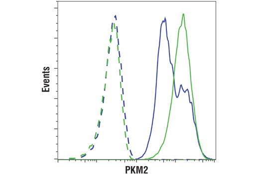
| PKM2 (D78A4) XP® Rabbit mAb 4053 | 20 µl |
|
H M R Mk | 60 | Rabbit IgG |
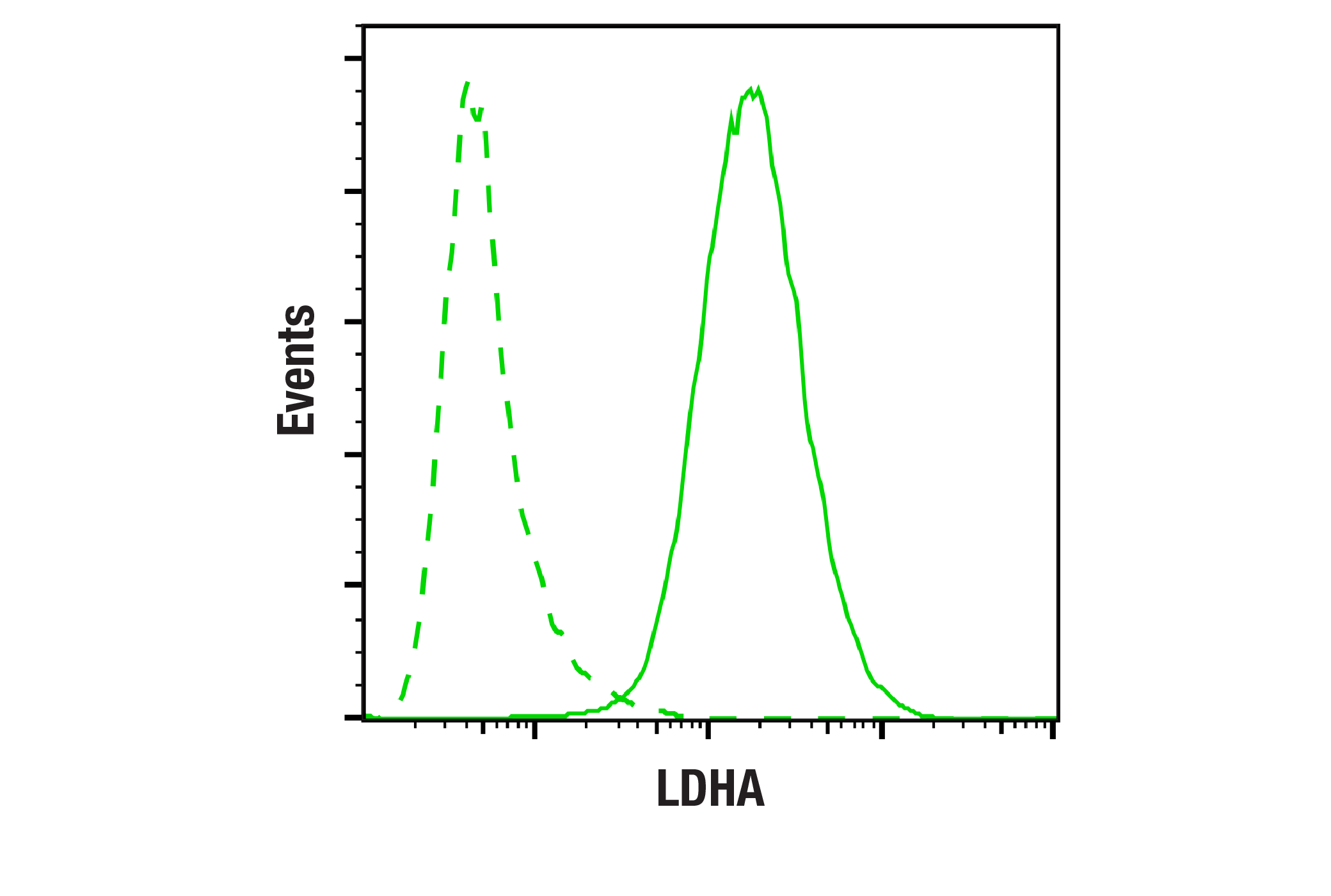
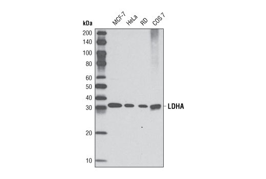
| LDHA (C4B5) Rabbit mAb 3582 | 20 µl |
|
H Mk | 37 | Rabbit IgG |
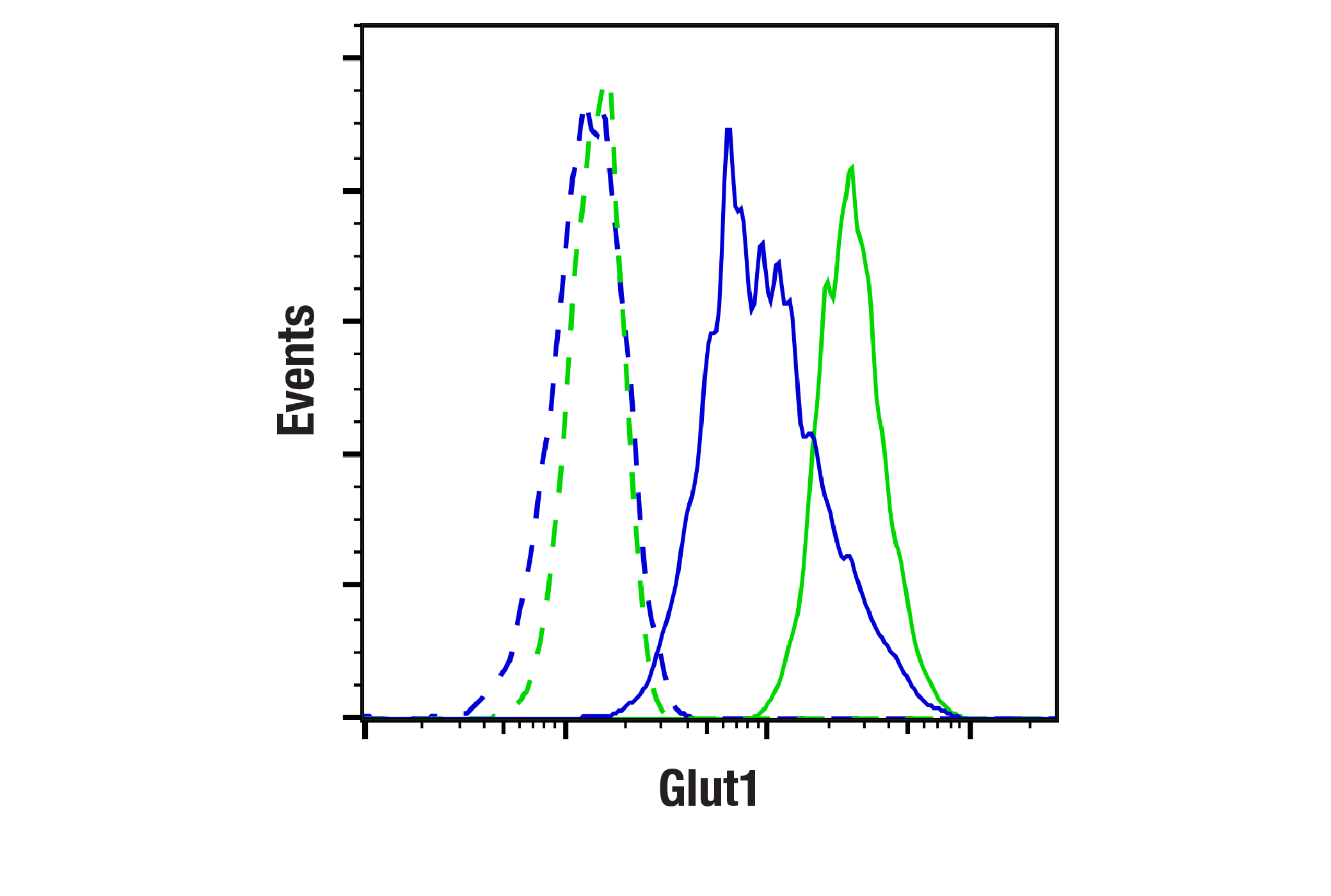
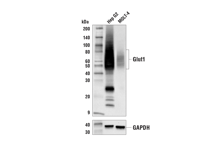
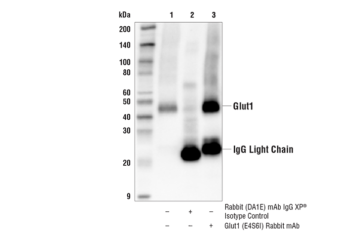
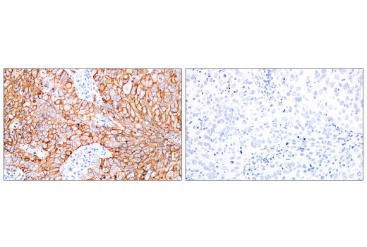
| Glut1 (E4S6I) Rabbit mAb 73015 | 20 µl |
|
H M R Mk | 45-60 | Rabbit IgG |
Product Information
Monoclonal antibodies are produced by immunizing animals with synthetic peptides corresponding to residues surrounding Ala475 of human HIF-1α protein, Ile479 of human HIF-1β/ARNT protein, Ser406 of human PKM2 protein, near the carboxy terminus of human p300 protein and human Glut1 protein, and corresponding to the sequence of human LDHA protein.
Monoclonal antibodies are also produced by immunizing animals with recombinant proteins specific to human VHL protein, representing the central region of human SirT1 protein, and specific to the carboxy terminus of human GSK-3β protein.
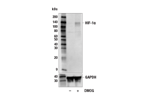
Hypoxia-inducible factor 1 (HIF1) is a heterodimeric transcription factor that plays a critical role in the cellular response to hypoxia (1). The HIF1 complex consists of two subunits, HIF-1α and HIF-1β, which are basic helix-loop-helix proteins of the PAS (Per, ARNT, Sim) family (2). HIF1 regulates the transcription of a broad range of genes that facilitate responses to the hypoxic environment, including genes regulating angiogenesis, erythropoiesis, cell cycle, metabolism, and apoptosis. The widely expressed HIF-1α is typically degraded rapidly in normoxic cells by the ubiquitin/proteasomal pathway. Under normoxic conditions, HIF-1α is proline hydroxylated leading to a conformational change that promotes binding to the von Hippel-Lindau protein (VHL) E3 ligase complex; ubiquitination and proteasomal degradation follows (3,4). Both hypoxic conditions and chemical hydroxylase inhibitors (such as desferrioxamine and cobalt) inhibit HIF-1α degradation and lead to its stabilization. In addition, HIF-1α can be induced in an oxygen-independent manner by various cytokines through the PI3K-AKT-mTOR pathway (5-7). HIF-1β is also known as AhR nuclear translocator (ARNT) due to its ability to partner with the aryl hydrocarbon receptor (AhR) to form a heterodimeric transcription factor complex (8). Together with AhR, HIF-1β plays an important role in xenobiotics metabolism (8). In addition, a chromosomal translocation leading to a TEL-ARNT fusion protein is associated with acute myeloblastic leukemia (9). Studies also found that ARNT/HIF-1β expression levels decrease significantly in pancreatic islets from patients with type 2 diabetes, suggesting that HIF-1β plays an important role in pancreatic β-cell function (10). CBP (CREB-binding protein) and p300 are highly conserved and functionally related transcriptional co-activators that associate with transcriptional regulators and signaling molecules, integrating multiple signal transduction pathways with the transcriptional machinery (11,12). CBP/p300 also contain histone acetyltransferase (HAT) activity, allowing them to acetylate histones and other proteins (12). The Silent Information Regulator (SIR2) family of genes is a highly conserved group of genes that encode nicotinamide adenine dinucleotide (NAD)-dependent protein deacetylases, also known as class III histone deacetylases. The first discovered and best characterized of these genes is Saccharomyces cerevisiae SIR2, which is involved in silencing of mating type loci, telomere maintenance, DNA damage response, and cell aging (13). SirT1, the mammalian ortholog of Sir2, is a nuclear protein implicated in the regulation of many cellular processes, including apoptosis, cellular senescence, endocrine signaling, glucose homeostasis, aging, and longevity. Targets of SirT1 include acetylated p53 (14,15), p300 (16), Ku70 (17), forkhead (FoxO) transcription factors (17,18), PPARγ (19), and the PPARγ coactivator-1α (PGC-1α) protein (20). Glycogen synthase kinase-3 (GSK-3) was initially identified as an enzyme that regulates glycogen synthesis in response to insulin (21). GSK-3 is a ubiquitously expressed serine/threonine protein kinase that phosphorylates and inactivates glycogen synthase. GSK-3 is a critical downstream element of the PI3K/Akt cell survival pathway whose activity can be inhibited by Akt-mediated phosphorylation at Ser21 of GSK-3α and Ser9 of GSK-3β (22,23). Pyruvate kinase is a glycolytic enzyme that catalyzes the conversion of phosphoenolpyruvate to pyruvate. In mammals, the M2 isoform (PKM2) is expressed during embryonic development (24). Lactate dehydrogenase (LDH) catalyzes the interconversion of pyruvate and NADH to lactate and NAD+. The major form of LDH found in muscle cells is the A (LDHA) isozyme (25). Glucose transporter 1 (Glut1, SLC2A1) is a widely expressed transport protein that transports a number of different aldose sugars into cells (26,27).
Explore pathways related to this product.
STRING - Known and Predicted Protein-Protein Interactions.
Except as otherwise expressly agreed in a writing signed by a legally authorized representative of CST, the following terms apply to Products provided by CST, its affiliates or its distributors. Any Customer's terms and conditions that are in addition to, or different from, those contained herein, unless separately accepted in writing by a legally authorized representative of CST, are rejected and are of no force or effect.
Products are labeled with For Research Use Only or a similar labeling statement and have not been approved, cleared, or licensed by the FDA or other regulatory foreign or domestic entity, for any purpose. Customer shall not use any Product for any diagnostic or therapeutic purpose, or otherwise in any manner that conflicts with its labeling statement. Products sold or licensed by CST are provided for Customer as the end-user and solely for research and development uses. Any use of Product for diagnostic, prophylactic or therapeutic purposes, or any purchase of Product for resale (alone or as a component) or other commercial purpose, requires a separate license from CST. Customer shall (a) not sell, license, loan, donate or otherwise transfer or make available any Product to any third party, whether alone or in combination with other materials, or use the Products to manufacture any commercial products, (b) not copy, modify, reverse engineer, decompile, disassemble or otherwise attempt to discover the underlying structure or technology of the Products, or use the Products for the purpose of developing any products or services that would compete with CST products or services, (c) not alter or remove from the Products any trademarks, trade names, logos, patent or copyright notices or markings, (d) use the Products solely in accordance with CST Product Terms of Sale and any applicable documentation, and (e) comply with any license, terms of service or similar agreement with respect to any third party products or services used by Customer in connection with the Products.