Revision 1
#15792
Store at -20C
Hypoxia Pathway Antibody Sampler Kit
1 Kit
(7 x 20 microliters)
877-616-CELL (2355)
877-678-TECH (8324)
3 Trask Lane | Danvers | Massachusetts | 01923 | USA
For Research Use Only. Not for Use in Diagnostic Procedures.
| Product Includes | Product # | Quantity | Mol. Wt | Isotype/Source |
|---|---|---|---|---|
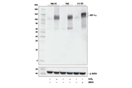
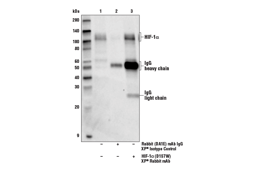
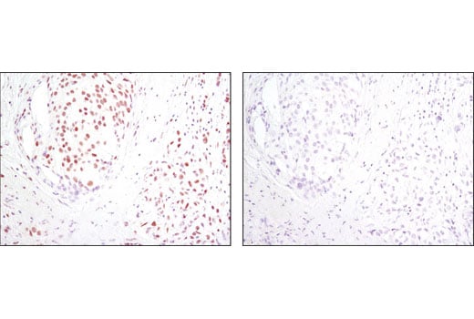
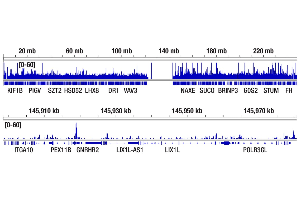
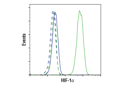
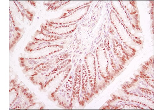
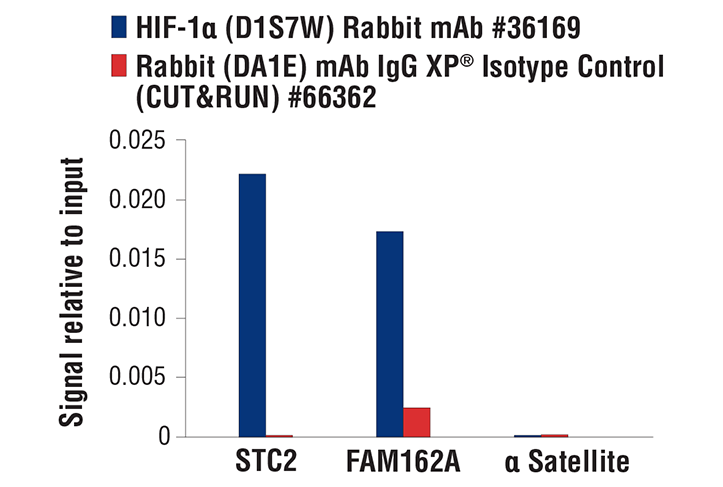
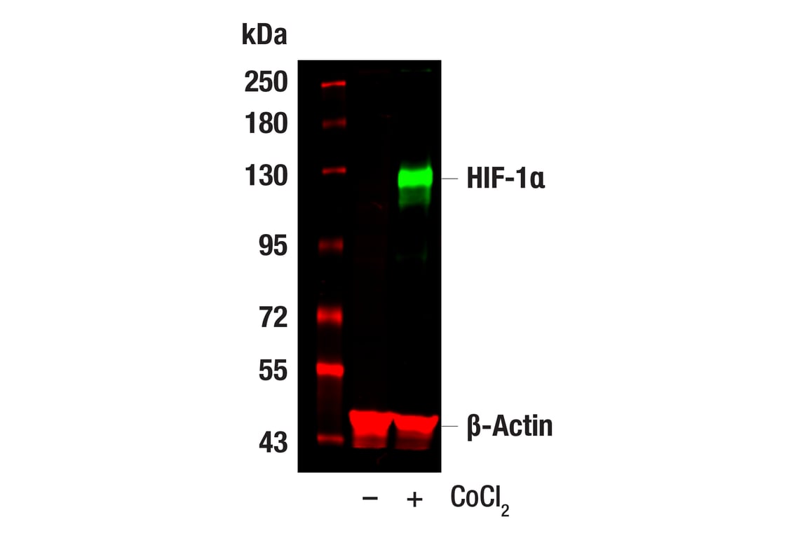
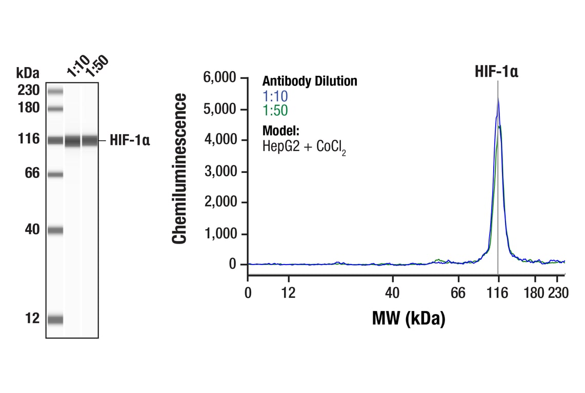
| HIF-1 alpha (D1S7W) Rabbit Monoclonal Antibody | 36169 | 20 µl | 120 kDa | Rabbit IgG |
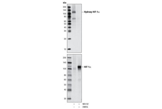
| Hydroxy-HIF-1 alpha (Pro564) (D43B5) Rabbit Monoclonal Antibody | 3434 | 20 µl | 120 kDa | Rabbit IgG |
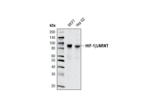
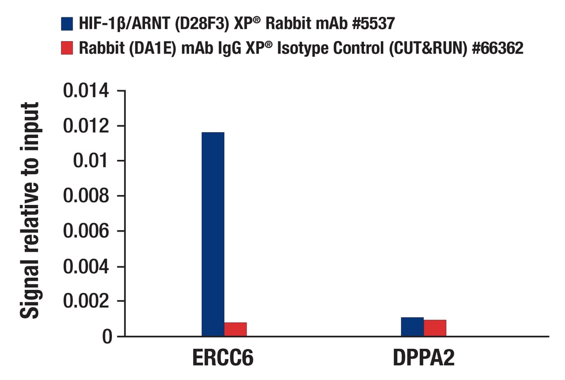
| HIF-1 beta/ARNT (D28F3) Rabbit Monoclonal Antibody | 5537 | 20 µl | 87 kDa | Rabbit IgG |
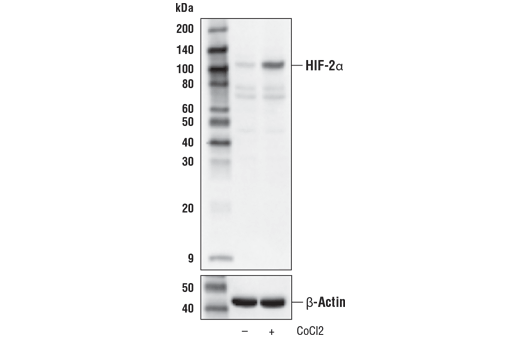
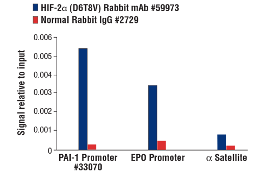
| HIF-2 alpha (D6T8V) Rabbit Monoclonal Antibody | 59973 | 20 µl | 120 kDa | Rabbit IgG |
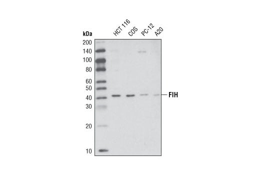
| FIH (D19B3) Rabbit Monoclonal Antibody | 4426 | 20 µl | 42 kDa | Rabbit IgG |
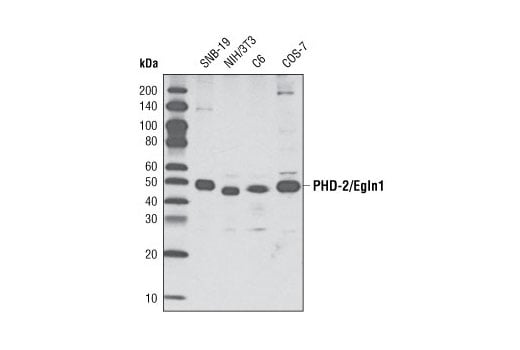
| PHD-2/Egln1 (D31E11) Rabbit Monoclonal Antibody | 4835 | 20 µl | 50 kDa | Rabbit IgG |
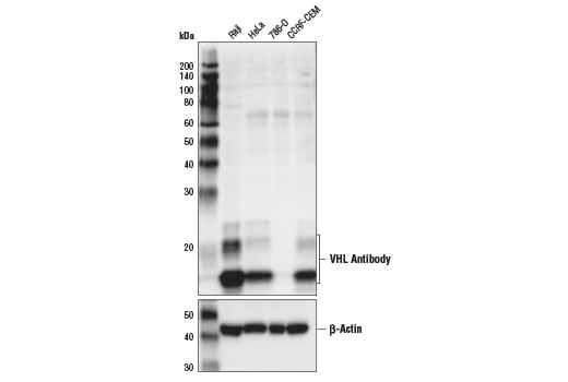
| VHL Antibody | 68547 | 20 µl | 18-22 kDa | Rabbit |
| Anti-rabbit IgG, HRP-linked Antibody | 7074 | 100 µl | Goat |
Please visit cellsignal.com for individual component applications, species cross-reactivity, dilutions, protocols, and additional product information.
Description
Storage
Background
HIF-1β is also known as AhR nuclear translocator (ARNT) due to its ability to partner with the aryl hydrocarbon receptor (AhR) to form a heterodimeric transcription factor complex (8). Together with AhR, HIF-1β plays an important role in xenobiotics metabolism (8). In addition, a chromosomal translocation leading to a TEL-ARNT fusion protein is associated with acute myeloblastic leukemia (9). Studies also found that ARNT/HIF-1β expression levels decrease significantly in pancreatic islets from patients with type 2 diabetes, suggesting that HIF-1β plays an important role in pancreatic β-cell function (10).
Hypoxia-inducible factor (HIF) is essential for the cellular response to hypoxia (11,12). There are several isoforms of the HIF α subunit. Studies have found that HIF-1α and HIF-2α expression is increased in some human cancers. HIF-1α has both pro- and anti-proliferative activities, whereas HIF-2α does not possess anti-proliferative activity (12). Therefore, HIF-2α likely plays an important role in tumorigenesis (12,13).
FIH (Factor inhibiting HIF-1, HIF asparagine hydroxylase) is a dioxygen-dependent asparaginyl hydroxylase that modifies target protein function by hydroxylating target protein asparagine residues (14-16). HIF, a transcriptional activator involved in control of cell cycle in response to hypoxic conditions, is an important target for FIH regulation. FIH functions as an oxygen sensor that regulates HIF function by hydroxylating at Asn803 in the carboxy-terminal transactivation domain (CAD) of HIF (17,18). During normoxia, FIH uses cellular oxygen to hydroxylate HIF-1 and prevent interaction of HIF-1 with transcriptional coactivators, including the CBP/p300-interacting transactivator. Under hypoxic conditions, FIH remains inactive and does not inhibit HIF, allowing the activator to regulate transcription of genes in response to low oxygen conditions (17-19). FIH activity is regulated through interaction with proteins, including Siah-1, which targets FIH for proteasomal degradation (20). The Cut-like homeodomain protein CDP can bind the FIH promoter region to regulate FIH expression at the transcriptional level (21). Phosphorylation of HIF at Thr796 also can prevent FIH hydroxylation on Asn803 (22). Potential FIH substrates also include proteins with ankyrin repeat domains, such as Iκ-B, Notch, and ASB4 (23-25).
PHD1 (Egln2), PHD-2 (Egln1), and PHD3 (Egln3) are members of the Egln family of proline hydroxylases. They function as oxygen sensors that catalyze the hydroxylation of HIF on prolines 564 and 402, initiating the first step of HIF degradation through the VHL/ubiquitin pathway (26,27).
Background References
- Sharp, F.R. and Bernaudin, M. (2004) Nat Rev Neurosci 5, 437-48.
- Wang, G.L. et al. (1995) Proc Natl Acad Sci U S A 92, 5510-4.
- Jaakkola, P. et al. (2001) Science 292, 468-72.
- Maxwell, P.H. et al. (1999) Nature 399, 271-5.
- Fukuda, R. et al. (2002) J Biol Chem 277, 38205-11.
- Jiang, B.H. et al. (2001) Cell Growth Differ 12, 363-9.
- Laughner, E. et al. (2001) Mol Cell Biol 21, 3995-4004.
- Walisser, J.A. et al. (2004) Proc Natl Acad Sci U S A 101, 16677-82.
- Salomon-Nguyen, F. et al. (2000) Proc Natl Acad Sci U S A 97, 6757-62.
- Gunton, J.E. et al. (2005) Cell 122, 337-49.
- Kaelin, W.G. (2005) Biochem Biophys Res Commun 338, 627-38.
- Toschi, A. et al. (2008) J Biol Chem 283, 34495-9.
- Gordan, J.D. and Simon, M.C. (2007) Curr Opin Genet Dev 17, 71-7.
- Koivunen, P. et al. (2004) J Biol Chem 279, 9899-904.
- Linke, S. et al. (2004) J Biol Chem 279, 14391-7.
- Lisy, K. and Peet, D.J. (2008) Cell Death Differ 15, 642-9.
- Mahon, P.C. et al. (2001) Genes Dev 15, 2675-86.
- Lando, D. et al. (2002) Genes Dev 16, 1466-71.
- Lando, D. et al. (2002) Science 295, 858-61.
- Fukuba, H. et al. (2007) Biochem Biophys Res Commun 353, 324-9.
- Li, J. et al. (2007) Mol Cell Biol 27, 7345-53.
- Lancaster, D.E. et al. (2004) Biochem J 383, 429-37.
- Ferguson, J.E. et al. (2007) Mol Cell Biol 27, 6407-19.
- Cockman, M.E. et al. (2006) Proc Natl Acad Sci U S A 103, 14767-72.
- Cockman, M.E. et al. (2009) Mol Cell Proteomics 8, 535-46.
- Freeman, R.S. et al. (2003) Mol Cells 16, 1-12.
- Villar, D. et al. (2007) Biochem J 408, 231-40.
Trademarks and Patents
Cell Signaling Technology is a trademark of Cell Signaling Technology, Inc.
All other trademarks are the property of their respective owners. Visit cellsignal.com/trademarks for more information.
Limited Uses
Except as otherwise expressly agreed in a writing signed by a legally authorized representative of CST, the following terms apply to Products provided by CST, its affiliates or its distributors. Any Customer's terms and conditions that are in addition to, or different from, those contained herein, unless separately accepted in writing by a legally authorized representative of CST, are rejected and are of no force or effect.
Products are labeled with For Research Use Only or a similar labeling statement and have not been approved, cleared, or licensed by the FDA or other regulatory foreign or domestic entity, for any purpose. Customer shall not use any Product for any diagnostic or therapeutic purpose, or otherwise in any manner that conflicts with its labeling statement. Products sold or licensed by CST are provided for Customer as the end-user and solely for research and development uses. Any use of Product for diagnostic, prophylactic or therapeutic purposes, or any purchase of Product for resale (alone or as a component) or other commercial purpose, requires a separate license from CST. Customer shall (a) not sell, license, loan, donate or otherwise transfer or make available any Product to any third party, whether alone or in combination with other materials, or use the Products to manufacture any commercial products, (b) not copy, modify, reverse engineer, decompile, disassemble or otherwise attempt to discover the underlying structure or technology of the Products, or use the Products for the purpose of developing any products or services that would compete with CST products or services, (c) not alter or remove from the Products any trademarks, trade names, logos, patent or copyright notices or markings, (d) use the Products solely in accordance with CST Product Terms of Sale and any applicable documentation, and (e) comply with any license, terms of service or similar agreement with respect to any third party products or services used by Customer in connection with the Products.
Revision 1
Revision 1
Revision 1


Revision 1
Revision 1
Revision 1
Revision 1
Revision 1

Revision 1
Revision 1
Revision 1
