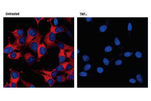
View Featured Offers >>
9240
PhosphoPlus® IκBα (Ser32/36) Antibody Kit
Primary Antibodies
PhosphoPlus Antibody Kit

PhosphoPlus® IκBα (Ser32/36) Antibody Kit #9240

Citations (0)
Western blot analysis of extracts from various cell lines using IκBα (L35A5) Mouse mAb (Amino-terminal Antigen) #4814 (upper) or β-Actin (D6A8) Rabbit mAb #8457 (lower).
After the primary antibody is bound to the target protein, a complex with HRP-linked secondary antibody is formed. The LumiGLO® is added and emits light during enzyme catalyzed decomposition.
After the primary antibody is bound to the target protein, a complex with HRP-linked secondary antibody is formed. The LumiGLO* is added and emits light during enzyme catalyzed decomposition.
Western blot analysis of NF-κB Control Cell Extracts #9243, using IKKα Antibody #2682 (upper), IKKβ (2C8) Rabbit mAb #2370 (middle) and Phospho-IKKα/β (Ser176/180) (16A6) Rabbit mAb #2697 (lower).
Western blot analysis of extracts from NIH/3T3 cells, untreated or TNF-α-treated (#8902, 20 ng/ml) for 5 minutes, using Phospho-IκBα (Ser32/36) (5A5) Mouse mAb #9246 (upper) or IκBα (L35A5) Mouse mAb (Amino-terminal Antigen) #4814, and β-Actin (D6A8) Rabbit mAb #8457 (lower).
Western blot analysis of extracts from THP-1 cells, differentiated with TPA (#9905, 80 nM for 24h) and treated with 1 μg/ml LPS for the indicated times, using Phospho-IκBα (Ser32/36) (5A5) Mouse mAb #9246 (upper) and IκBα (L35A5) Mouse mAb (Amino-terminal Antigen) (lower).
Western blot analysis of NF-κB Control Cell Extracts #9243, using IκBα (44D4) Rabbit mAb #4812 (left) and Phospho-IκBα (Ser32) (14D4) Rabbit mAb #2859 (right).
Western blot analysis of extracts from various cell lines, untreated or treated with IFNa (#36000, 20 ng/mL, 5 min); using Phospho-IκBα (Ser32/36) (5A5) Mouse mAb #9246 (upper), IκBα (L35A5) Mouse mAb (Amino-terminal Antigen) #4814 (middle), and β-Actin (D6A8) Rabbit mAb #8457 (lower).
Immunoprecipitation of IkBa from HeLa cell extracts. Lane 1 is 10% input, lane 2 is precipitated with Mouse (G3A1) mAb IgG1 Isotype Control #5415, and lane 3 is IκBα (L35A5) Mouse mAb (Amino-terminal Antigen), #4814. Western blot was performed using IκBα Antibody #9242.
Western blot analysis of extracts from THP-1 cells, differentiated with TPA (#9905, 80 nM for 24 h) and treated with 1 μg/ml LPS for the indicated times, using Phospho-IκBα (Ser32/36) (5A5) Mouse mAb (upper) and IκBα (L35A5) Mouse mAb (Amino-terminal Antigen) #4814 (lower).
Immunohistochemical analysis of paraffin-embedded human leiomyoma, using IκBα (L35A5) Mouse mAb (Amino-terminal Antigen).
Immunohistochemical analysis of paraffin-embedded human lung carcinoma, using IκBα (L35A5) Mouse mAb (Amino-terminal Antigen).
Immunohistochemical analysis of paraffin-embedded human renal adenocarcinoma, using IκBα (L35A5) Mouse mAb (Amino-terminal Antigen).
Confocal immunofluorescent analysis of HeLa cells, untreated (left), or TNF-α-treated (#8902, 10 ng/ml for 15 min, right) using IκBα (L35A5) Mouse mAb (Amino-terminal Antigen) (red). Blue pseudocolor = DRAQ5® #4084 (fluorescent DNA dye).
Flow cytometric analysis of NIH/3T3 cells, treated with TNF-α (#8902, 10 ng/ml for 5 min; blue, negative) or untreated (green, positive) using IκBα (L35A5) Mouse mAb (Amino-terminal Antigen) (solid lines) or concentration-matched Mouse (G3A1) mAb IgG1 Isotype Control #5415 (dashed lines). Anti-mouse IgG (H+L), F(ab')2 Fragment (Alexa Fluor® 488 Conjugate) #4408 was used as a secondary antibody.
Inquiry Info.# 9240

Product Description

The PhosphoPlus® IκBα (Ser32/36) Antibody Kit provides reagents and protocols for the rapid analysis of IκBα phosphorylation at Ser32/36. The kit contains a total IκBα antibody, a phospho-specific antibody, and positive/negative control whole cell lysates, along with secondary antibodies and reagents for Western blotting.

Specificity / Sensitivity

IκBα (L35A5) Mouse mAb (Amino-terminal Antigen) #4814 detects endogenous levels of total IκBα protein. Phospho-IκBα (Ser32/36) (5A5) Mouse mAb #9246 detects endogenous levels of IκBα only when phosphorylated at Ser32/36. Neither antibody cross-reacts with other IκB family members at physiological levels.

Source / Purification

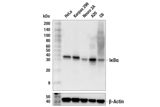
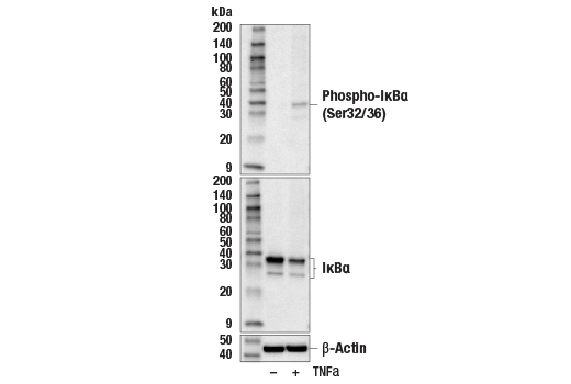
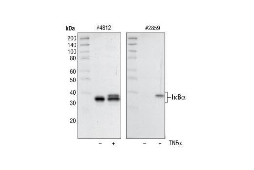
IκBα (L35A5) Mouse mAb (Amino-terminal Antigen) #4814 is produced by immunizing mice with a GST-IκBα fusion protein corresponding the amino-terminus of human IκBα. Phospho-IκBα (Ser32/36) (5A5) Mouse mAb #9246 is produced by immunizing mice with a synthetic phosphopeptide corresponding to residues surrounding Ser32/36 of human IκBα. Total cell extracts from HeLa cells prepared without treatment serve as a negative control. Supplied in SDS Sample Buffer. Total cell extracts from HeLa cells prepared with TNF-α treatment (#2169, 20 ng/ml for 5 minutes) serve as a positive control. Supplied in SDS Sample Buffer.

Background

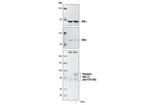
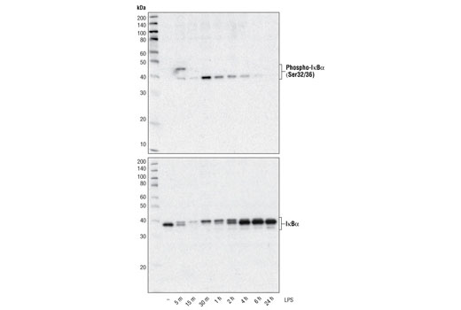
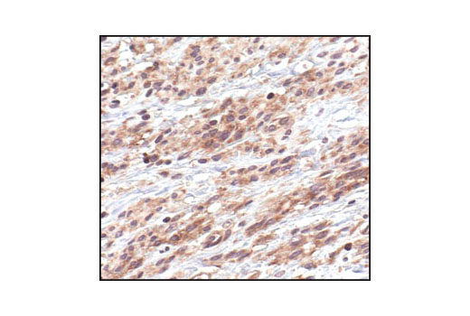
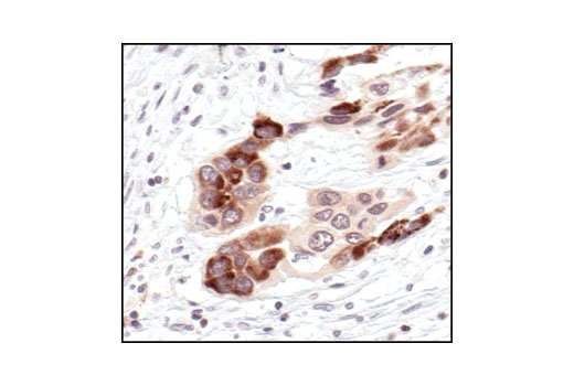
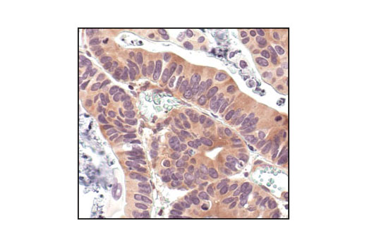
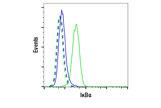
The NF-κB/Rel transcription factors are present in the cytosol in an inactive state complexed with the inhibitory IκB proteins (1-3). Activation occurs via phosphorylation of IκBα at Ser32 and Ser36 followed by proteasome-mediated degradation that results in the release and nuclear translocation of active NF-κB (3-7). IκBα phosphorylation and resulting Rel-dependent transcription are activated by a highly diverse group of extracellular signals including inflammatory cytokines, growth factors, and chemokines. Kinases that phosphorylate IκB at these activating sites have been identified (8).

Pathways

Explore pathways related to this product.

Limited Uses

Except as otherwise expressly agreed in a writing signed by a legally authorized representative of CST, the following terms apply to Products provided by CST, its affiliates or its distributors. Any Customer's terms and conditions that are in addition to, or different from, those contained herein, unless separately accepted in writing by a legally authorized representative of CST, are rejected and are of no force or effect.

Products are labeled with For Research Use Only or a similar labeling statement and have not been approved, cleared, or licensed by the FDA or other regulatory foreign or domestic entity, for any purpose. Customer shall not use any Product for any diagnostic or therapeutic purpose, or otherwise in any manner that conflicts with its labeling statement. Products sold or licensed by CST are provided for Customer as the end-user and solely for research and development uses. Any use of Product for diagnostic, prophylactic or therapeutic purposes, or any purchase of Product for resale (alone or as a component) or other commercial purpose, requires a separate license from CST. Customer shall (a) not sell, license, loan, donate or otherwise transfer or make available any Product to any third party, whether alone or in combination with other materials, or use the Products to manufacture any commercial products, (b) not copy, modify, reverse engineer, decompile, disassemble or otherwise attempt to discover the underlying structure or technology of the Products, or use the Products for the purpose of developing any products or services that would compete with CST products or services, (c) not alter or remove from the Products any trademarks, trade names, logos, patent or copyright notices or markings, (d) use the Products solely in accordance with CST Product Terms of Sale and any applicable documentation, and (e) comply with any license, terms of service or similar agreement with respect to any third party products or services used by Customer in connection with the Products.

For Research Use Only. Not for Use in Diagnostic Procedures.
Cell Signaling Technology is a trademark of Cell Signaling Technology, Inc.
PhosphoPlus is a trademark of Cell Signaling Technology, Inc.
All other trademarks are the property of their respective owners. Visit our Trademark Information page.