Revision 4
#12675
Store at -20C
Initiator Caspases Antibody Sampler Kit
1 Kit
(5 x 20 microliters)
877-616-CELL (2355)
877-678-TECH (8324)
3 Trask Lane | Danvers | Massachusetts | 01923 | USA
For Research Use Only. Not for Use in Diagnostic Procedures.
| Product Includes | Product # | Quantity | Mol. Wt | Isotype/Source |
|---|---|---|---|---|
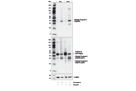
| Cleaved Caspase-9 (Asp330) (E5Z7N) Rabbit Monoclonal Antibody | 52873 | 20 µl | 37 kDa | Rabbit IgG |
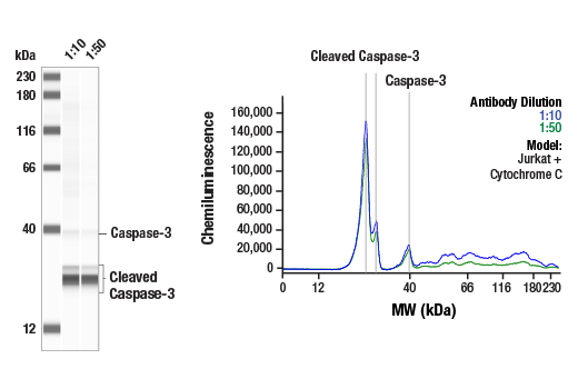
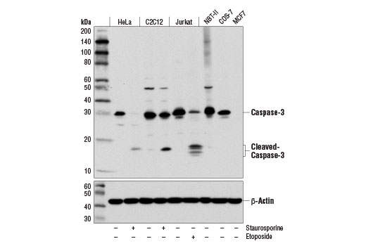
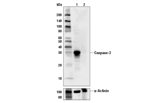
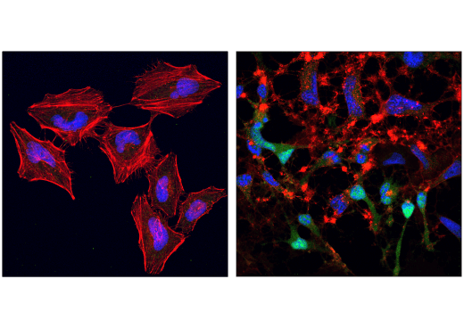
| Caspase-3 (D3R6Y) Rabbit Monoclonal Antibody | 14220 | 20 µl | 35, 19, 17 kDa | Rabbit IgG |
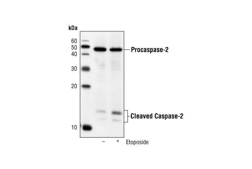
| Caspase-2 (C2) Mouse Monoclonal Antibody | 2224 | 20 µl | 12, 14, 48 kDa | Mouse IgG1 |
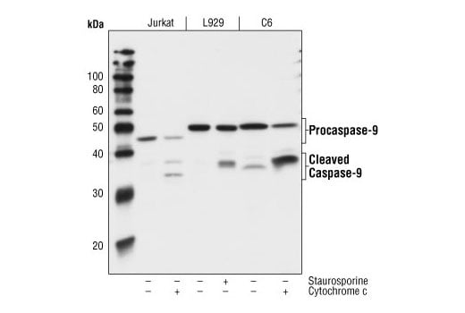
| Caspase-9 (C9) Mouse Monoclonal Antibody | 9508 | 20 µl | 47/37/35 (H). 49/39/37 (M). 51/40/38 (R). kDa | Mouse IgG1 |
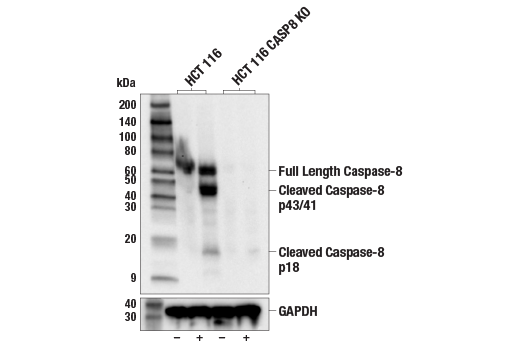
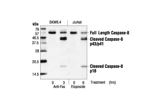
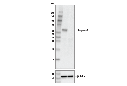
| Caspase-8 (1C12) Mouse Monoclonal Antibody | 9746 | 20 µl | 18, 43, 57 kDa | Mouse IgG1 |
| Anti-rabbit IgG, HRP-linked Antibody | 7074 | 100 µl | Goat | |
| Anti-mouse IgG, HRP-linked Antibody | 7076 | 100 µl | Horse |
Please visit cellsignal.com for individual component applications, species cross-reactivity, dilutions, protocols, and additional product information.
Description
Storage
Background
Formation of a death-inducing signaling complex (DISC) around the receptors for death factors, including FasL and TNF-α, is essential for receptor-mediated apoptosis (3). Upon ligand activation, Fas and TNF-R1 associate with death domain (DD) containing adaptor proteins FADD (Fas associated death domain) (4,5) and TRADD (TNF-R1 associated death domain) (6). In addition to a carboxy-terminal DD, FADD contains an amino-terminal death effector domain (DED) that binds to DEDs and activates initiator caspase 8 (FLICE, Mch5, MACH) and caspase 10 (FLICE2, Mch4) (7-12). TRADD does not contain a DED and therefore must associate with FADD in response to TNF-R1 driven apoptosis (13).
Caspase-9 (ICE-LAP6, Mch6) is activated through the mitochondrial-mediated pathway. Cytochrome c released from mitochondria associates with procaspase-9 (47 kDa)/Apaf-1. Apaf-1 mediated activation of caspase-9 involves proteolytic processing resulting in cleavage at Asp315 and producing a p35 subunit. Another cleavage occurs at Asp330 producing a p37 subunit that can amplify the apoptotic response (14-17).
Caspase-2 (Nedd2/ICH-1) is the nuclear apoptotic respondent to cellular genotoxic stress or mitotic catastrophe. The procaspase is cleaved at Asp316, producing a 14 kDa fragment and a 32 kDa prodomain/large subunit. Subsequent processing at Asp152 and Asp330 produces an 18 kDa large subunit and a 12 kDa small fragment (18). Activation occurs upon recruitment to a complex containing a p53-induced death domain protein, PIDD (19). This suggests that caspase-2 can be a nuclear initiator caspase with a requirement for caspase-9 and caspase-3 activation in downstream apoptotic events (20,22). In apoptotic pathways resulting from UV-induced DNA damage, processing of caspase-2 occurs downstream of mitochondrial dysfunction and of caspase-9 and caspase-3 activation, extending a possible role for caspase-2 as a parallel effector caspase (22).
Caspase-3 (CPP-32, Apoptain, Yama, SCA-1) is a critical executioner of apoptosis and caspase-3 cleavage is a key indicator of initiator caspase activation. Caspase-3 is either partially or totally responsible for the proteolytic cleavage of many key proteins including the nuclear enzyme poly (ADP-ribose) polymerase (PARP) (23). Activation of caspase-3 requires proteolytic processing of its inactive zymogen into activated p17 and p12 fragments (24).
Background References
- Budihardjo, I. et al. (1999) Annu Rev Cell Dev Biol 15, 269-90.
- Cohen, G.M. (1997) Biochem J 326 ( Pt 1), 1-16.
- Nagata, S. (1997) Cell 88, 355-65.
- Chinnaiyan, A.M. et al. (1995) Cell 81, 505-12.
- Boldin, M.P. et al. (1995) J Biol Chem 270, 7795-8.
- Hsu, H. et al. (1995) Cell 81, 495-504.
- Muzio, M. et al. (1996) Cell 85, 817-27.
- Boldin, M.P. et al. (1996) Cell 85, 803-15.
- Vincenz, C. and Dixit, V.M. (1997) J Biol Chem 272, 6578-83.
- Fernandes-Alnemri, T. et al. (1996) Proc Natl Acad Sci U S A 93, 7464-9.
- Kischkel, F.C. et al. (2001) J Biol Chem 276, 46639-46.
- Wang, J. et al. (2001) Proc Natl Acad Sci U S A 98, 13884-8.
- Hsu, H. et al. (1996) Cell 84, 299-308.
- Liu, X. et al. (1996) Cell 86, 147-57.
- Li, P. et al. (1997) Cell 91, 479-89.
- Zou, H. et al. (1999) J Biol Chem 274, 11549-56.
- Srinivasula, S.M. et al. (1998) Mol Cell 1, 949-57.
- Li, H. et al. (1997) J Biol Chem 272, 21010-7.
- Tinel, A. and Tschopp, J. (2004) Science 304, 843-6.
- Dirsch, V.M. et al. (2004) Oncogene 23, 1586-93.
- Castedo, M. et al. (2004) Oncogene 23, 4362-70.
- Paroni, G. et al. (2001) J Biol Chem 276, 21907-15.
- Fernandes-Alnemri, T. et al. (1994) J Biol Chem 269, 30761-4.
- Nicholson, D.W. et al. (1995) Nature 376, 37-43.
Trademarks and Patents
Cell Signaling Technology is a trademark of Cell Signaling Technology, Inc.
All other trademarks are the property of their respective owners. Visit cellsignal.com/trademarks for more information.
Limited Uses
Except as otherwise expressly agreed in a writing signed by a legally authorized representative of CST, the following terms apply to Products provided by CST, its affiliates or its distributors. Any Customer's terms and conditions that are in addition to, or different from, those contained herein, unless separately accepted in writing by a legally authorized representative of CST, are rejected and are of no force or effect.
Products are labeled with For Research Use Only or a similar labeling statement and have not been approved, cleared, or licensed by the FDA or other regulatory foreign or domestic entity, for any purpose. Customer shall not use any Product for any diagnostic or therapeutic purpose, or otherwise in any manner that conflicts with its labeling statement. Products sold or licensed by CST are provided for Customer as the end-user and solely for research and development uses. Any use of Product for diagnostic, prophylactic or therapeutic purposes, or any purchase of Product for resale (alone or as a component) or other commercial purpose, requires a separate license from CST. Customer shall (a) not sell, license, loan, donate or otherwise transfer or make available any Product to any third party, whether alone or in combination with other materials, or use the Products to manufacture any commercial products, (b) not copy, modify, reverse engineer, decompile, disassemble or otherwise attempt to discover the underlying structure or technology of the Products, or use the Products for the purpose of developing any products or services that would compete with CST products or services, (c) not alter or remove from the Products any trademarks, trade names, logos, patent or copyright notices or markings, (d) use the Products solely in accordance with CST Product Terms of Sale and any applicable documentation, and (e) comply with any license, terms of service or similar agreement with respect to any third party products or services used by Customer in connection with the Products.
Revision 4
Revision 4


Revision 4
Revision 4