Revision 4
#87841
Store at -20C
877-616-CELL (2355)
877-678-TECH (8324)
3 Trask Lane | Danvers | Massachusetts | 01923 | USA
For Research Use Only. Not for Use in Diagnostic Procedures.
Applications:
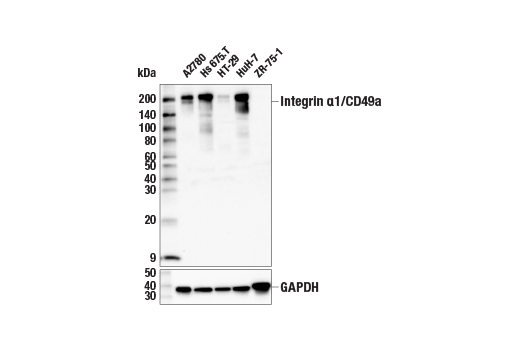
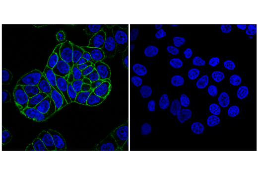
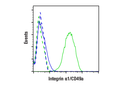
W, IF-IC, FC-FP, FC-L
Reactivity:
H
Sensitivity:
Endogenous
MW (kDa):
200
Source/Isotype:
Rabbit IgG
UniProt ID:
#P56199
Entrez-Gene Id:
3672
Product Usage Information
| Application | Dilution |
|---|---|
| Western Blotting | 1:1000 |
| Immunofluorescence (Immunocytochemistry) | 1:200 - 1:800 |
| Flow Cytometry (Fixed/Permeabilized) | 1:200 - 1:800 |
| Flow Cytometry (Live) | 1:100 - 1:400 |
Storage
Specificity/Sensitivity
Source / Purification
Background
Integrin α1 (CD49a, VLA-1, ITGA1) pairs with integrin β1 to form an α1β1 dimer on the cell surface. This integrin dimer plays important roles in colorectal cancer and pancreatic cancer cell proliferation, migration, and metastasis (4-6). The cytoplasmic tail of integrin α1 activates ERK and FAK/Src signaling to promote these biological processes (7,8). In immune systems, integrin α1 expression defines a tissue-resident cytotoxic T cell population contributing to both cancer immunity and vitiligo, an autoimmune disease (9,10).
Background References
- Liu, S. et al. (2000) J Cell Sci 113 (Pt 20), 3563-71.
- Hood, J.D. and Cheresh, D.A. (2002) Nat Rev Cancer 2, 91-100.
- Hogervorst, F. et al. (1993) J Cell Biol 121, 179-91.
- Gharibi, A. et al. (2017) Sci Rep 7, 10060.
- Boudjadi, S. et al. (2017) Cancers (Basel) 9, pii: E96. doi: 10.3390/cancers9080096.
- Shang, L. et al. (2017) Oncotarget 8, 76816-31.
- Abair, T.D. et al. (2008) Blood 112, 3242-54.
- Van Slambrouck, S. et al. (2007) Int J Oncol 31, 1501-8.
- Cheuk, S. et al. (2017) Immunity 46, 287-300.
- Molodtsov, A. and Turk, M.J. (2018) Front Immunol 9, 2810.
Species Reactivity
Species reactivity is determined by testing in at least one approved application (e.g., western blot).
Western Blot Buffer
IMPORTANT: For western blots, incubate membrane with diluted primary antibody in 5% w/v BSA, 1X TBS, 0.1% Tween® 20 at 4°C with gentle shaking, overnight.
Applications Key
W: Western Blotting IF-IC: Immunofluorescence (Immunocytochemistry) FC-FP: Flow Cytometry (Fixed/Permeabilized) FC-L: Flow Cytometry (Live)
Cross-Reactivity Key
H: Human
Trademarks and Patents
Cell Signaling Technology is a trademark of Cell Signaling Technology, Inc.
Alexa Fluor is a registered trademark of Life Technologies Corporation.
All other trademarks are the property of their respective owners. Visit cellsignal.com/trademarks for more information.
Limited Uses
Except as otherwise expressly agreed in a writing signed by a legally authorized representative of CST, the following terms apply to Products provided by CST, its affiliates or its distributors. Any Customer's terms and conditions that are in addition to, or different from, those contained herein, unless separately accepted in writing by a legally authorized representative of CST, are rejected and are of no force or effect.
Products are labeled with For Research Use Only or a similar labeling statement and have not been approved, cleared, or licensed by the FDA or other regulatory foreign or domestic entity, for any purpose. Customer shall not use any Product for any diagnostic or therapeutic purpose, or otherwise in any manner that conflicts with its labeling statement. Products sold or licensed by CST are provided for Customer as the end-user and solely for research and development uses. Any use of Product for diagnostic, prophylactic or therapeutic purposes, or any purchase of Product for resale (alone or as a component) or other commercial purpose, requires a separate license from CST. Customer shall (a) not sell, license, loan, donate or otherwise transfer or make available any Product to any third party, whether alone or in combination with other materials, or use the Products to manufacture any commercial products, (b) not copy, modify, reverse engineer, decompile, disassemble or otherwise attempt to discover the underlying structure or technology of the Products, or use the Products for the purpose of developing any products or services that would compete with CST products or services, (c) not alter or remove from the Products any trademarks, trade names, logos, patent or copyright notices or markings, (d) use the Products solely in accordance with CST Product Terms of Sale and any applicable documentation, and (e) comply with any license, terms of service or similar agreement with respect to any third party products or services used by Customer in connection with the Products.
Revision 4
Revision 4