Revision 4
#8478
Store at -20C
877-616-CELL (2355)
877-678-TECH (8324)
3 Trask Lane | Danvers | Massachusetts | 01923 | USA
For Research Use Only. Not for Use in Diagnostic Procedures.
Applications:
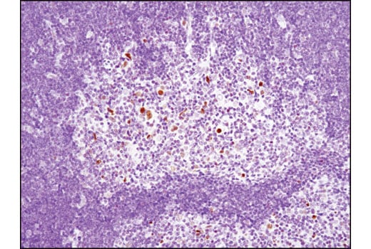
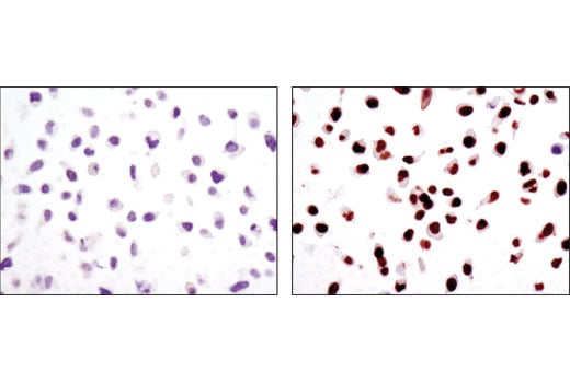
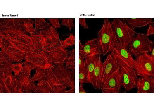
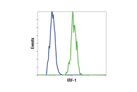
W, IP, IHC-Bond, IHC-P, IF-IC, FC-FP, C&R
Reactivity:
H M R
Sensitivity:
Endogenous
MW (kDa):
45-48
Source/Isotype:
Rabbit IgG
UniProt ID:
#P10914
Entrez-Gene Id:
3659
Product Usage Information
| Application | Dilution |
|---|---|
| Western Blotting | 1:1000 |
| Immunoprecipitation | 1:50 |
| IHC Leica Bond | 1:50 |
| Immunohistochemistry (Paraffin) | 1:100 |
| Immunofluorescence (Immunocytochemistry) | 1:200 |
| Flow Cytometry (Fixed/Permeabilized) | 1:50 |
| CUT&RUN | 1:50 |
Storage
Specificity/Sensitivity
Source / Purification
Background
The IRF-1 transcription factor was originally identified as a regulator of virus-inducible enhancer-like elements of the IFN-β gene (3). IRF-1 is widely expressed and upregulated by viral infection or stimulation with IFN or other cytokines. IRF-1 is serine-phosphorylated by casein kinase II (CKII) at two clustered sites, one in the DNA-binding domain (amino acids 138-150) and another in the transactivation domain (amino acids 219-231) (4). Mutation analysis of the latter site suggests that these phosphorylation sites help regulate IRF-1 activity. Tyrosine phosphorylation has also been shown to be important in IFN-γ-mediated differentiation of myeloid cell lines (5). C-terminal SUMOylated IRF-1 inhibits apoptosis in tumor cells by repression of its transcriptional activity (6).
Background References
- Taniguchi, T. et al. (2001) Annu Rev Immunol 19, 623-55.
- Honda, K. and Taniguchi, T. (2006) Nat Rev Immunol 6, 644-58.
- Fujita, T. et al. (1988) EMBO J 7, 3397-405.
- Lin, R. and Hiscott, J. (1999) Mol Cell Biochem 191, 169-80.
- Kautz, B. et al. (2001) J Biol Chem 276, 37868-78.
- Park, J. et al. (2007) Proc Natl Acad Sci USA 104, 17028-33.
Species Reactivity
Species reactivity is determined by testing in at least one approved application (e.g., western blot).
Western Blot Buffer
IMPORTANT: For western blots, incubate membrane with diluted primary antibody in 5% w/v BSA, 1X TBS, 0.1% Tween® 20 at 4°C with gentle shaking, overnight.
Applications Key
W: Western Blotting IP: Immunoprecipitation IHC-Bond: IHC Leica Bond IHC-P: Immunohistochemistry (Paraffin) IF-IC: Immunofluorescence (Immunocytochemistry) FC-FP: Flow Cytometry (Fixed/Permeabilized) C&R: CUT&RUN
Cross-Reactivity Key
H: Human M: Mouse R: Rat
Trademarks and Patents
Cell Signaling Technology is a trademark of Cell Signaling Technology, Inc.
All other trademarks are the property of their respective owners. Visit cellsignal.com/trademarks for more information.
Limited Uses
Except as otherwise expressly agreed in a writing signed by a legally authorized representative of CST, the following terms apply to Products provided by CST, its affiliates or its distributors. Any Customer's terms and conditions that are in addition to, or different from, those contained herein, unless separately accepted in writing by a legally authorized representative of CST, are rejected and are of no force or effect.
Products are labeled with For Research Use Only or a similar labeling statement and have not been approved, cleared, or licensed by the FDA or other regulatory foreign or domestic entity, for any purpose. Customer shall not use any Product for any diagnostic or therapeutic purpose, or otherwise in any manner that conflicts with its labeling statement. Products sold or licensed by CST are provided for Customer as the end-user and solely for research and development uses. Any use of Product for diagnostic, prophylactic or therapeutic purposes, or any purchase of Product for resale (alone or as a component) or other commercial purpose, requires a separate license from CST. Customer shall (a) not sell, license, loan, donate or otherwise transfer or make available any Product to any third party, whether alone or in combination with other materials, or use the Products to manufacture any commercial products, (b) not copy, modify, reverse engineer, decompile, disassemble or otherwise attempt to discover the underlying structure or technology of the Products, or use the Products for the purpose of developing any products or services that would compete with CST products or services, (c) not alter or remove from the Products any trademarks, trade names, logos, patent or copyright notices or markings, (d) use the Products solely in accordance with CST Product Terms of Sale and any applicable documentation, and (e) comply with any license, terms of service or similar agreement with respect to any third party products or services used by Customer in connection with the Products.
Revision 4
Revision 4
Revision 4
Revision 4